Ciri dan struktur
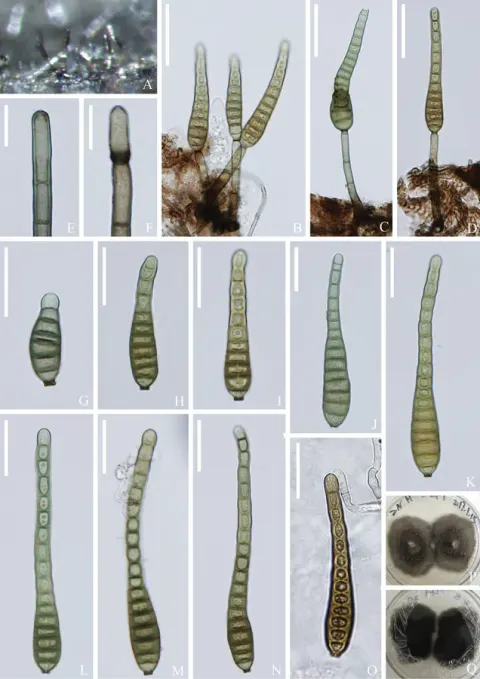
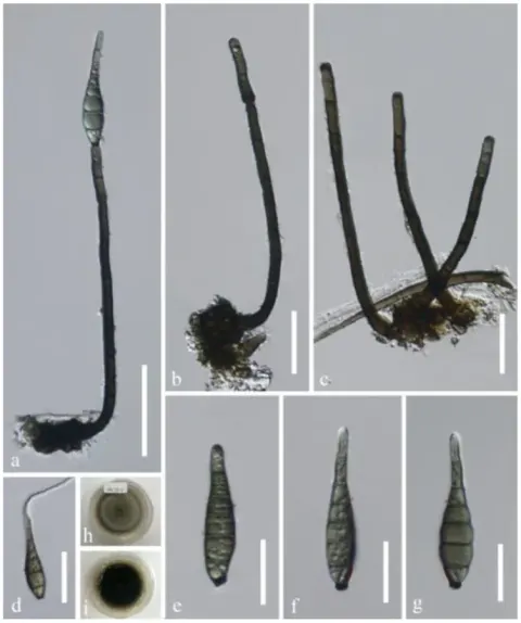
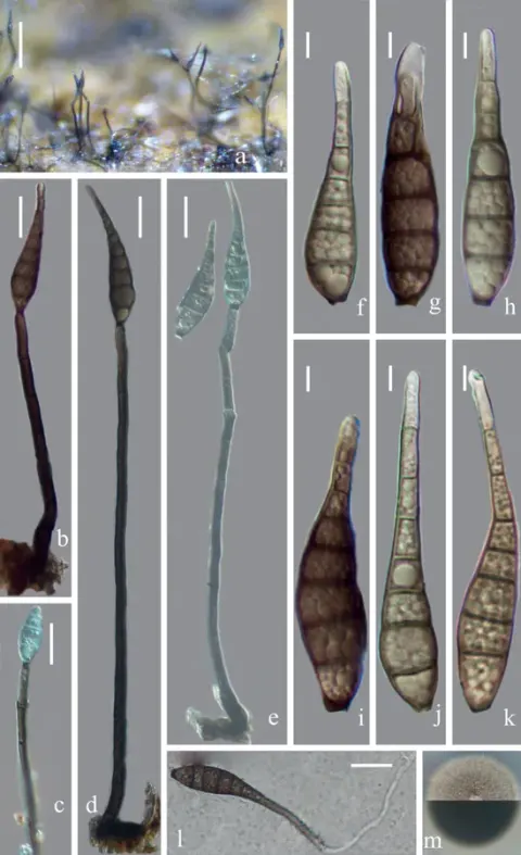
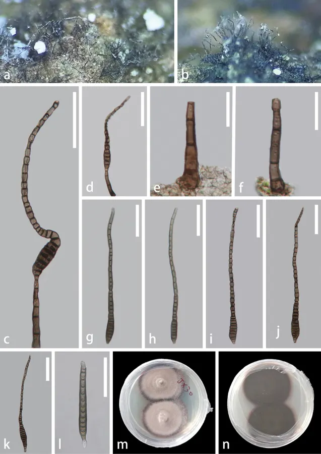
Saprobic on dead twigs of Edgeworthia chrysantha. Sexual morph: Undetermined. Asexual morph: Colonies on natural substrate abundant, superficial, dark brown, hairy. Conidiophores 35–80 μm × 4–7.5 μm (x– = 58 × 5.5 μm, n = 20), macronematous, mononematous, simple, erect to slightly curved, solitary, pale brown to dark brown, cylindrical, 2–4-septate, slightly constricted at the septa, unbranched, thick-walled. Conidiogenous cells 6–10 μm × 3.5–6.5 μm (x– = 8 × 4.5 μm, n = 20), holoblastic, monoblastic, integrated, terminal, cylindrical, slightly tapering towards the apex, brown to reddish brown, percurrent. Conidia 190–255 μm × 9.5–16 μm (x– = 220 μm × 13 μm, n = 20), 5–16 μm (x– = 13 μm, n = 20) wide at the protruding truncate base; 4.5–8 μm (x– = 6.5 μm, n = 20) wide in the tapering part, acrogenous, solitary, obclavate, elongate, rostrate, straight or curved, tapering towards the apex, 9–39-distoseptate, olivaceous-green when young, dark reddish brown at maturity, verrucose. Conidia germinating on PDA within 24 h, colonies circular, dense, umbonate, spreading, fluffy. The surface is slightly rough with reddish-gray mycelium, colonies somewhat raised in the middle, and with a filiform edge. The reverse side is dark gray with a circular, pale reddish-gray edge, not pigmented.
Habitat dan peran ekologis
Tectona grandis (Thailand, Hyde et al. 2016), on dead stems (Thailand, Sun et al. 2020), on dead, submerged, decaying wood of unidentified plants (China & Thailand, Luo et al. 2019; Dong et al. 2021; Zhang et al. 2022), and dead twig and branch of Edgeworthia chrysantha (China, this study).
- Habitat atau konteks temuan
- Informasi belum tersedia pada sumber rilis ini.
- Substrat
- Informasi belum tersedia pada sumber rilis ini.
- Peran atau konteks
- Belum dikategorikan pada rilis ini.
Reproduksi
Saprobic on dead twigs of Edgeworthia chrysantha. Sexual morph: Undetermined. Asexual morph: Colonies on natural substrate abundant, superficial, dark brown, hairy. Conidiophores 35–80 μm × 4–7.5 μm (x– = 58 × 5.5 μm, n = 20), macronematous, mononematous, simple, erect to slightly curved, solitary, pale brown to dark brown, cylindrical, 2–4-septate, slightly constricted at the septa, unbranched, thick-walled. Conidiogenous cells 6–10 μm × 3.5–6.5 μm (x– = 8 × 4.5 μm, n = 20), holoblastic, monoblastic, integrated, terminal, cylindrical, slightly tapering towards the apex, brown to reddish brown, percurrent. Conidia 190–255 μm × 9.5–16 μm (x– = 220 μm × 13 μm, n = 20), 5–16 μm (x– = 13 μm, n = 20) wide at the protruding truncate base; 4.5–8 μm (x– = 6.5 μm, n = 20) wide in the tapering part, acrogenous, solitary, obclavate, elongate, rostrate, straight or curved, tapering towards the apex, 9–39-distoseptate, olivaceous-green when young, dark reddish brown at maturity, verrucose.
Catatan identitas dan cakupan
- Nama diterima mengikuti Catalogue of Life Base2026-09-11; uraian mempertahankan konsep dan nama sumber aslinya.
- Preserve original treatment localities, hosts and substrate scope. Materials examined describe documented specimens, not the full global range.
- Impor dan terjemahan otomatis belum diperiksa editorial per profil. Struktur reproduktif yang disebutkan tidak membuktikan seluruh siklus hidup diketahui.
- Pengukuran pada uraian mengikuti spesimen, kultur, dan metode sumber; angka tidak diperluas ke semua populasi.
Catatan distribusi dalam narasi mengikuti sumbernya. Dokumentasi spesimen atau foto mengikuti catatan sumber. Bukti wilayah Indonesia dan penilaian konservasi tidak disimpulkan dari gambar.
Melihat dengan konteks
Distoseptispora tectonae (GZAAS 22-2046) a, b colonies on substrate c, d conidiophores and conidia e, f conidiophores g–k conidia l germinated conidium m, n colonies on PDA (m from front n from reverse) Scale bars: 50 μm (c, d, g–l); 20 μm (e, f).
- Jenis
- Pelat ilmiah
- Kredit
- Xue-Mei Chen, Xia Tang, Jian Ma, Ning-Guo Liu, Saowaluck Tibpromma, Samantha C. Karunarathna, Yuan-Pin Xiao, Yong-Zhong Lu
- Lisensi
- CC BY 4.0
- Sumber media
- Buka entri sumber
Dokumentasi tambahan
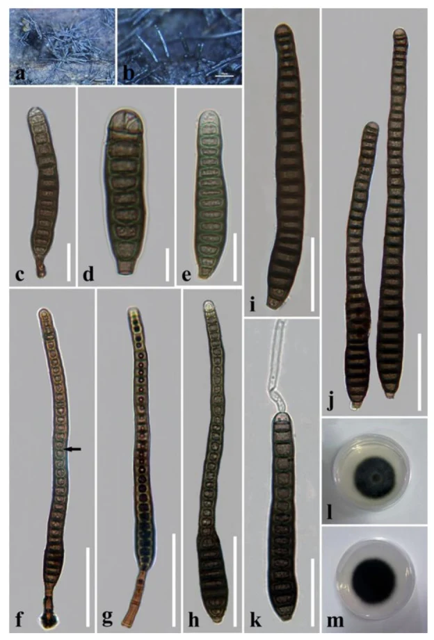
Distoseptispora tectonae. (a, b) Colonies on natural substrate. (c, f, g) Conidiophores with conidia. The arrow in f points to a gap in the middle of the septum, which indicates the distosepta. (d, e, h – j) Conidia. (k) Germinated conidium. (l) Colony on PDA (from front). (m) Colony on PDA (from reverse). Scale bars: (c, e) 20 μm; (d) 10 μm; (f – h) 50 μm; (i, k) 30 μm; (j) 40 μm.
Kredit dan penggunaan
© 2022 by the authors.
Ukuran dan format gambar disesuaikan tanpa pemotongan, retus, atau penambahan skala. Lisensi sumber dipertahankan.
Sumber profil
- [1] Identification of two new species and a new host record of Distoseptispora (Distoseptisporaceae, Distoseptisporales, Sordariomycetes) from terrestrial and freshwater habitats in Southern China — Distoseptispora tectonae
- [2] Catalogue of Life Base2026-09-11 — Distoseptispora tectonae
- [3] Dokumentasi visual — Distoseptispora tectonae
- [4] Dokumentasi visual — Distoseptispora tectonae
Rujukan halaman ini
Fungi. Distoseptispora tectonae. Rilis data fungi-2026-10-12-expansion-2. https://fungi-atlas.mgadib.chatgpt.site/jamur/distoseptispora-tectonae/.
Status: impor sumber; pemeriksaan editorial per profil belum selesai. Untuk klaim biologis, gunakan rujukan pada bagian terkait dan periksa konteks sumber aslinya.