Saprobic on puncak mati dari Thysanolaena maxima. Morph seksual tidak ditentukan. Transformasi aseksual Hyphomycetous. Koloni bermunculan, coklat sampai coklat gelap, berambut atau berambut, berasal dari subiculum, dengan tuft. Mycelium partly superficial, terdiri dari bercabang, bersepta, halus, cokelat hifa. konidiofor 30–80 × 3.5–5.5 μm (\(\bar{x} \) = 52 × 4.5 μm, n = 30), makronemat, mononemat, cokelat terang hingga gelap, 2–8-bersepta, halus, biasanya lentur atau kadang lurus, tidak bercabang, silinder, bulat di puncak.
Profil ini diimpor dari uraian spesies pada sumber berlisensi dan sebagian teks diterjemahkan otomatis. Pemeriksaan editorial per profil belum selesai. Bagian yang belum diterjemahkan atau belum lolos pemeriksaan terjemahan tetap ditampilkan dalam bahasa sumber. Angka dan nama ilmiah dipertahankan.
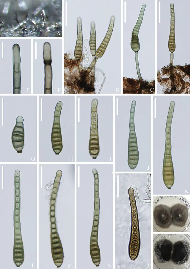
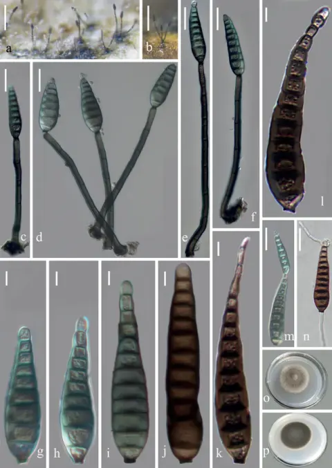
Distoseptispora thysanolaenae (KUN-HKAS 112710) A colonies on the substratum B-D conidiophores with conidia E, F conidiogenous cells G-N conidia O germinating conidium P, Q culture on PDA Scale bars: 30 μm (B-D); 10 μm (E, F); 20 μm (G-O). Hong-Wei Shen, Dan-Feng Bao, Kevin D. Hyde, Hong-Yan Su, Darbhe J. Bhat, Zong-Long Luo · CC BY 4.0 · Ukuran dan format gambar disesuaikan; lisensi asli dipertahankan.
01 / Morfologi
Ciri dan struktur
Saprobic on dead culms of Thysanolaena maxima. Sexual morph Undetermined. Asexual morph Hyphomycetous. Colonies effuse, brown to dark brown, hairy or fluffy, arising from subiculum, with tufts. Mycelium partly superficial, composed of branched, septate, smooth, brown hyphae. Conidiophores 30–80 × 3.5–5.5 μm (\(\bar{x} \) = 52 × 4.5 μm, n = 30), macronematous, mononematous, light to dark brown, 2–8-septate, smooth, usually flexuous or sometimes straight, unbranched, cylindrical, rounded at the apex. Conidiogenous cells monoblastic, integrated, terminal, determinate, hyaline to light brown, cylindrical. Conidia 21.5–80 × 6.5–12.8 μm (\(\bar{x} \) = 53 × 9.5 μm, n = 30), acrogenous, solitary, narrow and elongated obclavate, slightly curved, 8–14-distoseptate, thick-walled, light to dark brown, paler at the apex, tapering towards a rounded, flat apex, truncate with flat base, with conspicuous spore attachment loci, guttulate, smooth-walled. Culture characteristics: Colonies on PDA reaching 43–45 mm diam. after 4 weeks at 20–25 °C, colonies dense, circular, flat, surface slightly rough, rugose with edge entire, floccose to velvety; colony from above, greenish grey at the margin, brown-grey at the centre, from below dark grey at the margin, black at the centre; not producing pigmentation in PDA. Sporulation on PDA after two months. Conidiophores 10–30(–47) × 3–5 μm (\(\bar{x} \) = 21.4 × 4.6 μm, n = 25), macronematous, micronematous, mononematous, light to dark brown, 1–3-septate, smooth, usually flexuous or sometimes straight, unbranched, cylindrical, rounded at the apex. Conidiogenous cells monoblastic, integrated, terminal, determinate, hyaline to light brown, cylindrical.
Sebagian uraian ditampilkan dalam bahasa sumber; terjemahannya belum tersedia atau belum lolos pemeriksaan.
Pengukuran mengikuti objek, spesimen atau kultur, dan metode yang disebutkan sumber. Baca uraian asli untuk konteks lengkap.
02 / Kehidupan
Habitat dan peran ekologis
Saprobic on dead culms of Thysanolaena maxima. Sexual morph Undetermined. Asexual morph Hyphomycetous. Colonies effuse, brown to dark brown, hairy or fluffy, arising from subiculum, with tufts. Mycelium partly superficial, composed of branched, septate, smooth, brown hyphae. Conidiophores 30–80 × 3.5–5.5 μm (\(\bar{x} \) = 52 × 4.5 μm, n = 30), macronematous, mononematous, light to dark brown, 2–8-septate, smooth, usually flexuous or sometimes straight, unbranched, cylindrical, rounded at the apex. Conidiogenous cells monoblastic, integrated, terminal, determinate, hyaline to light brown, cylindrical. Conidia 21.5–80 × 6.5–12.8 μm (\(\bar{x} \) = 53 × 9.5 μm, n = 30), acrogenous, solitary, narrow and elongated obclavate, slightly curved, 8–14-distoseptate, thick-walled, light to dark brown, paler at the apex, tapering towards a rounded, flat apex, truncate with flat base, with conspicuous spore attachment loci, guttulate, smooth-walled. Culture characteristics: Colonies on PDA reaching 43–45 mm diam. after 4 weeks at 20–25 °C, colonies dense, circular, flat, surface slightly rough, rugose with edge entire, floccose to velvety; colony from above, greenish grey at the margin, brown-grey at the centre, from below dark grey at the margin, black at the centre; not producing pigmentation in PDA.
Sebagian uraian ditampilkan dalam bahasa sumber; terjemahannya belum tersedia atau belum lolos pemeriksaan.
Saprobic on dead culms of Thysanolaena maxima. Sexual morph Undetermined. Asexual morph Hyphomycetous. Colonies effuse, brown to dark brown, hairy or fluffy, arising from subiculum, with tufts. Mycelium partly superficial, composed of branched, septate, smooth, brown hyphae. Conidiophores 30–80 × 3.5–5.5 μm (\(\bar{x} \) = 52 × 4.5 μm, n = 30), macronematous, mononematous, light to dark brown, 2–8-septate, smooth, usually flexuous or sometimes straight, unbranched, cylindrical, rounded at the apex. Conidiogenous cells monoblastic, integrated, terminal, determinate, hyaline to light brown, cylindrical. Conidia 21.5–80 × 6.5–12.8 μm (\(\bar{x} \) = 53 × 9.5 μm, n = 30), acrogenous, solitary, narrow and elongated obclavate, slightly curved, 8–14-distoseptate, thick-walled, light to dark brown, paler at the apex, tapering towards a rounded, flat apex, truncate with flat base, with conspicuous spore attachment loci, guttulate, smooth-walled.
Sebagian uraian ditampilkan dalam bahasa sumber; terjemahannya belum tersedia atau belum lolos pemeriksaan.
Nama diterima mengikuti Catalogue of Life Base2026-09-11; uraian mempertahankan konsep dan nama sumber aslinya.
Preserve original monograph treatment localities, hosts and specimen/substrate scope. Materials examined document specimens, not the full global range.
Impor dan terjemahan otomatis belum diperiksa editorial per profil. Struktur reproduktif yang disebutkan tidak membuktikan seluruh siklus hidup diketahui.
Pengukuran pada uraian mengikuti spesimen, kultur, dan metode sumber; angka tidak diperluas ke semua populasi.
Catatan distribusi dalam narasi mengikuti sumbernya. Dokumentasi spesimen atau foto mengikuti catatan sumber. Bukti wilayah Indonesia dan penilaian konservasi tidak disimpulkan dari gambar.
05 / Dokumentasi
Melihat dengan konteks
Distoseptispora thysanolaenae (KUN-HKAS 112710) A colonies on the substratum B-D conidiophores with conidia E, F conidiogenous cells G-N conidia O germinating conidium P, Q culture on PDA Scale bars: 30 μm (B-D); 10 μm (E, F); 20 μm (G-O).
Jenis
Pelat ilmiah
Kredit
Hong-Wei Shen, Dan-Feng Bao, Kevin D. Hyde, Hong-Yan Su, Darbhe J. Bhat, Zong-Long Luo
Gambar menunjukkan objek menurut identifikasi sumber, bukan verifikasi independen spesimen oleh Fungi. Tidak ada skala baru yang ditambahkan.
Dokumentasi tambahan
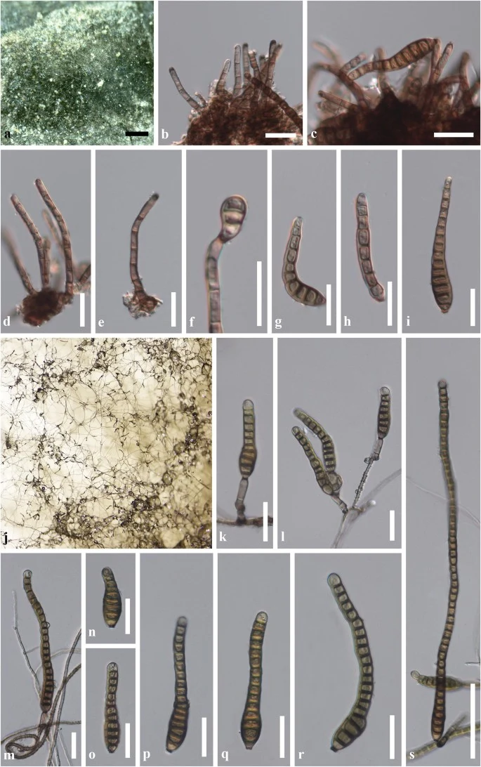
Distoseptispora thysanolaenae (KUN-HKAS 102247, holotype). On host substrate: a Appearance of colonies on host surface. b, d, e Conidiophores. c, f Conidiophores with attached conidia. g – i Variable shapes of conidia. In vitro: j Sporulation of conidia on PDA after 4 weeks. k – m Conidiophores with attached conidia. n – s Variable shapes of conidia. Scale bars a = 500 μm, b–i, k–r = 20 μm, s = 50 μm
[3] Dokumentasi visual — Distoseptispora thysanolaenaeBagian sumber: Identitas mengikuti sumber dokumentasi; bukan penentuan spesimen independen. Tanggal penghimpunan sumber: 2026-10-17 Media sumber: CC BY 4.0 · Hong-Wei Shen, Dan-Feng Bao, Kevin D. Hyde, Hong-Yan Su, Darbhe J. Bhat, Zong-Long Luo
[4] Dokumentasi visual — Distoseptispora thysanolaenaeBagian sumber: Identitas mengikuti sumber dokumentasi; bukan penentuan spesimen independen. Tanggal penghimpunan sumber: 2026-10-17 Teks sumber: CC BY 4.0 · Penulis dan sumber artikel: Phookamsak, Rungtiwa; Hyde, Kevin D.; Jeewon, Rajesh; Bhat, D. Jayarama; Jones, E. B. Gareth; Maharachchikumbura, Sajeewa S. N.; Raspé, Olivier; Karunarathna, Samantha C.; Wanasinghe, Dhanushka N.; Hongsanan, Sinang; Doilom, Mingkwan; Tennakoon, Danushka S.; Machado, Alexandre R.; Firmino, André L.; Ghosh, Aniket; Karunarathna, Anuruddha; Mešić, Armin; Dutta, Arun Kumar; Thongbai, Benjarong; Devadatha, Bandarupalli; Norphanphoun, Chada; Senwanna, Chanokned; Wei, Deping; Pem, Dhandevi; Ackah, Frank Kwekucher; Wang, Gen-Nuo; Jiang, Hong-Bo; Madrid, Hugo; Lee, Hyang Burm; Goonasekara, Ishani D.; Manawasinghe, Ishara S.; Kušan, Ivana; Cano, Josep; Gené, Josepa; Li, Junfu; Das, Kanad; Acharya, Krishnendu; Raj, K. N. Anil; Latha, K. P. Deepna; Chethana, K. W. Thilini; He, Mao-Qiang; Dueñas, Margarita; Jadan, Margita; Martín, María P.; Samarakoon, Milan C.; Dayarathne, Monika C.; Raza, Mubashar; Park, Myung Soo; Telleria, M. Teresa; Chaiwan, Napalai; Matočec, Neven; de Silva, Nimali I.; Pereira, Olinto L.; Singh, Paras Nath; Manimohan, Patinjareveettil; Uniyal, Priyanka; Shang, Qiu-Ju; Bhatt, Rajendra P.; Perera, Rekhani H.; Alvarenga, Renato Lúcio Mendes; Nogal-Prata, Sandra; Singh, Sanjay K.; Vadthanarat, Santhiti; Oh, Seung-Yoon; Huang, Shi-Ke; Rana, Shiwali; Konta, Sirinapa; Paloi, Soumitra; Jayasiri, Subashini C.; Jeon, Sun Jeong; Mehmood, Tahir; Gibertoni, Tatiana Baptista; Nguyen, Thuong T. T.; Singh, Upendra; Thiyagaraja, Vinodhini; Sarma, V. Venkateswara; Dong, Wei; Yu, Xian-Dong; Lu, Yong-Zhong; Lim, Young Woon; Chen, Yun; Tkalčec, Zdenko; Zhang, Zhi-Feng; Luo, Zong-Long; Daranagama, Dinushani A.; Thambugala, Kasun M.; Tibpromma, Saowaluck; Camporesi, Erio; Bulgakov, Timur S.; Dissanayake, Asha J.; Senanayake, Indunil C.; Dai, Dong Qin; Tang, Li-Zhou; Khan, Sehroon; Zhang, Huang; Promputtha, Itthayakorn; Cai, Lei; Chomnunti, Putarak; Zhao, Rui-Lin; Lumyong, Saisamorn; Boonmee, Saranyaphat; Wen, Ting-Chi; Mortimer, Peter E.; Xu, Jianchu
Adaptasi teks: CC BY 4.0. Phookamsak, Rungtiwa; Hyde, Kevin D.; Jeewon, Rajesh; Bhat, D. Jayarama; Jones, E. B. Gareth; Maharachchikumbura, Sajeewa S. N.; Raspé, Olivier; Karunarathna, Samantha C.; Wanasinghe, Dhanushka N.; Hongsanan, Sinang; Doilom, Mingkwan; Tennakoon, Danushka S.; Machado, Alexandre R.; Firmino, André L.; Ghosh, Aniket; Karunarathna, Anuruddha; Mešić, Armin; Dutta, Arun Kumar; Thongbai, Benjarong; Devadatha, Bandarupalli; Norphanphoun, Chada; Senwanna, Chanokned; Wei, Deping; Pem, Dhandevi; Ackah, Frank Kwekucher; Wang, Gen-Nuo; Jiang, Hong-Bo; Madrid, Hugo; Lee, Hyang Burm; Goonasekara, Ishani D.; Manawasinghe, Ishara S.; Kušan, Ivana; Cano, Josep; Gené, Josepa; Li, Junfu; Das, Kanad; Acharya, Krishnendu; Raj, K. N. Anil; Latha, K. P. Deepna; Chethana, K. W. Thilini; He, Mao-Qiang; Dueñas, Margarita; Jadan, Margita; Martín, María P.; Samarakoon, Milan C.; Dayarathne, Monika C.; Raza, Mubashar; Park, Myung Soo; Telleria, M. Teresa; Chaiwan, Napalai; Matočec, Neven; de Silva, Nimali I.; Pereira, Olinto L.; Singh, Paras Nath; Manimohan, Patinjareveettil; Uniyal, Priyanka; Shang, Qiu-Ju; Bhatt, Rajendra P.; Perera, Rekhani H.; Alvarenga, Renato Lúcio Mendes; Nogal-Prata, Sandra; Singh, Sanjay K.; Vadthanarat, Santhiti; Oh, Seung-Yoon; Huang, Shi-Ke; Rana, Shiwali; Konta, Sirinapa; Paloi, Soumitra; Jayasiri, Subashini C.; Jeon, Sun Jeong; Mehmood, Tahir; Gibertoni, Tatiana Baptista; Nguyen, Thuong T. T.; Singh, Upendra; Thiyagaraja, Vinodhini; Sarma, V. Venkateswara; Dong, Wei; Yu, Xian-Dong; Lu, Yong-Zhong; Lim, Young Woon; Chen, Yun; Tkalčec, Zdenko; Zhang, Zhi-Feng; Luo, Zong-Long; Daranagama, Dinushani A.; Thambugala, Kasun M.; Tibpromma, Saowaluck; Camporesi, Erio; Bulgakov, Timur S.; Dissanayake, Asha J.; Senanayake, Indunil C.; Dai, Dong Qin; Tang, Li-Zhou; Khan, Sehroon; Zhang, Huang; Promputtha, Itthayakorn; Cai, Lei; Chomnunti, Putarak; Zhao, Rui-Lin; Lumyong, Saisamorn; Boonmee, Saranyaphat; Wen, Ting-Chi; Mortimer, Peter E.; Xu, Jianchu — Fungal diversity notes 929–1035: taxonomic and phylogenetic contributions on genera and species of fungi — Distoseptispora thysanolaenae
Saprobic on dead culms of Thysanolaena maxima.
Sexual morph Undetermined.
Asexual morph Hyphomycetous.
Colonies effuse, brown to dark brown, hairy or fluffy, arising from subiculum, with tufts.
Mycelium partly superficial, composed of branched, septate, smooth, brown hyphae.
Conidiophores 30–80 × 3.5–5.5 μm (\(\bar{x} \) = 52 × 4.5 μm, n = 30), macronematous, mononematous, light to dark brown, 2–8-septate, smooth, usually flexuous or sometimes straight, unbranched, cylindrical, rounded at the apex.
Saprobic on dead culms of Thysanolaena maxima.
Sexual morph Undetermined.
Asexual morph Hyphomycetous.
Colonies effuse, brown to dark brown, hairy or fluffy, arising from subiculum, with tufts.
Mycelium partly superficial, composed of branched, septate, smooth, brown hyphae.
Conidiophores 30–80 × 3.5–5.5 μm (\(\bar{x} \) = 52 × 4.5 μm, n = 30), macronematous, mononematous, light to dark brown, 2–8-septate, smooth, usually flexuous or sometimes straight, unbranched, cylindrical, rounded at the apex.
Conidiogenous cells monoblastic, integrated, terminal, determinate, hyaline to light brown, cylindrical.
Conidia 21.5–80 × 6.5–12.8 μm (\(\bar{x} \) = 53 × 9.5 μm, n = 30), acrogenous, solitary, narrow and elongated obclavate, slightly curved, 8–14-distoseptate, thick-walled, light to dark brown, paler at the apex, tapering towards a rounded, flat apex, truncate with flat base, with conspicuous spore attachment loci, guttulate, smooth-walled.
Culture characteristics: Colonies on PDA reaching 43–45 mm diam. after 4 weeks at 20–25 °C, colonies dense, circular, flat, surface slightly rough, rugose with edge entire, floccose to velvety; colony from above, greenish grey at the margin, brown-grey at the centre, from below dark grey at the margin, black at the centre; not producing pigmentation in PDA.
Sporulation on PDA after two months.
Conidiophores 10–30(–47) × 3–5 μm (\(\bar{x} \) = 21.4 × 4.6 μm, n = 25), macronematous, micronematous, mononematous, light to dark brown, 1–3-septate, smooth, usually flexuous or sometimes straight, unbranched, cylindrical, rounded at the apex.
Conidiogenous cells monoblastic, integrated, terminal, determinate, hyaline to light brown, cylindrical.
Saprobic on dead culms of Thysanolaena maxima.
Sexual morph Undetermined.
Asexual morph Hyphomycetous.
Colonies effuse, brown to dark brown, hairy or fluffy, arising from subiculum, with tufts.
Mycelium partly superficial, composed of branched, septate, smooth, brown hyphae.
Conidiophores 30–80 × 3.5–5.5 μm (\(\bar{x} \) = 52 × 4.5 μm, n = 30), macronematous, mononematous, light to dark brown, 2–8-septate, smooth, usually flexuous or sometimes straight, unbranched, cylindrical, rounded at the apex.
Conidiogenous cells monoblastic, integrated, terminal, determinate, hyaline to light brown, cylindrical.
Conidia 21.5–80 × 6.5–12.8 μm (\(\bar{x} \) = 53 × 9.5 μm, n = 30), acrogenous, solitary, narrow and elongated obclavate, slightly curved, 8–14-distoseptate, thick-walled, light to dark brown, paler at the apex, tapering towards a rounded, flat apex, truncate with flat base, with conspicuous spore attachment loci, guttulate, smooth-walled.
Culture characteristics: Colonies on PDA reaching 43–45 mm diam. after 4 weeks at 20–25 °C, colonies dense, circular, flat, surface slightly rough, rugose with edge entire, floccose to velvety; colony from above, greenish grey at the margin, brown-grey at the centre, from below dark grey at the margin, black at the centre; not producing pigmentation in PDA.
Saprobic on dead culms of Thysanolaena maxima.
Sexual morph Undetermined.
Asexual morph Hyphomycetous.
Colonies effuse, brown to dark brown, hairy or fluffy, arising from subiculum, with tufts.
Mycelium partly superficial, composed of branched, septate, smooth, brown hyphae.
Conidiophores 30–80 × 3.5–5.5 μm (\(\bar{x} \) = 52 × 4.5 μm, n = 30), macronematous, mononematous, light to dark brown, 2–8-septate, smooth, usually flexuous or sometimes straight, unbranched, cylindrical, rounded at the apex.
Conidiogenous cells monoblastic, integrated, terminal, determinate, hyaline to light brown, cylindrical.
Conidia 21.5–80 × 6.5–12.8 μm (\(\bar{x} \) = 53 × 9.5 μm, n = 30), acrogenous, solitary, narrow and elongated obclavate, slightly curved, 8–14-distoseptate, thick-walled, light to dark brown, paler at the apex, tapering towards a rounded, flat apex, truncate with flat base, with conspicuous spore attachment loci, guttulate, smooth-walled.
Status: impor sumber; pemeriksaan editorial per profil belum selesai. Untuk klaim biologis, gunakan rujukan pada bagian terkait dan periksa konteks sumber aslinya.
Catatan: Dalam analisis filogenetik, Distoseptispora verrucosa dikelompokkan dengan D. lancangjiangensis, D. suoluoensis, dan D. bambusae. Penempatan Their systematic berkorelasi dengan karakter morfologi yang sangat mirip dengan memiliki…
Saprobic on puncak bambu yang membusuk tenggelam di habitat air tawar. Morph seksual: tidak ditentukan. Transformasi aseksual: Hipomiketous. Koloni bermunculan, berwarna zaitun atau coklat gelap, berambut, berambut. Mycelium mostly…