01 / Morfologi

Ciri dan struktur

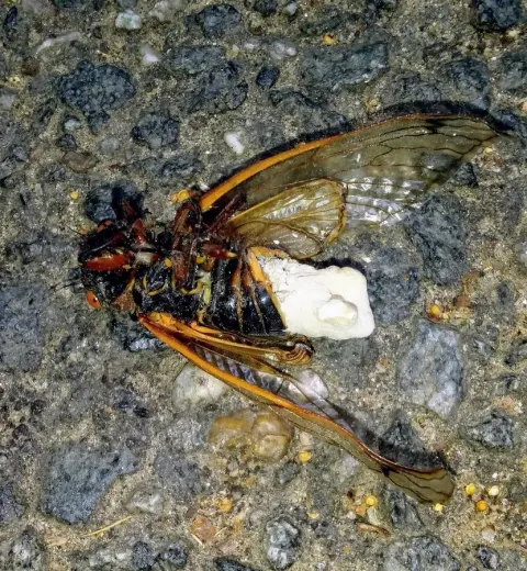
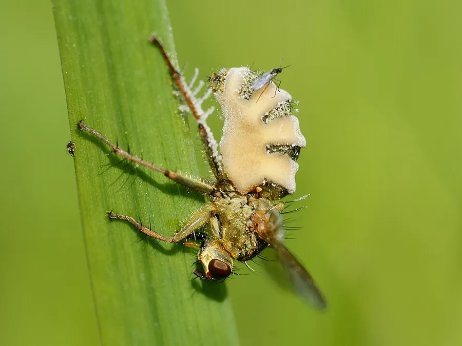
Soon after a fly dies from infection with this pathogenic fungus, large primary conidia are produced at the apex of a conidiophore which emerge from the intersegmental membranes. When the spores are mature they are forcibly ejected and may fall onto flies resting nearby. If no hosts are available for infection, a smaller secondary conidium may develop. Once on a fly, the conidia germinate within a few hours and a germ tube begins to penetrate the insect's cuticle. Once this reaches the haemocoel, the protoplast flows through the tube and into the fly's haemolymph. The mycelium of the fungus may grow into an area of the brain that controls the behaviour of the fly, forcing it to land on a surface and crawl upwards. The hyphae gradually grow through the whole of the body, digesting the guts, and the fly dies in about five to seven days. When it is critically ill, it tends to crawl to a high point, straighten its hind legs and open its wings, a behaviour that ensures that the fungal spores are dispersed as widely as possible. Some three hours later, conidiophores start to develop and a new shower of conidia is initiated. There are technical difficulties in controlling flies with Entomophthora muscae. The fungus is sensitive to temperature and when the air temperature is high, the prevalence of the disease decreases to very low levels. Houseflies infected with E. muscae were able to rid themselves of the infection by resting at temperatures that inhibited the growth of the fungus. Storage of the organism in a viable state is difficult and it cannot be easily cultured in the laboratory.

Sebagian uraian ditampilkan dalam bahasa sumber; terjemahannya belum tersedia atau belum lolos pemeriksaan.

Rujukan: [1]

Pengukuran mengikuti objek, spesimen atau kultur, dan metode yang disebutkan sumber. Baca uraian asli untuk konteks lengkap.

02 / Kehidupan

Habitat dan peran ekologis

Selain lalat rumah Musca domestica, infeksi telah diamati pada lalat dewasa dalam keluarga Calliphoridae, Culicidae, Drosophilidae, Muscidae, Sarcophagidae, Scathophagidae, Syrphidae and Tachinidae. Infeksi Outbreaks of dengan Entomophthora muscae cenderung terjadi pada musim semi dan musim gugur. Jamur ini ditemukan di sebagian besar wilayah yang sedang dan sporulasi biasanya terjadi dalam kondisi dingin dan lembab di daerah di mana lalat berkumpul dan beristirahat. Di rumah, mayat lalat sering terlihat melekat pada jendela atau bingkai jendela, tempat di mana lalat lain dapat dengan mudah terinfeksi. Di luar ruangan, mereka dapat terlihat melekat di bawah daun, di pagar dan dinding, di bangunan pertanian dan rumah ayam.

Rujukan: [1]

Habitat atau konteks temuan
Informasi belum tersedia pada sumber rilis ini.
Substrat
Informasi belum tersedia pada sumber rilis ini.
Peran atau konteks
Belum dikategorikan pada rilis ini.
Temukan hubungan serupa
03 / Siklus hidup

Reproduksi

Soon after a fly dies from infection with this pathogenic fungus, large primary conidia are produced at the apex of a conidiophore which emerge from the intersegmental membranes. When the spores are mature they are forcibly ejected and may fall onto flies resting nearby. If no hosts are available for infection, a smaller secondary conidium may develop. Once on a fly, the conidia germinate within a few hours and a germ tube begins to penetrate the insect's cuticle. Once this reaches the haemocoel, the protoplast flows through the tube and into the fly's haemolymph. The mycelium of the fungus may grow into an area of the brain that controls the behaviour of the fly, forcing it to land on a surface and crawl upwards.

Sebagian uraian ditampilkan dalam bahasa sumber; terjemahannya belum tersedia atau belum lolos pemeriksaan.

Rujukan: [1]

Sinonim menurut acuan taksonomi rilis: Empusa muscae; Myiophyton cohnii; Sporendonema muscae
04 / Batas informasi

Catatan identitas dan cakupan

  • Nama diterima mengikuti Catalogue of Life Base2026-09-11; uraian mempertahankan konsep dan nama sumber aslinya.
  • Original geographic, specimen and life-cycle scope retained; source import awaits individual editorial review.
  • Impor dan terjemahan otomatis belum diperiksa editorial per profil. Struktur reproduktif yang disebutkan tidak membuktikan seluruh siklus hidup diketahui.
  • Pengukuran pada uraian mengikuti spesimen, kultur, dan metode sumber; angka tidak diperluas ke semua populasi.

Catatan distribusi dalam narasi mengikuti sumbernya. Dokumentasi spesimen atau foto mengikuti catatan sumber. Bukti wilayah Indonesia dan penilaian konservasi tidak disimpulkan dari gambar.

05 / Dokumentasi

Melihat dengan konteks

Dokumentasi Entomophthora muscae dari Wikimedia Commons. Fly fungus on Common yellow dung fly at Oostende, Belgium Identifikasi mengikuti record berkas sumber.

Jenis
Fotografi
Kredit
Hans Hillewaert — © Hans Hillewaert
Sumber media
Buka entri sumber

Gambar menunjukkan objek menurut identifikasi sumber, bukan verifikasi independen spesimen oleh Fungi. Tidak ada skala baru yang ditambahkan.

06 / Rujukan

Sumber profil

  1. [1] Entomophthora muscae — Wikipedia (en)Bagian sumber: Frozen revision 1306718743
    Tanggal penghimpunan sumber: 2026-10-12
    Teks sumber: CC BY-SA4.0 · Wikipedia contributors
  2. [2] Catalogue of Life Base2026-09-11 — Entomophthora muscaeBagian sumber: ColDP NameUsage.tsv ID 6FG2S; accepted species; source 2073
    Tanggal penghimpunan sumber: 2026-10-12
  3. [3] Dokumentasi visual — Entomophthora muscaeBagian sumber: Individual media record; identification follows the source, not independent determination
    Tanggal penghimpunan sumber: 2026-10-12
    Media sumber: CC BY-SA 4.0 · Hans Hillewaert — © Hans Hillewaert

Adaptasi teks: CC BY-SA 4.0. Wikipedia contributors — Entomophthora muscae — Wikipedia (en)

Uraian asli dan riwayat sumber

Riwayat atau halaman sumber

Ringkasan sumber

Soon after a fly dies from infection with this pathogenic fungus, large primary conidia are produced at the apex of a conidiophore which emerge from the intersegmental membranes. When the spores are mature they are forcibly ejected and may fall onto flies resting nearby. If no hosts are available for infection, a smaller secondary conidium may develop. Once on a fly, the conidia germinate within a few hours and a germ tube begins to penetrate the insect's cuticle. Once this reaches the haemocoel, the protoplast flows through the tube and into the fly's haemolymph.

Revisi sumber

Morfologi sumber

Soon after a fly dies from infection with this pathogenic fungus, large primary conidia are produced at the apex of a conidiophore which emerge from the intersegmental membranes. When the spores are mature they are forcibly ejected and may fall onto flies resting nearby. If no hosts are available for infection, a smaller secondary conidium may develop. Once on a fly, the conidia germinate within a few hours and a germ tube begins to penetrate the insect's cuticle. Once this reaches the haemocoel, the protoplast flows through the tube and into the fly's haemolymph. The mycelium of the fungus may grow into an area of the brain that controls the behaviour of the fly, forcing it to land on a surface and crawl upwards. The hyphae gradually grow through the whole of the body, digesting the guts, and the fly dies in about five to seven days. When it is critically ill, it tends to crawl to a high point, straighten its hind legs and open its wings, a behaviour that ensures that the fungal spores are dispersed as widely as possible. Some three hours later, conidiophores start to develop and a new shower of conidia is initiated. There are technical difficulties in controlling flies with Entomophthora muscae. The fungus is sensitive to temperature and when the air temperature is high, the prevalence of the disease decreases to very low levels. Houseflies infected with E. muscae were able to rid themselves of the infection by resting at temperatures that inhibited the growth of the fungus. Storage of the organism in a viable state is difficult and it cannot be easily cultured in the laboratory.

Revisi sumber

Ekologi sumber

As well as the housefly Musca domestica, infection has been observed in adult flies in the families Calliphoridae, Culicidae, Drosophilidae, Muscidae, Sarcophagidae, Scathophagidae, Syrphidae and Tachinidae. Outbreaks of infection with Entomophthora muscae tend to occur in the spring and autumn. The fungus is found in most temperate regions and sporulation usually takes place in cool, humid conditions in areas where flies congregate and rest. In houses, the corpses of flies are frequently seen attached to windows or window frames, a place where other flies may easily become infected. In the open, they may be seen attached to the underneath of leaves, on fences and walls, in agricultural buildings and poultry houses.

Revisi sumber

Struktur reproduksi sumber

Soon after a fly dies from infection with this pathogenic fungus, large primary conidia are produced at the apex of a conidiophore which emerge from the intersegmental membranes. When the spores are mature they are forcibly ejected and may fall onto flies resting nearby. If no hosts are available for infection, a smaller secondary conidium may develop. Once on a fly, the conidia germinate within a few hours and a germ tube begins to penetrate the insect's cuticle. Once this reaches the haemocoel, the protoplast flows through the tube and into the fly's haemolymph. The mycelium of the fungus may grow into an area of the brain that controls the behaviour of the fly, forcing it to land on a surface and crawl upwards.

Revisi sumber

Rujukan halaman ini

Fungi. Entomophthora muscae. Rilis data fungi-2026-10-12-expansion-2. https://fungi-atlas.mgadib.chatgpt.site/jamur/entomophthora-muscae/.

Status: impor sumber; pemeriksaan editorial per profil belum selesai. Untuk klaim biologis, gunakan rujukan pada bagian terkait dan periksa konteks sumber aslinya.