01 / Morfologi

Ciri dan struktur

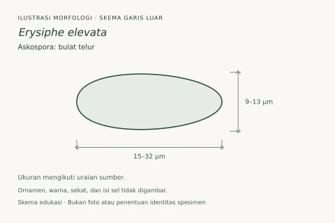
Erysiphe cruciferarum menunjukkan karakteristik bubuk lendir khas, muncul sebagai koloni kecil berpencar, difus dari mycelium putih permukaan di permukaan daun; biasanya kedua sisi daun menunjukkan pertumbuhan jamur putih, bubuk. Similarly to sebagian besar Erysiphaceae species, E. cruciferarum sangat spesifik dengan rentang host yang terdiri dari hanya tiga genus. Akibatnya, spesies ini tidak mungkin menyebabkan masalah pada tanaman atau tanaman kebun atau rumah kaca. Hal ini berbeda dengan Erysiphe radulescui, yang menginfeksi banyak genus di Brassicaceae including tanaman dan tanaman liar, termasuk beberapa yang merupakan rumput biasa yang dapat diusahakan dari mana ia berpotensi melompat ke individu yang dibudidayakan. Mycelium putih atau abu-abu, biasanya ditemukan di kedua sisi daun, dan secara teratur di batang atau bahkan kapsul. Appressoria hifa berbentuk payudara hingga lobis sedang, dan terjadi secara solitaris. Conidophores arise dari permukaan atas sel induk mereka. They are berdiri dengan sel kaki silinder yang lurus atau terkadang melengkung. konidiofor menghasilkan tunggal konidia. Primal konidia adalah obovoid. Secondary conidia berbentuk tong. Cachmothecia (badan buah) memiliki banyak lampiran myceloid terutama di bagian bawah. They may become pigmented at the base. Peridium chasmothecia memiliki sel bentuk tidak teratur. Erysiphe cruciferarum memiliki tiga sampai tujuh spora per askus yang kira-kira elipsoid dan tidak berwarna. askus adalah sakkat dan hampir sessile atau pendek.

Sebagian uraian ditampilkan dalam bahasa sumber; terjemahannya belum tersedia atau belum lolos pemeriksaan.

Rujukan: [1]

Pengukuran mengikuti objek, spesimen atau kultur, dan metode yang disebutkan sumber. Baca uraian asli untuk konteks lengkap.

02 / Kehidupan

Habitat dan peran ekologis

Mycelium putih atau abu-abu, biasanya ditemukan di kedua sisi daun, dan secara teratur di batang atau bahkan kapsul. Appressoria hifa berbentuk payudara hingga lobis sedang, dan terjadi secara solitaris. Conidophores arise dari permukaan atas sel induk mereka. They are berdiri dengan sel kaki silinder yang lurus atau terkadang melengkung. konidiofor menghasilkan tunggal konidia. Primal konidia adalah obovoid. Secondary conidia berbentuk tong. Cachmothecia (badan buah) memiliki banyak lampiran myceloid terutama di bagian bawah. They may become pigmented at the base. Peridium chasmothecia memiliki sel bentuk tidak teratur. Erysiphe cruciferarum memiliki tiga sampai tujuh spora per askus yang kira-kira elipsoid dan tidak berwarna. askus adalah sakkat dan hampir sessile atau pendek.

Sebagian uraian ditampilkan dalam bahasa sumber; terjemahannya belum tersedia atau belum lolos pemeriksaan.

Rujukan: [1]

Habitat atau konteks temuan
Informasi belum tersedia pada sumber rilis ini.
Substrat
Informasi belum tersedia pada sumber rilis ini.
Peran atau konteks
Belum dikategorikan pada rilis ini.
Temukan hubungan serupa
03 / Siklus hidup

Reproduksi

Mycelium putih atau abu-abu, biasanya ditemukan di kedua sisi daun, dan secara teratur di batang atau bahkan kapsul. Appressoria hifa berbentuk payudara hingga lobis sedang, dan terjadi secara solitaris. Conidophores arise dari permukaan atas sel induk mereka. They are berdiri dengan sel kaki silinder yang lurus atau terkadang melengkung. konidiofor menghasilkan tunggal konidia. Primal konidia adalah obovoid. Secondary conidia berbentuk tong. Cachmothecia (badan buah) memiliki banyak lampiran myceloid terutama di bagian bawah. They may become pigmented at the base. Peridium chasmothecia memiliki sel bentuk tidak teratur. Erysiphe cruciferarum memiliki tiga sampai tujuh spora per askus yang kira-kira elipsoid dan tidak berwarna.

Sebagian uraian ditampilkan dalam bahasa sumber; terjemahannya belum tersedia atau belum lolos pemeriksaan.

Rujukan: [1]

Sinonim menurut acuan taksonomi rilis: Albigo communis; Alphitomorpha communis; Erysiphe cruciferarum var. longispora; Erysiphe pisi var. cruciferarum; Erysiphe radulescui; Tigria leveillei
04 / Batas informasi

Catatan identitas dan cakupan

  • Nama diterima mengikuti Catalogue of Life Base2026-09-11; uraian mempertahankan konsep dan nama sumber aslinya.
  • Original geographic, specimen and life-cycle scope retained; source import awaits individual editorial review.
  • Impor dan terjemahan otomatis belum diperiksa editorial per profil. Struktur reproduktif yang disebutkan tidak membuktikan seluruh siklus hidup diketahui.
  • Pengukuran pada uraian mengikuti spesimen, kultur, dan metode sumber; angka tidak diperluas ke semua populasi.

Catatan distribusi dalam narasi mengikuti sumbernya. Dokumentasi spesimen atau foto mengikuti catatan sumber. Bukti wilayah Indonesia dan penilaian konservasi tidak disimpulkan dari gambar.

05 / Dokumentasi

Melihat dengan konteks

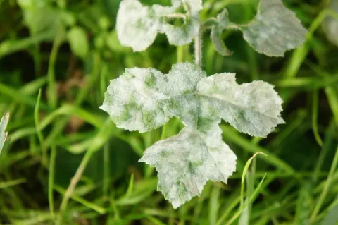
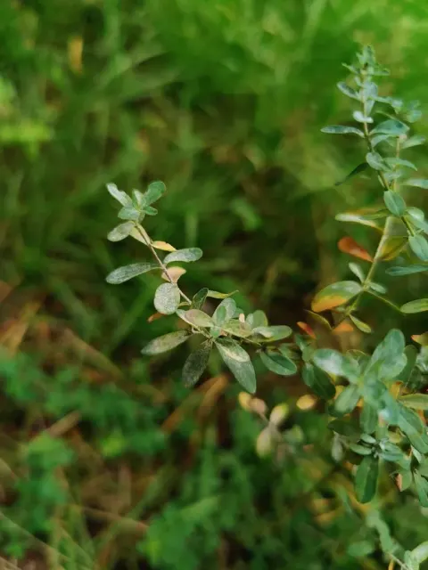
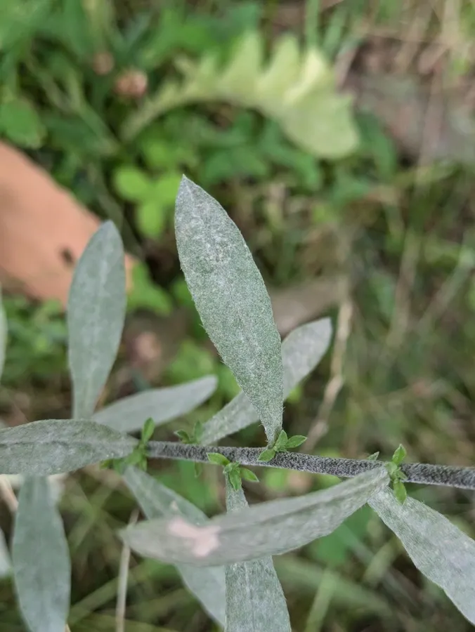
Dokumentasi Erysiphe cruciferarum menurut asosiasi foto pada record takson iNaturalist. Kredit: Alexis Tinker-Tsavalas. Identitas mengikuti sumber; bukan penentuan spesimen independen.

Jenis
Fotografi
Kredit
(c) Alexis Tinker-Tsavalas, some rights reserved (CC BY), uploaded by Alexis Tinker-Tsavalas
Lisensi
CC BY 4.0
Sumber media
Buka entri sumber

Gambar menunjukkan objek menurut identifikasi sumber, bukan verifikasi independen spesimen oleh Fungi. Tidak ada skala baru yang ditambahkan.

06 / Rujukan

Sumber profil

  1. [1] Erysiphe cruciferarum — Wikipedia (en)Bagian sumber: Frozen revision 1370556518
    Tanggal penghimpunan sumber: 2026-10-12
    Teks sumber: CC BY-SA4.0 · Wikipedia contributors
  2. [2] Catalogue of Life Base2026-09-11 — Erysiphe cruciferarumBagian sumber: ColDP NameUsage.tsv ID 3BBZ7; accepted species; source 2073
    Tanggal penghimpunan sumber: 2026-10-12
  3. [3] Dokumentasi visual — Erysiphe cruciferarumBagian sumber: Individual media record; identification follows the source, not independent determination
    Tanggal penghimpunan sumber: 2026-10-12
    Media sumber: CC BY 4.0 · (c) Alexis Tinker-Tsavalas, some rights reserved (CC BY), uploaded by Alexis Tinker-Tsavalas

Adaptasi teks: CC BY-SA 4.0. Wikipedia contributors — Erysiphe cruciferarum — Wikipedia (en)

Uraian asli dan riwayat sumber

Riwayat atau halaman sumber

Ringkasan sumber

Erysiphe cruciferarum exhibits typical powdery mildew characteristics, appearing as small radiating, diffuse colonies of superficial white mycelium on the surface of the leaf; usually both sides of the leaf show white, powdery fungal growth. Similarly to most Erysiphaceae species, E. cruciferarum is highly host-specific with a host range comprising just three genera. As a result, the species is unlikely to cause problems on crops or garden or greenhouse plants. This is in contrast to Erysiphe radulescui, which infects many genera in the Brassicaceae including crops and wild plants, including some which are regular arable weeds from which it has potential to jump to cultivated individuals.

Revisi sumber

Morfologi sumber

Erysiphe cruciferarum exhibits typical powdery mildew characteristics, appearing as small radiating, diffuse colonies of superficial white mycelium on the surface of the leaf; usually both sides of the leaf show white, powdery fungal growth. Similarly to most Erysiphaceae species, E. cruciferarum is highly host-specific with a host range comprising just three genera. As a result, the species is unlikely to cause problems on crops or garden or greenhouse plants. This is in contrast to Erysiphe radulescui, which infects many genera in the Brassicaceae including crops and wild plants, including some which are regular arable weeds from which it has potential to jump to cultivated individuals. The mycelium is white or greyish, commonly found on both sides of the leaf, and regularly on stems or even pods. The hyphal appressoria are nipple-shaped to moderately lobed, and occur solitarily. Conidophores arise from the upper surface of their mother cell. They are erect with cylindrical foot cells that are straight or occasionally curved. The conidiophores produce single conidia. The primary conidia are obovoid. Secondary conidia are barrel-shaped. The chasmothecia (fruiting bodies) have numerous myceloid appendages mostly in the lower half. They may become pigmented at the base. The peridium of the chasmothecia has cells of irregular shape. Erysiphe cruciferarum has three to seven spores per ascus which are roughly ellipsoid and colourless. The asci are saccate and are either almost sessile or short-stalked.

Revisi sumber

Ekologi sumber

The mycelium is white or greyish, commonly found on both sides of the leaf, and regularly on stems or even pods. The hyphal appressoria are nipple-shaped to moderately lobed, and occur solitarily. Conidophores arise from the upper surface of their mother cell. They are erect with cylindrical foot cells that are straight or occasionally curved. The conidiophores produce single conidia. The primary conidia are obovoid. Secondary conidia are barrel-shaped. The chasmothecia (fruiting bodies) have numerous myceloid appendages mostly in the lower half. They may become pigmented at the base. The peridium of the chasmothecia has cells of irregular shape. Erysiphe cruciferarum has three to seven spores per ascus which are roughly ellipsoid and colourless. The asci are saccate and are either almost sessile or short-stalked.

Revisi sumber

Struktur reproduksi sumber

The mycelium is white or greyish, commonly found on both sides of the leaf, and regularly on stems or even pods. The hyphal appressoria are nipple-shaped to moderately lobed, and occur solitarily. Conidophores arise from the upper surface of their mother cell. They are erect with cylindrical foot cells that are straight or occasionally curved. The conidiophores produce single conidia. The primary conidia are obovoid. Secondary conidia are barrel-shaped. The chasmothecia (fruiting bodies) have numerous myceloid appendages mostly in the lower half. They may become pigmented at the base. The peridium of the chasmothecia has cells of irregular shape. Erysiphe cruciferarum has three to seven spores per ascus which are roughly ellipsoid and colourless.

Revisi sumber

Rujukan halaman ini

Fungi. Erysiphe cruciferarum. Rilis data fungi-2026-10-12-expansion-2. https://fungi-atlas.mgadib.chatgpt.site/jamur/erysiphe-cruciferarum/.

Status: impor sumber; pemeriksaan editorial per profil belum selesai. Untuk klaim biologis, gunakan rujukan pada bagian terkait dan periksa konteks sumber aslinya.