Ciri dan struktur
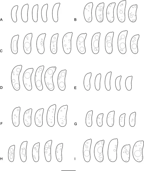
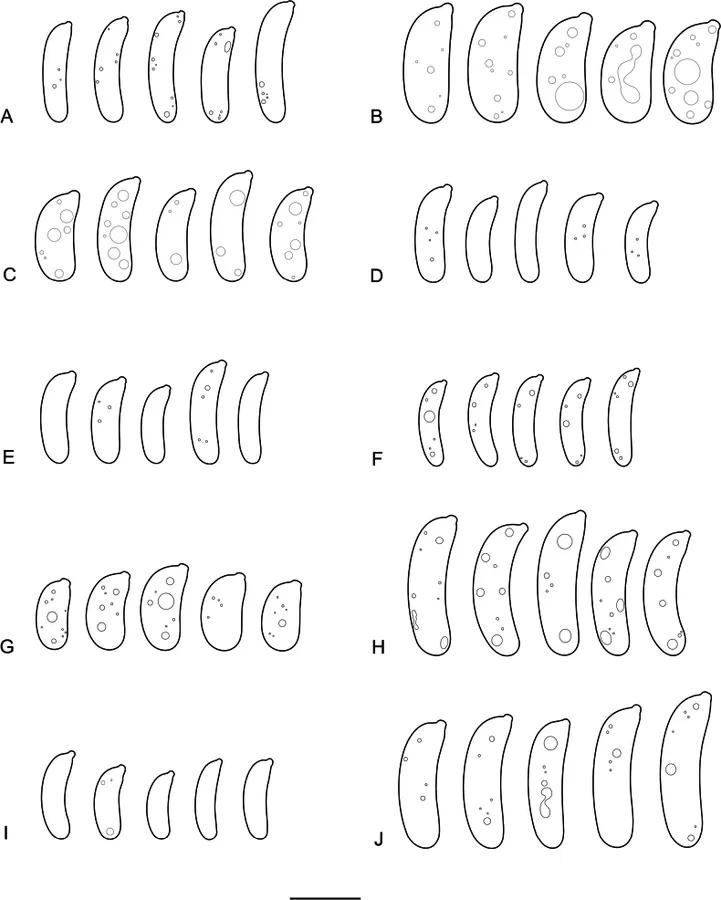
Basidiomata annual, at first adpressed-orbicular, narrowly attached, then turbinate or nearly cupulate, up to 1 cm in diam., up to 0.5 cm thick, sometimes partly fusing together and forming compound effused basidiomata, up to 3 cm in the longest dimension, gelatinous. Hymenial surface uneven or indistinctly folded, rarely minutely warted, brownish-black, in dry condition completely black. Abhymenial surface smooth, grayish-brown to brownish-black. Margin free, concolourous with hymenial surface, entire, even or lobate. Hyphal structure monomitic, hyphae clamped; context hyphae hyaline or brownish, predominantly thin-walled, interwoven, anastomosing, 1–5 μm in diam., subhymenial hyphae hyaline to brownish, thin-walled or with a distinct wall, ascending or interwoven, 1–4 μm in diam., basidia-bearing hyphae 2–6 μm in diam. Hyphidia with a hyaline or brownish hyphoid stem, 2–11 μm in diam., and hyaline or brownish apical branches, 0.5–2 μm in diam., forming a continuous layer up to 100 μm thick. Probasidia broadly clavate to tapering or subfusiform, 15–29 × 6–11.5 μm. Basidia four-celled, ellipsoid-ovoid to narrowly ovoid, thin-walled, (14–) 14.5–26 (–28.5) × (8.9–) 9.0–15.1 (–15.3) μm (n = 71/5), sterigmata up to 50 × 1.5–2.5 μm. Basidiospores narrowly cylindrical or cylindrical to allantoid, slightly to distinctly curved, (13.0–) 13.2–21.9 (–22.2) × (4.1–) 4.2–7.0 (–7.2) μm (n = 140/5), L = 16.49–18.68, W = 4.81–6.08, Q = 2.92–3.55.
Habitat dan peran ekologis
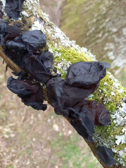
Exidia zelleri is a member of the E. glandulosa complex (Figs 1, 2, 8). Macroscopically, it is very different from the two other species of this group, E. glandulosa and E. truncata, due to its rather small basidiomata with smooth hymenial and abhymenial surfaces (Fig. 24). However, its large basidia and basidiospores are strongly reminiscent of those of the two aforementioned species. Exidia zelleri is widely distributed in the northern hemisphere but rarely collected, most likely due to its quite insignificant fructifications. It occurs predominantly on dead wood of Salicaceae and rarely also on other host trees (Acer and Alnus). Specimens of E. zelleri are often sterile. They can be nonetheless identified as belonging to this species due to the nearly smooth hymenial surface without spine-like projections, as well as the large basidia. Exidia zelleri was described by Lloyd (1920) from Oregon, USA. The host species was identified by the original collector as Sambucus. However, the later re-examination by H.C. Klett (herbarium note in NY from August 26 th, 1963) showed that the host tree was actually Acer sp. The isotype of E. zelleri in NY is still in a good condition, with rather numerous, turgid, allantoid or narrowly cylindrical spores, ca. 14–18 × 4–5.5 μm.
- Habitat atau konteks temuan
- Informasi belum tersedia pada sumber rilis ini.
- Substrat
- kayu
- Peran atau konteks
- Belum dikategorikan pada rilis ini.
Reproduksi
Basidiomata annual, at first adpressed-orbicular, narrowly attached, then turbinate or nearly cupulate, up to 1 cm in diam., up to 0.5 cm thick, sometimes partly fusing together and forming compound effused basidiomata, up to 3 cm in the longest dimension, gelatinous. Hymenial surface uneven or indistinctly folded, rarely minutely warted, brownish-black, in dry condition completely black. Abhymenial surface smooth, grayish-brown to brownish-black. Margin free, concolourous with hymenial surface, entire, even or lobate. Hyphal structure monomitic, hyphae clamped; context hyphae hyaline or brownish, predominantly thin-walled, interwoven, anastomosing, 1–5 μm in diam., subhymenial hyphae hyaline to brownish, thin-walled or with a distinct wall, ascending or interwoven, 1–4 μm in diam., basidia-bearing hyphae 2–6 μm in diam.
Catatan identitas dan cakupan
- Nama diterima mengikuti Catalogue of Life Base2026-09-11; uraian mempertahankan konsep dan nama sumber aslinya.
- Preserve original treatment localities, hosts and substrate scope. Materials examined describe documented specimens, not the full global range.
- Impor dan terjemahan otomatis belum diperiksa editorial per profil. Struktur reproduktif yang disebutkan tidak membuktikan seluruh siklus hidup diketahui.
- Pengukuran pada uraian mengikuti spesimen, kultur, dan metode sumber; angka tidak diperluas ke semua populasi.
Catatan distribusi dalam narasi mengikuti sumbernya. Dokumentasi spesimen atau foto mengikuti catatan sumber. Bukti wilayah Indonesia dan penilaian konservasi tidak disimpulkan dari gambar.
Melihat dengan konteks
Basidiospores: A Exidia recisa (epitype); B E. repleta (holotype); C E. saturata (holotype); D E. semiorbis (holotype); E E. spiculata (isotype of E. inflata); F E. straminea (epitype); G E. tanzanica (holotype); H E. truncata (neotype); I E. umbrinella (lectotype); J E. zelleri (Spirin 17780). Scale bar: 10 μm.
- Jenis
- Pelat ilmiah
- Kredit
- Viacheslav Spirin, Vera Malysheva, Nathan Schoutteten, Anton Savchenko, Bernard Rivoire, Jan Olsson, Thomas Læssøe, Leif Ryvarden, Django Grootmyers, Matti Kulju, Josef Vlasák, María Victoria Vignale, Ilya Viner
- Lisensi
- CC BY 4.0
- Sumber media
- Buka entri sumber
Dokumentasi tambahan
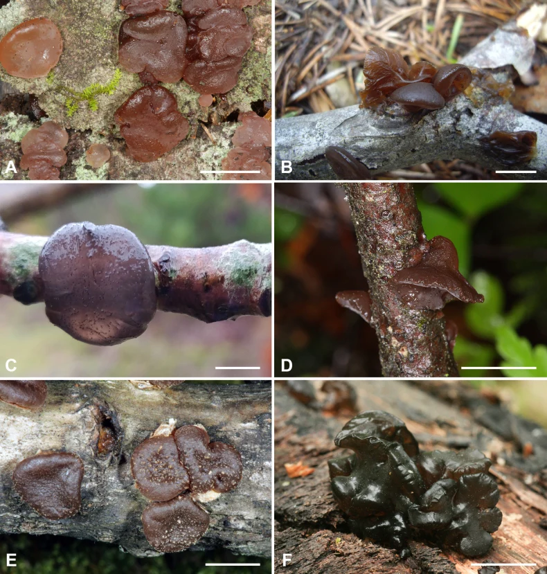
Pelat perbandingan tubuh buah. Exidia zelleri ditampilkan pada panel F; panel lainnya memuat spesies pembanding. Pembagian panel mengikuti keterangan sumber.
Kredit dan penggunaan
Viacheslav Spirin, Vera Malysheva, Nathan Schoutteten, Anton Savchenko, Bernard Rivoire, Jan Olsson, Thomas Læssøe, Leif Ryvarden, Django Grootmyers, Matti Kulju, Josef Vlasák, María Victoria Vignale, Ilya Viner
Ukuran dan format gambar disesuaikan tanpa pemotongan, retus, atau penambahan skala. Lisensi sumber dipertahankan.
Sumber profil
- [1] Exidia and Exidiopsis (Auriculariales, Basidiomycota): a reconsideration — Exidia zelleri
- [2] Catalogue of Life Base2026-09-11 — Exidia zelleri
- [3] Dokumentasi visual — Exidia zelleri
- [4] Dokumentasi visual — Exidia zelleri
Rujukan halaman ini
Fungi. Exidia zelleri. Rilis data fungi-2026-10-12-expansion-2. https://fungi-atlas.mgadib.chatgpt.site/jamur/exidia-zelleri/.
Status: impor sumber; pemeriksaan editorial per profil belum selesai. Untuk klaim biologis, gunakan rujukan pada bagian terkait dan periksa konteks sumber aslinya.