Ciri dan struktur
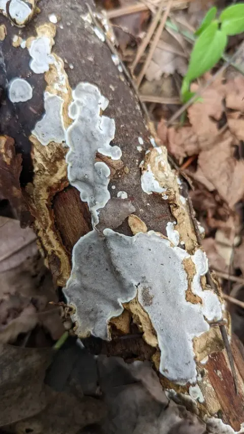
Basidiomata annual, effused, usually up to 10 cm in the longest dimension but occasionally exceeding 1 m long (while growing on dead standing logs), at first waxy-gelatinous and watery-grayish, semitranslucent, then more or less distinctly gelatinized, drying to a hardly visible, vernicose crust, up to 0.1 mm thick. Hymenial surface smooth or more often bearing randomly or rather regularly arranged spine-like projections up to 150 × 100 μm, 2–3 per mm, and at least partly covered by a grayish or pale lilac, easily removable pruina. Margin rather sharply delimited, adnate, concolorous with hymenial surface. Hyphal structure monomitic, hyphae clamped; subicular hyphae thin- or slightly thick-walled, gelatinized, mostly interwoven, 2–4 μm in diam., occasionally inflated up to 7–8 μm in diam., subhymenial hyphae thin-walled, predominantly ascending, often agglutinated, 1.5–4.5 μm in diam., occasionally inflated up to 5–6 μm in diam.; basidia-bearing hyphae thin-walled, 1.5–4 μm in diam., sometimes showing a zigzag-like branching pattern. Hyphidia thin-walled, with a hyaline, hyphoid or subfusiform stem, 20–40 × 2–6 μm in diam., and dendroid apical branches 1–1.5 μm in diam., forming a loose continuous layer up to 15 μm thick. Probasidia broadly clavate, 10–30 (–40) × 5–9 μm. Basidia narrowly ellipsoid-ovoid, four-celled, (10.5–) 12–21.5 (–23) × (8.0–) 8.1–12.3 (–13.2) μm (n = 80/7), thin-walled, embedded, often with a distinctly tapering stalk-like base, up to 7 × 4 μm, sterigmata up to 25 × 2–3 μm. Basidiospores thin-walled, narrowly cylindrical to cylindrical, often distinctly curved, (9.4–) 9.8–16.8 (–17.2) × (3.6–) 3.7–6.2 (–6.6) μm (n = 210/7), L = 11.40–14.26, W = 4.14–5.26, Q = 2.46–2.91.
Habitat dan peran ekologis
Basidiomata annual, effused, usually up to 10 cm in the longest dimension but occasionally exceeding 1 m long (while growing on dead standing logs), at first waxy-gelatinous and watery-grayish, semitranslucent, then more or less distinctly gelatinized, drying to a hardly visible, vernicose crust, up to 0.1 mm thick. Hymenial surface smooth or more often bearing randomly or rather regularly arranged spine-like projections up to 150 × 100 μm, 2–3 per mm, and at least partly covered by a grayish or pale lilac, easily removable pruina. Margin rather sharply delimited, adnate, concolorous with hymenial surface. Hyphal structure monomitic, hyphae clamped; subicular hyphae thin- or slightly thick-walled, gelatinized, mostly interwoven, 2–4 μm in diam., occasionally inflated up to 7–8 μm in diam., subhymenial hyphae thin-walled, predominantly ascending, often agglutinated, 1.5–4.5 μm in diam., occasionally inflated up to 5–6 μm in diam.; basidia-bearing hyphae thin-walled, 1.5–4 μm in diam., sometimes showing a zigzag-like branching pattern. Hyphidia thin-walled, with a hyaline, hyphoid or subfusiform stem, 20–40 × 2–6 μm in diam., and dendroid apical branches 1–1.5 μm in diam., forming a loose continuous layer up to 15 μm thick.
- Habitat atau konteks temuan
- Informasi belum tersedia pada sumber rilis ini.
- Substrat
- Informasi belum tersedia pada sumber rilis ini.
- Peran atau konteks
- Belum dikategorikan pada rilis ini.
Reproduksi
Basidiomata annual, effused, usually up to 10 cm in the longest dimension but occasionally exceeding 1 m long (while growing on dead standing logs), at first waxy-gelatinous and watery-grayish, semitranslucent, then more or less distinctly gelatinized, drying to a hardly visible, vernicose crust, up to 0.1 mm thick. Hymenial surface smooth or more often bearing randomly or rather regularly arranged spine-like projections up to 150 × 100 μm, 2–3 per mm, and at least partly covered by a grayish or pale lilac, easily removable pruina. Margin rather sharply delimited, adnate, concolorous with hymenial surface. Hyphal structure monomitic, hyphae clamped; subicular hyphae thin- or slightly thick-walled, gelatinized, mostly interwoven, 2–4 μm in diam., occasionally inflated up to 7–8 μm in diam., subhymenial hyphae thin-walled, predominantly ascending, often agglutinated, 1.5–4.5 μm in diam., occasionally inflated up to 5–6 μm in diam.; basidia-bearing hyphae thin-walled, 1.5–4 μm in diam., sometimes showing a zigzag-like branching pattern.
Catatan identitas dan cakupan
- Nama diterima mengikuti Catalogue of Life Base2026-09-11; uraian mempertahankan konsep dan nama sumber aslinya.
- Preserve original treatment localities, hosts and substrate scope. Materials examined describe documented specimens, not the full global range.
- Impor dan terjemahan otomatis belum diperiksa editorial per profil. Struktur reproduktif yang disebutkan tidak membuktikan seluruh siklus hidup diketahui.
- Pengukuran pada uraian mengikuti spesimen, kultur, dan metode sumber; angka tidak diperluas ke semua populasi.
Catatan distribusi dalam narasi mengikuti sumbernya. Dokumentasi spesimen atau foto mengikuti catatan sumber. Bukti wilayah Indonesia dan penilaian konservasi tidak disimpulkan dari gambar.
Melihat dengan konteks
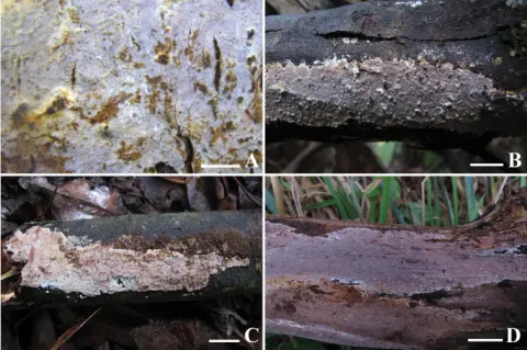
Pelat perbandingan tubuh buah. Exidiopsis effusa ditampilkan pada panel A; panel lainnya memuat spesies pembanding. Pembagian panel mengikuti keterangan sumber.
- Jenis
- Pelat ilmiah
- Kredit
- Viacheslav Spirin, Vera Malysheva, Nathan Schoutteten, Anton Savchenko, Bernard Rivoire, Jan Olsson, Thomas Læssøe, Leif Ryvarden, Django Grootmyers, Matti Kulju, Josef Vlasák, María Victoria Vignale, Ilya Viner
- Lisensi
- CC BY 4.0
- Sumber media
- Buka entri sumber
Dokumentasi tambahan
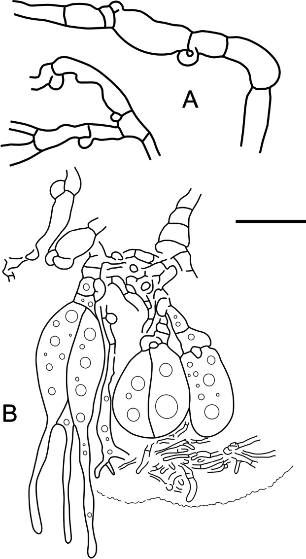
Microstructures of Exidiopsis effusa (epitype): A subicular hyphae; B subhymenial hyphae and hymenial cells. Scale bar: 10 μm.
Kredit dan penggunaan
Viacheslav Spirin, Vera Malysheva, Nathan Schoutteten, Anton Savchenko, Bernard Rivoire, Jan Olsson, Thomas Læssøe, Leif Ryvarden, Django Grootmyers, Matti Kulju, Josef Vlasák, María Victoria Vignale, Ilya Viner
Ukuran dan format gambar disesuaikan tanpa pemotongan, retus, atau penambahan skala. Lisensi sumber dipertahankan.
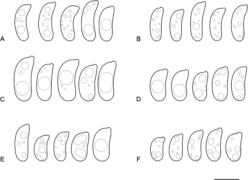
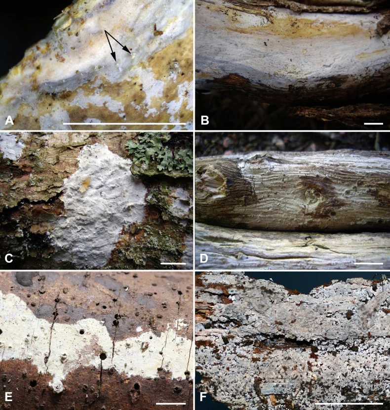
Pelat perbandingan basidiospora. Exidiopsis effusa ditampilkan pada panel A dan dua spora di sisi kanan B; panel lainnya memuat spesies pembanding. Pembagian panel mengikuti keterangan sumber.
Kredit dan penggunaan
Viacheslav Spirin, Vera Malysheva, Nathan Schoutteten, Anton Savchenko, Bernard Rivoire, Jan Olsson, Thomas Læssøe, Leif Ryvarden, Django Grootmyers, Matti Kulju, Josef Vlasák, María Victoria Vignale, Ilya Viner
Ukuran dan format gambar disesuaikan tanpa pemotongan, retus, atau penambahan skala. Lisensi sumber dipertahankan.
Sumber profil
- [1] Exidia and Exidiopsis (Auriculariales, Basidiomycota): a reconsideration — Exidiopsis effusa
- [2] Catalogue of Life Base2026-09-11 — Exidiopsis effusa
- [3] Dokumentasi visual — Exidiopsis effusa
- [4] Dokumentasi visual — Exidiopsis effusa
- [5] Dokumentasi visual — Exidiopsis effusa
Rujukan halaman ini
Fungi. Exidiopsis effusa. Rilis data fungi-2026-10-12-expansion-2. https://fungi-atlas.mgadib.chatgpt.site/jamur/exidiopsis-effusa/.
Status: impor sumber; pemeriksaan editorial per profil belum selesai. Untuk klaim biologis, gunakan rujukan pada bagian terkait dan periksa konteks sumber aslinya.