Saprobic on rachides dari Cocos nucifera. Morph seksual Ascomata 150–330 μm diamond, solitary atau gregarious, tenggelam di bawah clypeus, terlihat sebagai titik hitam pada substrat, dengan ostiolar atau papilla, di bagian vertikal 210–320 μm diamond, 190–220 μm tinggi, subglobosa atau globose, dengan dasar bulat atau datar, coriaceous. Ostiole central, periphasate. Peridium 9–18 µm lebar, terdiri dari beberapa lapisan sel tekstura angularis, lapisan luar sel cokelat dan lapisan antar hialin, lebih tipis di dasar.
Profil ini diimpor dari uraian spesies pada sumber berlisensi dan sebagian teks diterjemahkan otomatis. Pemeriksaan editorial per profil belum selesai. Bagian yang belum diterjemahkan atau belum lolos pemeriksaan terjemahan tetap ditampilkan dalam bahasa sumber. Angka dan nama ilmiah dipertahankan.
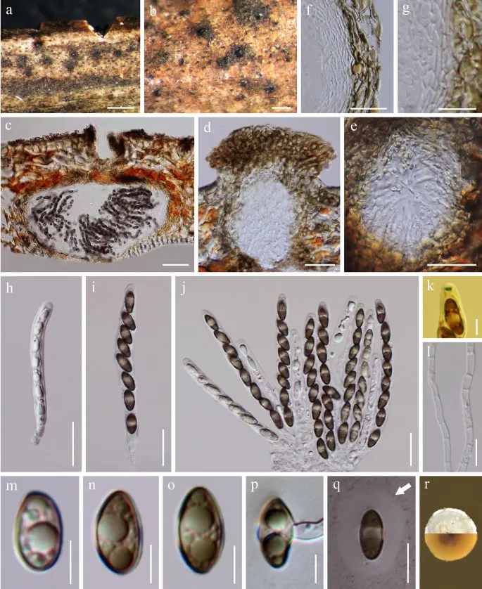
Fasciatispora cocoes (MFLU 18-1589, holotype). a, b Ascomata on substrate surface. c Vertical section through an ascoma. d, e Ostiole with periphyses. f, g Peridium (g = at the base). h – j Asci. k Ascus in Melzer’s reagent, with a J +, subapical ring. l Paraphyses. m – o Ascospores. p Germinated ascospore. q Ascospore in India ink presents the clear mucilaginous sheath. r Colony on PDA. Scale bars: a = 500 μm, b = 200 μm, c = 50 μm, d – f, h – j = 20 μm, g, l, q = 10 μm, k, m – p = 5 μm Penulis dan sumber artikel: Hyde, Kevin D.; Dong, Yang; Phookamsak, Rungtiwa; Jeewon, Rajesh; Bhat, D. Jayarama; Jones, E. B. Gareth; Liu, Ning-Guo; A… (kredit lengkap di bawah) · CC BY 4.0 · Ukuran dan format gambar disesuaikan; lisensi asli dipertahankan.
01 / Morfologi
Ciri dan struktur
Saprobic on rachides of Cocos nucifera. Sexual morph Ascomata 150–330 μm diam., solitary or gregarious, immersed under a clypeus, visible as black dots on substrate, with an ostiolar or papilla, in vertical section 210–320 μm diam., 190–220 μm high, subglobose or globose, with a rounded or flattened base, coriaceous. Ostiole central, periphysate. Peridium 9–18 µm wide, consisting several layers of cells of textura angularis, outside layers brown cells and inter layers hyaline, thinner at the base. Paraphyses 2–3 µm wide, hyphae-like, irregular, septate, rarely branched. Asci 78–110 × 6–12 µm (\(\bar{x} \) = 94.2 × 9 µm, n = 20), 8-spored, unitunicate, cylindrical, pedicellate, thin-walled, with a J +, wedge-shaped, subapical ring, 2–2.5 µm diam., 1 µm high. Ascospores 10–15 × 4.5–6 µm, (\(\bar{x} \) = 12.7 × 5.4 µm, n = 50), 1-seriate, pale brown to brown, ellipsoidal, unicellular, with a central pallid band, smooth-walled, surrounded by a mucilaginous sheath. Asexual morph Undetermined. Culture characteristics: Ascospores germinated on PDA within 24 h and transferred to MEA. Colonies growing on MEA, circular, with flat surface, edge entire or a little filamentous, creamy white, reached about a diam. of 20 mm after three weeks at 25–28 °C. Mycelium superficial, branched, septate, hyaline to pale brown, smooth.
Sebagian uraian ditampilkan dalam bahasa sumber; terjemahannya belum tersedia atau belum lolos pemeriksaan.
Pengukuran mengikuti objek, spesimen atau kultur, dan metode yang disebutkan sumber. Baca uraian asli untuk konteks lengkap.
02 / Kehidupan
Habitat dan peran ekologis
Saprobic on rachides of Cocos nucifera. Sexual morph Ascomata 150–330 μm diam., solitary or gregarious, immersed under a clypeus, visible as black dots on substrate, with an ostiolar or papilla, in vertical section 210–320 μm diam., 190–220 μm high, subglobose or globose, with a rounded or flattened base, coriaceous. Ostiole central, periphysate. Peridium 9–18 µm wide, consisting several layers of cells of textura angularis, outside layers brown cells and inter layers hyaline, thinner at the base. Paraphyses 2–3 µm wide, hyphae-like, irregular, septate, rarely branched. Asci 78–110 × 6–12 µm (\(\bar{x} \) = 94.2 × 9 µm, n = 20), 8-spored, unitunicate, cylindrical, pedicellate, thin-walled, with a J +, wedge-shaped, subapical ring, 2–2.5 µm diam., 1 µm high. Ascospores 10–15 × 4.5–6 µm, (\(\bar{x} \) = 12.7 × 5.4 µm, n = 50), 1-seriate, pale brown to brown, ellipsoidal, unicellular, with a central pallid band, smooth-walled, surrounded by a mucilaginous sheath. Asexual morph Undetermined. Culture characteristics: Ascospores germinated on PDA within 24 h and transferred to MEA. Colonies growing on MEA, circular, with flat surface, edge entire or a little filamentous, creamy white, reached about a diam. of 20 mm after three weeks at 25–28 °C.
Sebagian uraian ditampilkan dalam bahasa sumber; terjemahannya belum tersedia atau belum lolos pemeriksaan.
Saprobic on rachides of Cocos nucifera. Sexual morph Ascomata 150–330 μm diam., solitary or gregarious, immersed under a clypeus, visible as black dots on substrate, with an ostiolar or papilla, in vertical section 210–320 μm diam., 190–220 μm high, subglobose or globose, with a rounded or flattened base, coriaceous. Ostiole central, periphysate. Peridium 9–18 µm wide, consisting several layers of cells of textura angularis, outside layers brown cells and inter layers hyaline, thinner at the base. Paraphyses 2–3 µm wide, hyphae-like, irregular, septate, rarely branched. Asci 78–110 × 6–12 µm (\(\bar{x} \) = 94.2 × 9 µm, n = 20), 8-spored, unitunicate, cylindrical, pedicellate, thin-walled, with a J +, wedge-shaped, subapical ring, 2–2.5 µm diam., 1 µm high.
Sebagian uraian ditampilkan dalam bahasa sumber; terjemahannya belum tersedia atau belum lolos pemeriksaan.
Nama diterima mengikuti Catalogue of Life Base2026-09-11; uraian mempertahankan konsep dan nama sumber aslinya.
Preserve original monograph treatment localities, hosts and specimen/substrate scope. Materials examined document specimens, not the full global range.
Impor dan terjemahan otomatis belum diperiksa editorial per profil. Struktur reproduktif yang disebutkan tidak membuktikan seluruh siklus hidup diketahui.
Pengukuran pada uraian mengikuti spesimen, kultur, dan metode sumber; angka tidak diperluas ke semua populasi.
Catatan distribusi dalam narasi mengikuti sumbernya. Dokumentasi spesimen atau foto mengikuti catatan sumber. Bukti wilayah Indonesia dan penilaian konservasi tidak disimpulkan dari gambar.
05 / Dokumentasi
Melihat dengan konteks
Fasciatispora cocoes (MFLU 18-1589, holotype). a, b Ascomata on substrate surface. c Vertical section through an ascoma. d, e Ostiole with periphyses. f, g Peridium (g = at the base). h – j Asci. k Ascus in Melzer’s reagent, with a J +, subapical ring. l Paraphyses. m – o Ascospores. p Germinated ascospore. q Ascospore in India ink presents the clear mucilaginous sheath. r Colony on PDA. Scale bars: a = 500 μm, b = 200 μm, c = 50 μm, d – f, h – j = 20 μm, g, l, q = 10 μm, k, m – p = 5 μm
Jenis
Pelat ilmiah
Kredit
Penulis dan sumber artikel: Hyde, Kevin D.; Dong, Yang; Phookamsak, Rungtiwa; Jeewon, Rajesh; Bhat, D. Jayarama; Jones, E. B. Gareth; Liu, Ning-Guo; Abeywickrama, Pranami D.; Mapook, Ausana; Wei, Deping; Perera, Rekhani H.; Manawasinghe, Ishara S.; Pem, Dhandevi; Bundhun, Digvijayini; Karunarathna, Anuruddha; Ekanayaka, Anusha H.; Bao, Dan-Feng; Li, Junfu; Samarakoon, Milan C.; Chaiwan, Napalai; Lin, Chuan-Gen; Phutthacharoen, Kunthida; Zhang, Sheng-Nan; Senanayake, Indunil C.; Goonasekara, Ishani D.; Thambugala, Kasun M.; Phukhamsakda, Chayanard; Tennakoon, Danushka S.; Jiang, Hong-Bo; Yang, Jing; Zeng, Ming; Huanraluek, Naruemon; Liu, Jian-Kui (Jack); Wijesinghe, Subodini N.; Tian, Qing; Tibpromma, Saowaluck; Brahmanage, Rashika S.; Boonmee, Saranyaphat; Huang, Shi-Ke; Thiyagaraja, Vinodhini; Lu, Yong-Zhong; Jayawardena, Ruvishika S.; Dong, Wei; Yang, Er-Fu; Singh, Sanjay K.; Singh, Shiv Mohan; Rana, Shiwali; Lad, Sneha S.; Anand, Garima; Devadatha, Bandarupalli; Niranjan, M.; Sarma, V. Venkateswara; Liimatainen, Kare; Aguirre-Hudson, Begoña; Niskanen, Tuula; Overall, Andy; Alvarenga, Renato Lúcio Mendes; Gibertoni, Tatiana Baptista; Pfliegler, Walter P.; Horváth, Enikő; Imre, Alexandra; Alves, Amanda Lucia; da Silva Santos, Ana Carla; Tiago, Patricia Vieira; Bulgakov, Timur S.; Wanasinghe, Dhanushaka N.; Bahkali, Ali H.; Doilom, Mingkwan; Elgorban, Abdallah M.; Maharachchikumbura, Sajeewa S. N.; Rajeshkumar, Kunhiraman C.; Haelewaters, Danny; Mortimer, Peter E.; Zhao, Qi; Lumyong, Saisamorn; Xu, Jianchu; Sheng, Jun
Saprobic on rachides of Cocos nucifera.
Sexual morph Ascomata 150–330 μm diam., solitary or gregarious, immersed under a clypeus, visible as black dots on substrate, with an ostiolar or papilla, in vertical section 210–320 μm diam., 190–220 μm high, subglobose or globose, with a rounded or flattened base, coriaceous.
Ostiole central, periphysate.
Peridium 9–18 µm wide, consisting several layers of cells of textura angularis, outside layers brown cells and inter layers hyaline, thinner at the base.
Saprobic on rachides of Cocos nucifera. Sexual morph Ascomata 150–330 μm diam., solitary or gregarious, immersed under a clypeus, visible as black dots on substrate, with an ostiolar or papilla, in vertical section 210–320 μm diam., 190–220 μm high, subglobose or globose, with a rounded or flattened base, coriaceous. Ostiole central, periphysate. Peridium 9–18 µm wide, consisting several layers of cells of textura angularis, outside layers brown cells and inter layers hyaline, thinner at the base. Paraphyses 2–3 µm wide, hyphae-like, irregular, septate, rarely branched. Asci 78–110 × 6–12 µm (\(\bar{x} \) = 94.2 × 9 µm, n = 20), 8-spored, unitunicate, cylindrical, pedicellate, thin-walled, with a J +, wedge-shaped, subapical ring, 2–2.5 µm diam., 1 µm high. Ascospores 10–15 × 4.5–6 µm, (\(\bar{x} \) = 12.7 × 5.4 µm, n = 50), 1-seriate, pale brown to brown, ellipsoidal, unicellular, with a central pallid band, smooth-walled, surrounded by a mucilaginous sheath. Asexual morph Undetermined.
Culture characteristics: Ascospores germinated on PDA within 24 h and transferred to MEA. Colonies growing on MEA, circular, with flat surface, edge entire or a little filamentous, creamy white, reached about a diam. of 20 mm after three weeks at 25–28 °C. Mycelium superficial, branched, septate, hyaline to pale brown, smooth.
Saprobic on rachides of Cocos nucifera.
Sexual morph Ascomata 150–330 μm diam., solitary or gregarious, immersed under a clypeus, visible as black dots on substrate, with an ostiolar or papilla, in vertical section 210–320 μm diam., 190–220 μm high, subglobose or globose, with a rounded or flattened base, coriaceous.
Ostiole central, periphysate.
Peridium 9–18 µm wide, consisting several layers of cells of textura angularis, outside layers brown cells and inter layers hyaline, thinner at the base.
Paraphyses 2–3 µm wide, hyphae-like, irregular, septate, rarely branched.
Asci 78–110 × 6–12 µm (\(\bar{x} \) = 94.2 × 9 µm, n = 20), 8-spored, unitunicate, cylindrical, pedicellate, thin-walled, with a J +, wedge-shaped, subapical ring, 2–2.5 µm diam., 1 µm high.
Ascospores 10–15 × 4.5–6 µm, (\(\bar{x} \) = 12.7 × 5.4 µm, n = 50), 1-seriate, pale brown to brown, ellipsoidal, unicellular, with a central pallid band, smooth-walled, surrounded by a mucilaginous sheath.
Asexual morph Undetermined.
Culture characteristics: Ascospores germinated on PDA within 24 h and transferred to MEA.
Colonies growing on MEA, circular, with flat surface, edge entire or a little filamentous, creamy white, reached about a diam. of 20 mm after three weeks at 25–28 °C.
Saprobic on rachides of Cocos nucifera.
Sexual morph Ascomata 150–330 μm diam., solitary or gregarious, immersed under a clypeus, visible as black dots on substrate, with an ostiolar or papilla, in vertical section 210–320 μm diam., 190–220 μm high, subglobose or globose, with a rounded or flattened base, coriaceous.
Ostiole central, periphysate.
Peridium 9–18 µm wide, consisting several layers of cells of textura angularis, outside layers brown cells and inter layers hyaline, thinner at the base.
Paraphyses 2–3 µm wide, hyphae-like, irregular, septate, rarely branched.
Asci 78–110 × 6–12 µm (\(\bar{x} \) = 94.2 × 9 µm, n = 20), 8-spored, unitunicate, cylindrical, pedicellate, thin-walled, with a J +, wedge-shaped, subapical ring, 2–2.5 µm diam., 1 µm high.
Status: impor sumber; pemeriksaan editorial per profil belum selesai. Untuk klaim biologis, gunakan rujukan pada bagian terkait dan periksa konteks sumber aslinya.
Fistulina subhepatica adalah spesies jamur dalam keluarga Fistulinaceae. Hal ini ditemukan di Yunnan Province in subtropis China, di mana ia menyebabkan kutu cokelat di Castanopsis and Lithocarpus. Hal ini dijelaskan sebagai baru untuk…
Flavignomonia rhoigena dapat dibedakan dari spesies gnomoniaceus lainnya dengan pembentukan synnemata. Morph seksual: tidak diamati. Morph asexual: Conidiomata synnematal. Synnemata (650 ⁇ ) 750–1100 µm tinggi, 150–300 µm diameter,…
Saprobic or patogen pada batang udara mati dari Maclura pomifera. Morph seksual tidak ditentukan. Morph asexual konidiomata 40–47 × 40–69 μm (\(\bar{x} \) = 43.3 × 48 µm, n = 20), pycnidial, globose ke subglobosa, elipsoidal, atau tidak…