Ciri dan struktur
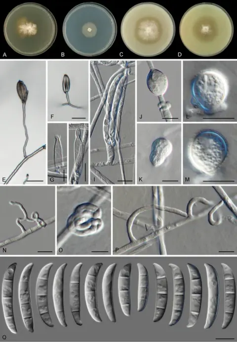
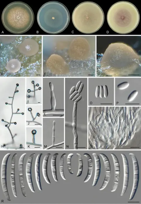
Associated with contaminated rubber latex. Sexual morph: Undetermined. Asexual morph on PDA: Conidiophores abundant on aerial and substrate mycelium, erect and simple, straight or flexuous, smooth- and thin-walled, bearing terminal or lateral, forming monophialides on the apices. Phialides (11–) 15 × 30 (–35) × 2.5–5 μm (x– = 22 × 4 μm, n = 30), subcylindrical to subulate, monophialidic, smooth- and thin-walled with a short flared apical collarette. Aerial conidia 5–11 × 2.5–5 μm (x– = 6.8 × 3.6 μm, n = 50), obovoidal, broadly ellipsoidal to ellipsoidal, straight or rarely curved, 0(–1)-septate, hyaline, smooth- and thin-walled, clustering abundantly in false heads at the tip of monophialides. Sporodochial conidiophores sparingly verticillately branched, bearing terminal whorls of 2–3-monophialides. Sporodochial phialides 11–20 × 3–4 μm (x– = 15 × 3.4 μm, n = 30), flask-shaped, subcylindrical to short subulate, smooth- and thin-walled, conidiogenous loci with inconspicuous periclinal thickening and collarette. Sporodochial conidia 22–38 × 4–6 μm (x– = 31 × 4.6 μm, n = 50), moderately to distinctly dorsiventrally curved narrowing gently toward base, dorsal line usually almost straight; apical cell often equal in length to adjacent cell, blunt and rounded with curved apex; basal cell often distinctly notched, 1–4-septate, hyaline, smooth- and thick-walled. Chlamydospores 6–10 μm diam., abundantly formed, globose to subglobose, smooth- and thick-walled, terminal or intercalary in hyphae or conidia, solitary, in chains or in clusters. Colonies on PDA at room temperature (20–25 °C) covered the entire 6 cm Petri dish within three days, white to pale yellow, with abundant aerial mycelium; reverse white to pale yellow.
Habitat dan peran ekologis
Associated with contaminated rubber latex. Sexual morph: Undetermined. Asexual morph on PDA: Conidiophores abundant on aerial and substrate mycelium, erect and simple, straight or flexuous, smooth- and thin-walled, bearing terminal or lateral, forming monophialides on the apices. Phialides (11–) 15 × 30 (–35) × 2.5–5 μm (x– = 22 × 4 μm, n = 30), subcylindrical to subulate, monophialidic, smooth- and thin-walled with a short flared apical collarette. Aerial conidia 5–11 × 2.5–5 μm (x– = 6.8 × 3.6 μm, n = 50), obovoidal, broadly ellipsoidal to ellipsoidal, straight or rarely curved, 0(–1)-septate, hyaline, smooth- and thin-walled, clustering abundantly in false heads at the tip of monophialides. Sporodochial conidiophores sparingly verticillately branched, bearing terminal whorls of 2–3-monophialides. Sporodochial phialides 11–20 × 3–4 μm (x– = 15 × 3.4 μm, n = 30), flask-shaped, subcylindrical to short subulate, smooth- and thin-walled, conidiogenous loci with inconspicuous periclinal thickening and collarette. Sporodochial conidia 22–38 × 4–6 μm (x– = 31 × 4.6 μm, n = 50), moderately to distinctly dorsiventrally curved narrowing gently toward base, dorsal line usually almost straight; apical cell often equal in length to adjacent cell, blunt and rounded with curved apex; basal cell often distinctly notched, 1–4-septate, hyaline, smooth- and thick-walled.
- Habitat atau konteks temuan
- kultur yang didokumentasikan
- Substrat
- Informasi belum tersedia pada sumber rilis ini.
- Peran atau konteks
- Belum dikategorikan pada rilis ini.
Reproduksi
Associated with contaminated rubber latex. Sexual morph: Undetermined. Asexual morph on PDA: Conidiophores abundant on aerial and substrate mycelium, erect and simple, straight or flexuous, smooth- and thin-walled, bearing terminal or lateral, forming monophialides on the apices. Phialides (11–) 15 × 30 (–35) × 2.5–5 μm (x– = 22 × 4 μm, n = 30), subcylindrical to subulate, monophialidic, smooth- and thin-walled with a short flared apical collarette. Aerial conidia 5–11 × 2.5–5 μm (x– = 6.8 × 3.6 μm, n = 50), obovoidal, broadly ellipsoidal to ellipsoidal, straight or rarely curved, 0(–1)-septate, hyaline, smooth- and thin-walled, clustering abundantly in false heads at the tip of monophialides.
Catatan identitas dan cakupan
- Nama diterima mengikuti Catalogue of Life Base2026-09-11; uraian mempertahankan konsep dan nama sumber aslinya.
- Preserve original treatment localities, hosts and substrate scope. Materials examined describe documented specimens, not the full global range.
- Impor dan terjemahan otomatis belum diperiksa editorial per profil. Struktur reproduktif yang disebutkan tidak membuktikan seluruh siklus hidup diketahui.
- Pengukuran pada uraian mengikuti spesimen, kultur, dan metode sumber; angka tidak diperluas ke semua populasi.
Catatan distribusi dalam narasi mengikuti sumbernya. Dokumentasi spesimen atau foto mengikuti catatan sumber. Bukti wilayah Indonesia dan penilaian konservasi tidak disimpulkan dari gambar.
Melihat dengan konteks
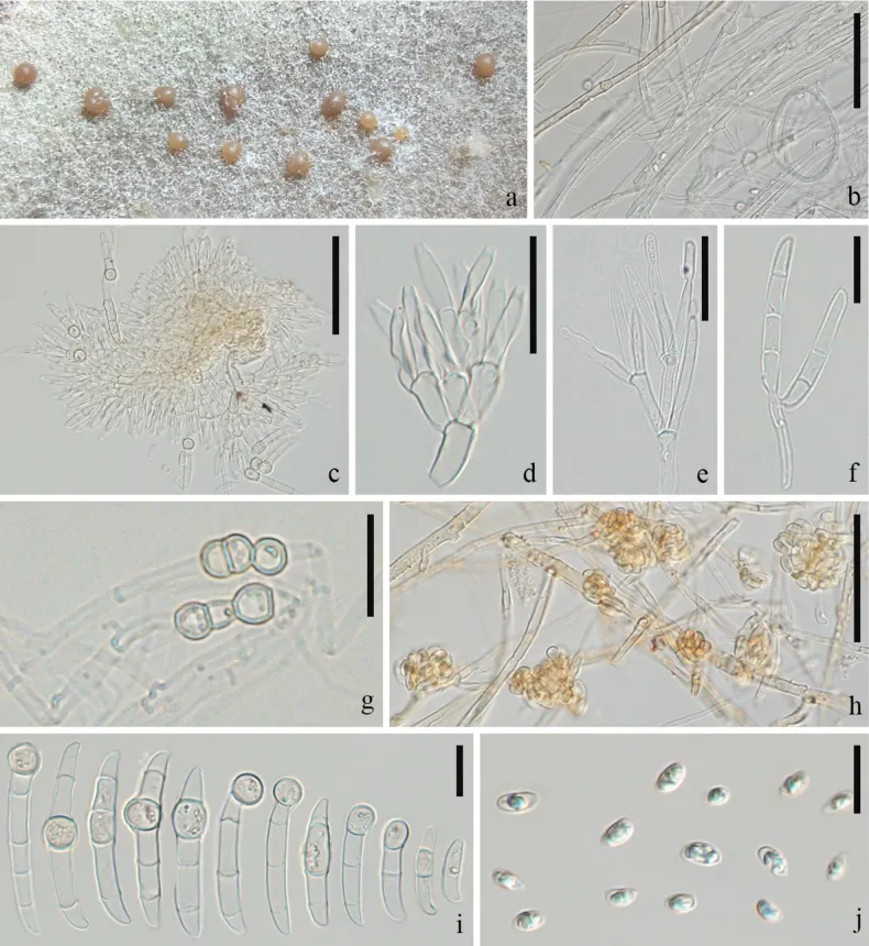
Neocosmospora bostrycoides (GMB-W1263). a. Sporodochia formed on the surface of PDA after 8 weeks; b. Mycelium; c–f. Sporodochial conidiophores; g. Chlamydospores; h. Aerial conidiophore with conidia; i. Sporodochial conidia with chlamydospores; j. Aerial microconidia. Scale bars: 50 μm (b, c, h), 20 μm (d, e, g), 10 μm (f, i, j).
- Jenis
- Pelat ilmiah
- Kredit
- Rui-Fang Xu, Samantha C. Karunarathna, Kevin D. Hyde, Chayanard Phukhamsakda, Sylvie Rapior, Dong-Qin Dai, Mei-Mei Wang, Sajad Ali, Jing-Ya Yang, Pattana Kakumyan, Saowaluck Tibpromma
- Lisensi
- CC BY 4.0
- Sumber media
- Buka entri sumber
Sumber profil
- [1] Screening and identification of fungal species associated with early-stage deterioration of natural rubber — Neocosmospora bostrycoides
- [2] Catalogue of Life Base2026-09-11 — Fusarium bostrycoides
- [3] Dokumentasi visual — Neocosmospora bostrycoides
Rujukan halaman ini
Fungi. Fusarium bostrycoides. Rilis data fungi-2026-10-12-expansion-2. https://fungi-atlas.mgadib.chatgpt.site/jamur/fusarium-bostrycoides/.
Status: impor sumber; pemeriksaan editorial per profil belum selesai. Untuk klaim biologis, gunakan rujukan pada bagian terkait dan periksa konteks sumber aslinya.