Ciri dan struktur
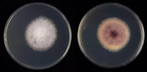
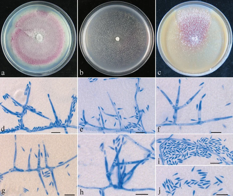
Mycelium not visible around ascomata or on host. Ascomata perithecial, crowded in group of 5 to 40, on basal stroma, oval, subglobose to globose, smooth, bright red when fresh, red brownish to dark red when dry, with a darker red ostiolar region, turning purple red in KOH and orange yellow in LA, 128–175 × 206–255 μm (n = 17). Perithecial wall consisting of a single layer, 15–25 μm thick, cells forming textura prismatica, 2–12 × 2–6 μm, walls 1–1.2 μm thick. Asci cylindrical, with a rounded apex, (4–7-)8-spored, 55–75 × 5–9 μm. Ascospores ellipsoidal to broadly ellipsoidal, equally 2-celled, slightly constricted at septum, smooth or finely verruculose, hyaline or pale brown, obliquely uniseriate in ascus often with ends overlapping, 7.5–12 × 4.5–5.5 μm. Colony on PDA 48 mm diam. after 7 d at 25 °C, surface cottony, aerial mycelium white, producing vinaceous pigment in medium. Colony on SNA 30 mm diam. after 7 d at 25 °C, surface slightly floccose, with sparse whitish aerial mycelium. Colony on CMD 56 mm diam. after 7 d at 25 °C, surface floccose, with sparse whitish aerial mycelium, producing vinaceous pigment in medium. Conidiophores with short simple branches. Conidiogenous cells monophialidic, cylindrical, tapering toward the tip, 12–63 × 1.5–3.5 μm. Conidia clavate, not in chains, hyaline, aseptate, 4–7 × 0.8–2 μm (n = 60). Macroconidia and chlamydospores not observed. Notes. Attempts were made to obtain macroconidia of the fungus in culture, but failed. Although the falcate macroconidia are lacking, the major phenotypic features of the fungus, such as occurrence on bark of Buxus sp., perithecia broadly ampulliform with a short neck, asci cylindrical with a rounded apex, ellipsoidal ascospores uniseptate and conidiophores monophialidic, fit well with the generic concept of Geejayessia.
Habitat dan peran ekologis
CHINA, Henan Province, Longyuwan, 33°40'45"N, 111°46'26"E, alt. 1500 m, on bark of Buxus sp., 17 September 2013, H.D. Zheng, Z.Q. Zeng & Z.X. Zhu 8728 (holotype: HMAS 275654), dried ex-type culture HMAS 248725. Colony on PDA 48 mm diam. after 7 d at 25 °C, surface cottony, aerial mycelium white, producing vinaceous pigment in medium. Colony on SNA 30 mm diam. after 7 d at 25 °C, surface slightly floccose, with sparse whitish aerial mycelium. Colony on CMD 56 mm diam. after 7 d at 25 °C, surface floccose, with sparse whitish aerial mycelium, producing vinaceous pigment in medium. Conidiophores with short simple branches. Conidiogenous cells monophialidic, cylindrical, tapering toward the tip, 12–63 × 1.5–3.5 μm. Conidia clavate, not in chains, hyaline, aseptate, 4–7 × 0.8–2 μm (n = 60). Macroconidia and chlamydospores not observed. Notes. Attempts were made to obtain macroconidia of the fungus in culture, but failed. Although the falcate macroconidia are lacking, the major phenotypic features of the fungus, such as occurrence on bark of Buxus sp., perithecia broadly ampulliform with a short neck, asci cylindrical with a rounded apex, ellipsoidal ascospores uniseptate and conidiophores monophialidic, fit well with the generic concept of Geejayessia.
- Habitat atau konteks temuan
- kultur yang didokumentasikan
- Substrat
- kulit kayu
- Peran atau konteks
- Belum dikategorikan pada rilis ini.
Reproduksi
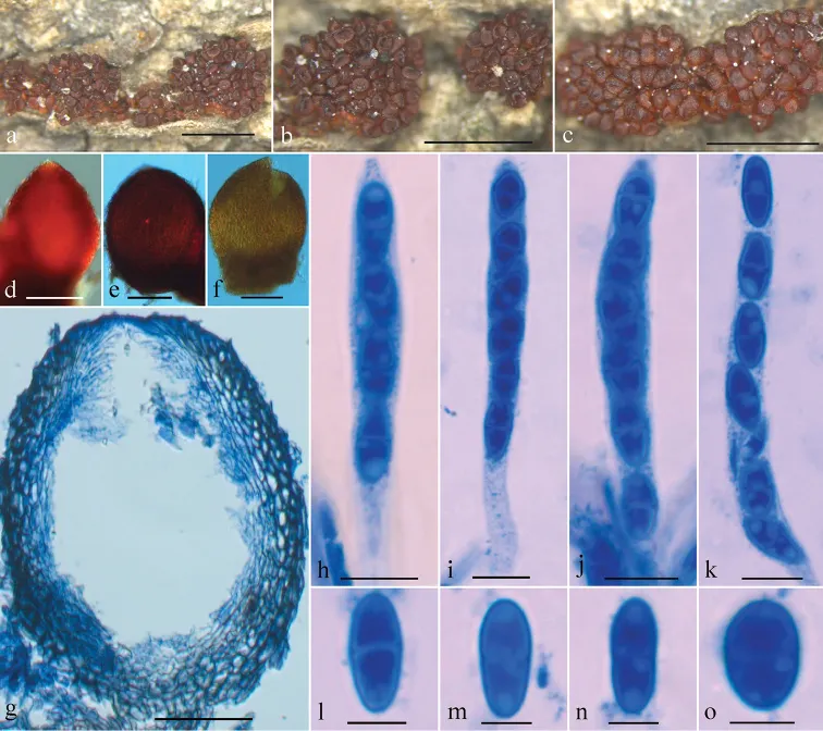
Mycelium not visible around ascomata or on host. Ascomata perithecial, crowded in group of 5 to 40, on basal stroma, oval, subglobose to globose, smooth, bright red when fresh, red brownish to dark red when dry, with a darker red ostiolar region, turning purple red in KOH and orange yellow in LA, 128–175 × 206–255 μm (n = 17). Perithecial wall consisting of a single layer, 15–25 μm thick, cells forming textura prismatica, 2–12 × 2–6 μm, walls 1–1.2 μm thick. Asci cylindrical, with a rounded apex, (4–7-)8-spored, 55–75 × 5–9 μm. Ascospores ellipsoidal to broadly ellipsoidal, equally 2-celled, slightly constricted at septum, smooth or finely verruculose, hyaline or pale brown, obliquely uniseriate in ascus often with ends overlapping, 7.5–12 × 4.5–5.5 μm.
Catatan identitas dan cakupan
- Nama diterima mengikuti Catalogue of Life Base2026-09-11; uraian mempertahankan konsep dan nama sumber aslinya.
- Preserve original treatment localities, hosts and substrate scope. Materials examined describe documented specimens, not the full global range.
- Impor dan terjemahan otomatis belum diperiksa editorial per profil. Struktur reproduktif yang disebutkan tidak membuktikan seluruh siklus hidup diketahui.
- Pengukuran pada uraian mengikuti spesimen, kultur, dan metode sumber; angka tidak diperluas ke semua populasi.
Catatan distribusi dalam narasi mengikuti sumbernya. Dokumentasi spesimen atau foto mengikuti catatan sumber. Bukti wilayah Indonesia dan penilaian konservasi tidak disimpulkan dari gambar.
Melihat dengan konteks
Geejayessia clavata sexual state (holotype, HMAS 275654): a–c Ascomata on natural substrate d–f colour of perithecium in water (d), 3% KOH (e) and 100% lactic acid (f) g median section through perithecium h–k Asci with ascospores l–o Ascospores. Scale bars: 1 mm (a–c); 100 μm (d–f); 50 μm (g); 10 μm (h–k), 5 μm (l–o).
- Jenis
- Pelat ilmiah
- Kredit
- Zhao-Qing Zeng, Wen-Ying Zhuang
- Lisensi
- CC BY 4.0
- Sumber media
- Buka entri sumber
Dokumentasi tambahan
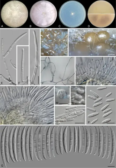
Pelat morfologi yang dicatat sumber sebagai Geejayessia clavata dan dipetakan ke Fusarium longyuwanense dalam backbone profil. Nama spesimen dan pembagian struktur mengikuti sumber.
Kredit dan penggunaan
Zhao-Qing Zeng, Wen-Ying Zhuang
Ukuran dan format gambar disesuaikan tanpa pemotongan, retus, atau penambahan skala. Lisensi sumber dipertahankan.
Sumber profil
- [1] Two new species of Geejayessia (Hypocreales) from Asia as evidenced by morphology and multi-gene analyses — Geejayessia clavata
- [2] Catalogue of Life Base2026-09-11 — Fusarium longyuwanense
- [3] Dokumentasi visual — Fusarium longyuwanense
- [4] Dokumentasi visual — Fusarium longyuwanense
Rujukan halaman ini
Fungi. Fusarium longyuwanense. Rilis data fungi-2026-10-12-expansion-2. https://fungi-atlas.mgadib.chatgpt.site/jamur/fusarium-longyuwanense/.
Status: impor sumber; pemeriksaan editorial per profil belum selesai. Untuk klaim biologis, gunakan rujukan pada bagian terkait dan periksa konteks sumber aslinya.