01 / Morfologi

Ciri dan struktur

Culture characteristics: Conidia germinated on WA within 24 h, and germ tubes produced from basal cell. Colonies growing on PDA reached 12−15 mm in diameter after one month at 25 °C, obverse olive to greyish green or dark greyish green in the inner, and light greyish green in the outer ring from above, reverse dark greyish green. Mycelium in culture up to 1–3 μm wide, subhyaline to brown, septate, branched. Conidiophores and conidiogenous cells indistinct. Chlamydospores are apparent in culture, globose to ellipsoidal or irregular shaped, hyaline at the beginning, becoming brown to black with ages, 10−21 × 5−19 μm (x ¯ = 16 × 12 μm, n = 30).

Sebagian uraian ditampilkan dalam bahasa sumber; terjemahannya belum tersedia atau belum lolos pemeriksaan.

Rujukan: [1]

Pengukuran mengikuti objek, spesimen atau kultur, dan metode yang disebutkan sumber. Baca uraian asli untuk konteks lengkap.

02 / Kehidupan

Habitat dan peran ekologis

Culture characteristics: Conidia germinated on WA within 24 h, and germ tubes produced from basal cell. Colonies growing on PDA reached 12−15 mm in diameter after one month at 25 °C, obverse olive to greyish green or dark greyish green in the inner, and light greyish green in the outer ring from above, reverse dark greyish green. Mycelium in culture up to 1–3 μm wide, subhyaline to brown, septate, branched. Conidiophores and conidiogenous cells indistinct. Chlamydospores are apparent in culture, globose to ellipsoidal or irregular shaped, hyaline at the beginning, becoming brown to black with ages, 10−21 × 5−19 μm (x ¯ = 16 × 12 μm, n = 30). Material examined: China, Guizhou Province, Guiyang City, Wudang District, Xiangsihe scenic spot, undisturbed forests with freshwater habitats, 26°26′51″ N, 106°37′53″ E, on decaying wood submerged in a freshwater stream, 22 February 2021, H.Z. Du, S99 (HKAS 122794, holotype); ex-holotype living culture CGMCC 3.20884; ibid., HUEST 22.0017, isotype, ex-isotype living culture UESTCC 22.0017.

Sebagian uraian ditampilkan dalam bahasa sumber; terjemahannya belum tersedia atau belum lolos pemeriksaan.

Rujukan: [1]

Habitat atau konteks temuan
hutan; kultur yang didokumentasikan
Substrat
kayu
Peran atau konteks
Belum dikategorikan pada rilis ini.
Temukan hubungan serupa
03 / Siklus hidup

Reproduksi

Culture characteristics: Conidia germinated on WA within 24 h, and germ tubes produced from basal cell. Colonies growing on PDA reached 12−15 mm in diameter after one month at 25 °C, obverse olive to greyish green or dark greyish green in the inner, and light greyish green in the outer ring from above, reverse dark greyish green. Mycelium in culture up to 1–3 μm wide, subhyaline to brown, septate, branched. Conidiophores and conidiogenous cells indistinct. Chlamydospores are apparent in culture, globose to ellipsoidal or irregular shaped, hyaline at the beginning, becoming brown to black with ages, 10−21 × 5−19 μm (x ¯ = 16 × 12 μm, n = 30).

Sebagian uraian ditampilkan dalam bahasa sumber; terjemahannya belum tersedia atau belum lolos pemeriksaan.

Rujukan: [1]

04 / Batas informasi

Catatan identitas dan cakupan

  • Nama diterima mengikuti Catalogue of Life Base2026-09-11; uraian mempertahankan konsep dan nama sumber aslinya.
  • Preserve original treatment localities, hosts and substrate scope. Materials examined describe documented specimens, not the full global range.
  • Impor dan terjemahan otomatis belum diperiksa editorial per profil. Struktur reproduktif yang disebutkan tidak membuktikan seluruh siklus hidup diketahui.
  • Pengukuran pada uraian mengikuti spesimen, kultur, dan metode sumber; angka tidak diperluas ke semua populasi.

Catatan distribusi dalam narasi mengikuti sumbernya. Dokumentasi spesimen atau foto mengikuti catatan sumber. Bukti wilayah Indonesia dan penilaian konservasi tidak disimpulkan dari gambar.

05 / Dokumentasi

Melihat dengan konteks

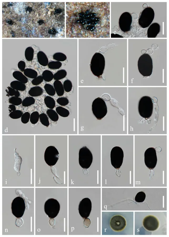
Fuscosporella guizhouensis (HKAS 122794, holotype). (a, b) Colony on submerged wood. (c, d) Conidiophores with conidia. (e – h) Conidia with apical appendages. (i − p) Conidiogenous cells and conidia. (q) Germinated conidium. (r, s) Colony on PDA (r from above, s from below). Scale bars: (c) = 30 µm, (d) = 40 µm, (e − p) = 30 µm, (q) = 40 µm.

Jenis
Pelat ilmiah
Kredit
© 2022 by the authors.
Lisensi
CC BY 4.0
Sumber media
Buka entri sumber

Gambar menunjukkan objek menurut identifikasi sumber, bukan verifikasi independen spesimen oleh Fungi. Tidak ada skala baru yang ditambahkan.

06 / Rujukan

Sumber profil

  1. [1] Morpho-Phylogenetic Evidence Reveals New Species of Fuscosporellaceae and Savoryellaceae from Freshwater Habitats in Guizhou Province, China — Fuscosporella guizhouensisBagian sumber: sec4-jof-08-01138_paragraph_1
    Tanggal penghimpunan sumber: 2026-10-12
    Teks sumber: CC BY 4.0 · Du HZ, Yang J, Liu NG, Cheewangkoon R, Liu JK.
  2. [2] Catalogue of Life Base2026-09-11 — Fuscosporella guizhouensisBagian sumber: ColDP NameUsage.tsv ID B2X2G; accepted species; source 2073
    Tanggal penghimpunan sumber: 2026-10-12
  3. [3] Dokumentasi visual — Fuscosporella guizhouensisBagian sumber: Individual media record; identification follows the source, not independent determination
    Tanggal penghimpunan sumber: 2026-10-12
    Media sumber: CC BY 4.0 · © 2022 by the authors.

Adaptasi teks: CC BY 4.0. Du HZ, Yang J, Liu NG, Cheewangkoon R, Liu JK. — Morpho-Phylogenetic Evidence Reveals New Species of Fuscosporellaceae and Savoryellaceae from Freshwater Habitats in Guizhou Province, China — Fuscosporella guizhouensis

Uraian asli dan riwayat sumber

Riwayat atau halaman sumber

Ringkasan sumber

Culture characteristics: Conidia germinated on WA within 24 h, and germ tubes produced from basal cell. Colonies growing on PDA reached 12−15 mm in diameter after one month at 25 °C, obverse olive to greyish green or dark greyish green in the inner, and light greyish green in the outer ring from above, reverse dark greyish green. Mycelium in culture up to 1–3 μm wide, subhyaline to brown, septate, branched. Conidiophores and conidiogenous cells indistinct. Chlamydospores are apparent in culture, globose to ellipsoidal or irregular shaped, hyaline at the beginning, becoming brown to black with ages, 10−21 × 5−19 μm (x ¯ = 16 × 12 μm, n = 30).

Revisi sumber

Morfologi sumber

Culture characteristics: Conidia germinated on WA within 24 h, and germ tubes produced from basal cell. Colonies growing on PDA reached 12−15 mm in diameter after one month at 25 °C, obverse olive to greyish green or dark greyish green in the inner, and light greyish green in the outer ring from above, reverse dark greyish green. Mycelium in culture up to 1–3 μm wide, subhyaline to brown, septate, branched. Conidiophores and conidiogenous cells indistinct. Chlamydospores are apparent in culture, globose to ellipsoidal or irregular shaped, hyaline at the beginning, becoming brown to black with ages, 10−21 × 5−19 μm (x ¯ = 16 × 12 μm, n = 30).

Revisi sumber

Ekologi sumber

Culture characteristics: Conidia germinated on WA within 24 h, and germ tubes produced from basal cell. Colonies growing on PDA reached 12−15 mm in diameter after one month at 25 °C, obverse olive to greyish green or dark greyish green in the inner, and light greyish green in the outer ring from above, reverse dark greyish green. Mycelium in culture up to 1–3 μm wide, subhyaline to brown, septate, branched. Conidiophores and conidiogenous cells indistinct. Chlamydospores are apparent in culture, globose to ellipsoidal or irregular shaped, hyaline at the beginning, becoming brown to black with ages, 10−21 × 5−19 μm (x ¯ = 16 × 12 μm, n = 30). Material examined: China, Guizhou Province, Guiyang City, Wudang District, Xiangsihe scenic spot, undisturbed forests with freshwater habitats, 26°26′51″ N, 106°37′53″ E, on decaying wood submerged in a freshwater stream, 22 February 2021, H.Z. Du, S99 (HKAS 122794, holotype); ex-holotype living culture CGMCC 3.20884; ibid., HUEST 22.0017, isotype, ex-isotype living culture UESTCC 22.0017.

Revisi sumber

Struktur reproduksi sumber

Culture characteristics: Conidia germinated on WA within 24 h, and germ tubes produced from basal cell. Colonies growing on PDA reached 12−15 mm in diameter after one month at 25 °C, obverse olive to greyish green or dark greyish green in the inner, and light greyish green in the outer ring from above, reverse dark greyish green. Mycelium in culture up to 1–3 μm wide, subhyaline to brown, septate, branched. Conidiophores and conidiogenous cells indistinct. Chlamydospores are apparent in culture, globose to ellipsoidal or irregular shaped, hyaline at the beginning, becoming brown to black with ages, 10−21 × 5−19 μm (x ¯ = 16 × 12 μm, n = 30).

Revisi sumber

Rujukan halaman ini

Fungi. Fuscosporella guizhouensis. Rilis data fungi-2026-10-12-expansion-2. https://fungi-atlas.mgadib.chatgpt.site/jamur/fuscosporella-guizhouensis/.

Status: impor sumber; pemeriksaan editorial per profil belum selesai. Untuk klaim biologis, gunakan rujukan pada bagian terkait dan periksa konteks sumber aslinya.