Saprobic on cabang mati dari Cornus sanguinea. Morph seksual Ascomata 100–130 μm tinggi, 160–220 μm berdiameter, sepi, tersebar ke kelompok, tenggelam, terlihat sebagai naik, bintik hitam di permukaan inang globose ke subglobosa, glabrous, uni-loculate, ostiole tengah dengan papilla kecil. Peridium 22–33 μm lebar, tipis hingga tebal dengan ketebalan yang sama, terdiri dari dua jenis lapisan sel pseudoparenkimat, lapisan dalam yang terdiri dari lapisan sel 3–5 dari sel datar, sel hialin, disusun dalam tekstura prismatik, lapisan luar yang terdiri dari beberapa lapisan sel cokelat gelap hingga hitam, disusun dalam tekstura angularis.
Profil ini diimpor dari uraian spesies pada sumber berlisensi dan sebagian teks diterjemahkan otomatis. Pemeriksaan editorial per profil belum selesai. Bagian yang belum diterjemahkan atau belum lolos pemeriksaan terjemahan tetap ditampilkan dalam bahasa sumber. Angka dan nama ilmiah dipertahankan.
Saprobic on dead branches of Cornus sanguinea. Sexual morph Ascomata 100–130 μm high, 160–220 μm diam., solitary, scattered to clustered, immersed, visible as raised, black spots on host surface globose to subglobose, glabrous, uni-loculate, ostiole central with minute papilla. Peridium 22–33 μm wide, thin to thick-walled with equal thickness, composed of two type layers of pseudoparenchymatous cells, inner layer comprising 3–5 cell layers of flattened, hyaline cells, arranged in a textura prismatica, outer layer comprising several layers of dark brown to black cells, arranged in a textura angularis. Hamathecium composed of dense, broad, 1.8–2.5 μm wide, filamentous, cellular pseudoparaphyses, with indistinct septa, not constricted at the septa, anastomosing at the apex, embedded in a hyaline gelatinous matrix. Asci (69–)80–90(–110) × (7–)8–9(–9.5) μm (\(\bar{x} \) = 86 × 8.5 μm, n = 20), 8-spored, bitunicate, fissitunicate, cylindric-clavate, short pedicellate, with furcate to obtuse end, apically rounded with well-developed ocular chamber. Ascospores (11.8–)13–15(–15.7) × (5.5–)6–6.5(–7.3) μm (\(\bar{x} \) = 13.9 × 6.3 μm, n = 40), overlapping, 1-seriate, ellipsoid to obovoid, initially hyaline to slightly brown, becoming brown at maturity, aseptate when young, becoming 1-septate, straight to slightly curved, smooth-walled. Asexual morph Undetermined. Culture characteristics: Colonies on PDA reaching 35–40 mm diam. after 5 weeks at 20–25 °C, colonies medium sparse, circular, convex, slightly rough surface with edge entire, well defined margin, cottony to fairly fluffy with sparse aspects, colony from above, white to cream at the margin, white to yellowish at the centre; from below, yellow to yellowish brown at the margin, light brown at the centre, mycelium white to cream with tufting, not producing pigmentation in PDA media agar.
Sebagian uraian ditampilkan dalam bahasa sumber; terjemahannya belum tersedia atau belum lolos pemeriksaan.
Pengukuran mengikuti objek, spesimen atau kultur, dan metode yang disebutkan sumber. Baca uraian asli untuk konteks lengkap.
02 / Kehidupan
Habitat dan peran ekologis
Saprobic on dead branches of Cornus sanguinea. Sexual morph Ascomata 100–130 μm high, 160–220 μm diam., solitary, scattered to clustered, immersed, visible as raised, black spots on host surface globose to subglobose, glabrous, uni-loculate, ostiole central with minute papilla. Peridium 22–33 μm wide, thin to thick-walled with equal thickness, composed of two type layers of pseudoparenchymatous cells, inner layer comprising 3–5 cell layers of flattened, hyaline cells, arranged in a textura prismatica, outer layer comprising several layers of dark brown to black cells, arranged in a textura angularis. Hamathecium composed of dense, broad, 1.8–2.5 μm wide, filamentous, cellular pseudoparaphyses, with indistinct septa, not constricted at the septa, anastomosing at the apex, embedded in a hyaline gelatinous matrix. Asci (69–)80–90(–110) × (7–)8–9(–9.5) μm (\(\bar{x} \) = 86 × 8.5 μm, n = 20), 8-spored, bitunicate, fissitunicate, cylindric-clavate, short pedicellate, with furcate to obtuse end, apically rounded with well-developed ocular chamber. Ascospores (11.8–)13–15(–15.7) × (5.5–)6–6.5(–7.3) μm (\(\bar{x} \) = 13.9 × 6.3 μm, n = 40), overlapping, 1-seriate, ellipsoid to obovoid, initially hyaline to slightly brown, becoming brown at maturity, aseptate when young, becoming 1-septate, straight to slightly curved, smooth-walled.
Sebagian uraian ditampilkan dalam bahasa sumber; terjemahannya belum tersedia atau belum lolos pemeriksaan.
Saprobic on dead branches of Cornus sanguinea. Sexual morph Ascomata 100–130 μm high, 160–220 μm diam., solitary, scattered to clustered, immersed, visible as raised, black spots on host surface globose to subglobose, glabrous, uni-loculate, ostiole central with minute papilla. Peridium 22–33 μm wide, thin to thick-walled with equal thickness, composed of two type layers of pseudoparenchymatous cells, inner layer comprising 3–5 cell layers of flattened, hyaline cells, arranged in a textura prismatica, outer layer comprising several layers of dark brown to black cells, arranged in a textura angularis. Hamathecium composed of dense, broad, 1.8–2.5 μm wide, filamentous, cellular pseudoparaphyses, with indistinct septa, not constricted at the septa, anastomosing at the apex, embedded in a hyaline gelatinous matrix.
Sebagian uraian ditampilkan dalam bahasa sumber; terjemahannya belum tersedia atau belum lolos pemeriksaan.
Nama diterima mengikuti Catalogue of Life Base2026-09-11; uraian mempertahankan konsep dan nama sumber aslinya.
Preserve original monograph treatment localities, hosts and specimen/substrate scope. Materials examined document specimens, not the full global range.
Impor dan terjemahan otomatis belum diperiksa editorial per profil. Struktur reproduktif yang disebutkan tidak membuktikan seluruh siklus hidup diketahui.
Pengukuran pada uraian mengikuti spesimen, kultur, dan metode sumber; angka tidak diperluas ke semua populasi.
Catatan distribusi dalam narasi mengikuti sumbernya. Dokumentasi spesimen atau foto mengikuti catatan sumber. Bukti wilayah Indonesia dan penilaian konservasi tidak disimpulkan dari gambar.
05 / Dokumentasi
Melihat dengan konteks
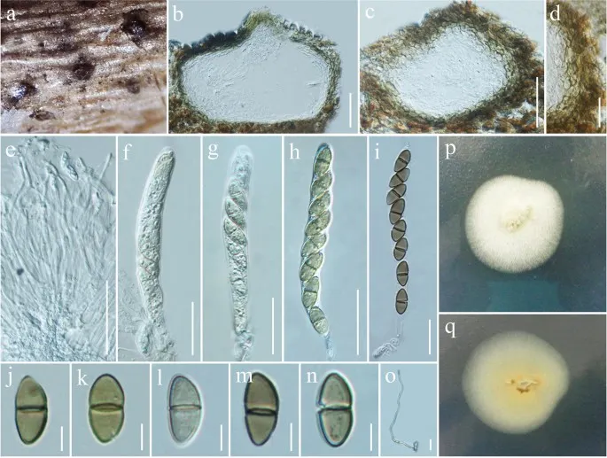
Fuscostagonospora camporesii (MFLU 16-1362, holotype). a Ascomata visible as black dots on host surface. b, c Vertical sections of ascomata. d Section through peridium. e Pseudoparaphyses. f – i Asci. j – n Ascospores. o Germinated ascospore. p Colony from above. q Colony from below. Scale bars: b, c = 50 µm, d – i = 20 µm, j – n = 5 µm, o = 10 µm
Jenis
Pelat ilmiah
Kredit
Penulis dan sumber artikel: Hyde, Kevin D.; Dong, Yang; Phookamsak, Rungtiwa; Jeewon, Rajesh; Bhat, D. Jayarama; Jones, E. B. Gareth; Liu, Ning-Guo; Abeywickrama, Pranami D.; Mapook, Ausana; Wei, Deping; Perera, Rekhani H.; Manawasinghe, Ishara S.; Pem, Dhandevi; Bundhun, Digvijayini; Karunarathna, Anuruddha; Ekanayaka, Anusha H.; Bao, Dan-Feng; Li, Junfu; Samarakoon, Milan C.; Chaiwan, Napalai; Lin, Chuan-Gen; Phutthacharoen, Kunthida; Zhang, Sheng-Nan; Senanayake, Indunil C.; Goonasekara, Ishani D.; Thambugala, Kasun M.; Phukhamsakda, Chayanard; Tennakoon, Danushka S.; Jiang, Hong-Bo; Yang, Jing; Zeng, Ming; Huanraluek, Naruemon; Liu, Jian-Kui (Jack); Wijesinghe, Subodini N.; Tian, Qing; Tibpromma, Saowaluck; Brahmanage, Rashika S.; Boonmee, Saranyaphat; Huang, Shi-Ke; Thiyagaraja, Vinodhini; Lu, Yong-Zhong; Jayawardena, Ruvishika S.; Dong, Wei; Yang, Er-Fu; Singh, Sanjay K.; Singh, Shiv Mohan; Rana, Shiwali; Lad, Sneha S.; Anand, Garima; Devadatha, Bandarupalli; Niranjan, M.; Sarma, V. Venkateswara; Liimatainen, Kare; Aguirre-Hudson, Begoña; Niskanen, Tuula; Overall, Andy; Alvarenga, Renato Lúcio Mendes; Gibertoni, Tatiana Baptista; Pfliegler, Walter P.; Horváth, Enikő; Imre, Alexandra; Alves, Amanda Lucia; da Silva Santos, Ana Carla; Tiago, Patricia Vieira; Bulgakov, Timur S.; Wanasinghe, Dhanushaka N.; Bahkali, Ali H.; Doilom, Mingkwan; Elgorban, Abdallah M.; Maharachchikumbura, Sajeewa S. N.; Rajeshkumar, Kunhiraman C.; Haelewaters, Danny; Mortimer, Peter E.; Zhao, Qi; Lumyong, Saisamorn; Xu, Jianchu; Sheng, Jun
Saprobic on dead branches of Cornus sanguinea.
Sexual morph Ascomata 100–130 μm high, 160–220 μm diam., solitary, scattered to clustered, immersed, visible as raised, black spots on host surface globose to subglobose, glabrous, uni-loculate, ostiole central with minute papilla.
Peridium 22–33 μm wide, thin to thick-walled with equal thickness, composed of two type layers of pseudoparenchymatous cells, inner layer comprising 3–5 cell layers of flattened, hyaline cells, arranged in a textura prismatica, outer layer comprising several layers of dark brown to black cells, arranged in a textura angularis.
Saprobic on dead branches of Cornus sanguinea.
Sexual morph Ascomata 100–130 μm high, 160–220 μm diam., solitary, scattered to clustered, immersed, visible as raised, black spots on host surface globose to subglobose, glabrous, uni-loculate, ostiole central with minute papilla.
Peridium 22–33 μm wide, thin to thick-walled with equal thickness, composed of two type layers of pseudoparenchymatous cells, inner layer comprising 3–5 cell layers of flattened, hyaline cells, arranged in a textura prismatica, outer layer comprising several layers of dark brown to black cells, arranged in a textura angularis.
Hamathecium composed of dense, broad, 1.8–2.5 μm wide, filamentous, cellular pseudoparaphyses, with indistinct septa, not constricted at the septa, anastomosing at the apex, embedded in a hyaline gelatinous matrix.
Asci (69–)80–90(–110) × (7–)8–9(–9.5) μm (\(\bar{x} \) = 86 × 8.5 μm, n = 20), 8-spored, bitunicate, fissitunicate, cylindric-clavate, short pedicellate, with furcate to obtuse end, apically rounded with well-developed ocular chamber.
Ascospores (11.8–)13–15(–15.7) × (5.5–)6–6.5(–7.3) μm (\(\bar{x} \) = 13.9 × 6.3 μm, n = 40), overlapping, 1-seriate, ellipsoid to obovoid, initially hyaline to slightly brown, becoming brown at maturity, aseptate when young, becoming 1-septate, straight to slightly curved, smooth-walled.
Asexual morph Undetermined.
Culture characteristics: Colonies on PDA reaching 35–40 mm diam. after 5 weeks at 20–25 °C, colonies medium sparse, circular, convex, slightly rough surface with edge entire, well defined margin, cottony to fairly fluffy with sparse aspects, colony from above, white to cream at the margin, white to yellowish at the centre; from below, yellow to yellowish brown at the margin, light brown at the centre, mycelium white to cream with tufting, not producing pigmentation in PDA media agar.
Saprobic on dead branches of Cornus sanguinea.
Sexual morph Ascomata 100–130 μm high, 160–220 μm diam., solitary, scattered to clustered, immersed, visible as raised, black spots on host surface globose to subglobose, glabrous, uni-loculate, ostiole central with minute papilla.
Peridium 22–33 μm wide, thin to thick-walled with equal thickness, composed of two type layers of pseudoparenchymatous cells, inner layer comprising 3–5 cell layers of flattened, hyaline cells, arranged in a textura prismatica, outer layer comprising several layers of dark brown to black cells, arranged in a textura angularis.
Hamathecium composed of dense, broad, 1.8–2.5 μm wide, filamentous, cellular pseudoparaphyses, with indistinct septa, not constricted at the septa, anastomosing at the apex, embedded in a hyaline gelatinous matrix.
Asci (69–)80–90(–110) × (7–)8–9(–9.5) μm (\(\bar{x} \) = 86 × 8.5 μm, n = 20), 8-spored, bitunicate, fissitunicate, cylindric-clavate, short pedicellate, with furcate to obtuse end, apically rounded with well-developed ocular chamber.
Ascospores (11.8–)13–15(–15.7) × (5.5–)6–6.5(–7.3) μm (\(\bar{x} \) = 13.9 × 6.3 μm, n = 40), overlapping, 1-seriate, ellipsoid to obovoid, initially hyaline to slightly brown, becoming brown at maturity, aseptate when young, becoming 1-septate, straight to slightly curved, smooth-walled.
Saprobic on dead branches of Cornus sanguinea.
Sexual morph Ascomata 100–130 μm high, 160–220 μm diam., solitary, scattered to clustered, immersed, visible as raised, black spots on host surface globose to subglobose, glabrous, uni-loculate, ostiole central with minute papilla.
Peridium 22–33 μm wide, thin to thick-walled with equal thickness, composed of two type layers of pseudoparenchymatous cells, inner layer comprising 3–5 cell layers of flattened, hyaline cells, arranged in a textura prismatica, outer layer comprising several layers of dark brown to black cells, arranged in a textura angularis.
Hamathecium composed of dense, broad, 1.8–2.5 μm wide, filamentous, cellular pseudoparaphyses, with indistinct septa, not constricted at the septa, anastomosing at the apex, embedded in a hyaline gelatinous matrix.
Status: impor sumber; pemeriksaan editorial per profil belum selesai. Untuk klaim biologis, gunakan rujukan pada bagian terkait dan periksa konteks sumber aslinya.
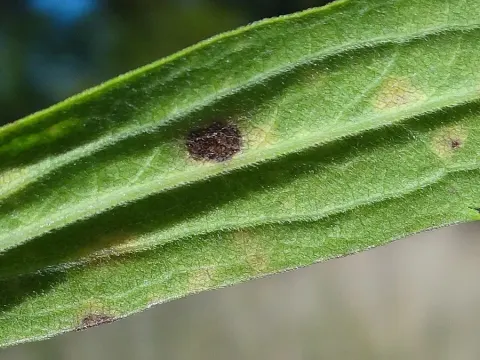
Fusicladium virgaureae Ondřej – gatunek grzybów z klasy Dothideomycetes. Jamur mikroskopis parasit pada tumbuhan jenis kaldu (Solidago). Itu menyebabkan noda daun.
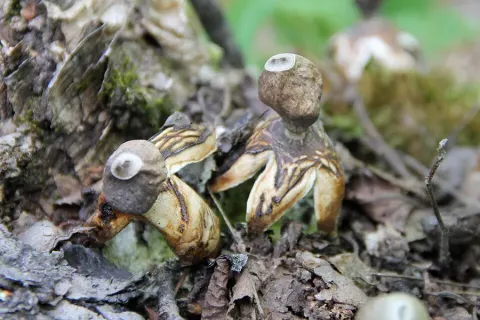
Geastrum quadrifidum, yang biasa dikenal sebagai bintang bumi berbunyi atau bintang bumi berempat kaki, adalah spesies jamur yang tidak dapat dimakan yang berasal dari genus Geastrum, atau jamur bintang bumi. First described scientifically…