Ciri dan struktur
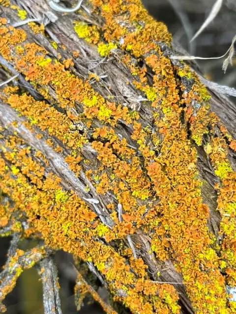
Gallowayella weberi is a foliose lichen that forms small thalli up to 4 mm wide, which adhere to surfaces ranging from closely to somewhat loosely attached. The of the thallus are flat, ranging from horizontal to slightly ascending, and measure between 0.3 and 0.5 mm in width. The tips of these lobes often curve inward, and the outermost parts of mature lobes are truncated or cut off squarely. The upper surface of Gallowayella weberi has a yellow to orange color and is smooth to the touch. Its lower surface contrasts with a white color. The cortex, or the outer layer of the lichen, is made of tightly packed cells and is colorless, except for the presence of external anthraquinone crystals, which are chemical compounds often found in lichens. The (photosynthetic partner) is a unicellular green alga from the genus Trebouxia. The medulla, which is the inner tissue beneath the cortex, has a net-like structure made of short cells. Rhizines, which are root-like structures helping the lichen to anchor to its , are mostly present and are white in color, turning yellow when exposed. They can be either attached to the substrate or free. Soralia, the structures for asexual reproduction, are found at the lobe tips and often form small structures resembling bird nests, with the soredia (powdery reproductive propagules) being yellow in color. Apothecia (fruiting bodies) are rare in this species and range from 0.7 to 2.0 mm in width. The measure between 12.7 and 14.0 by 7.0 to 7.7 μm, with a septum thickness of 2.9 to 4.6 μm. Pycnidia, an asexual reproductive structure, are common on the surface () and are orange in color, measuring about 0.1 to 0.2 mm in diameter.
Habitat dan peran ekologis
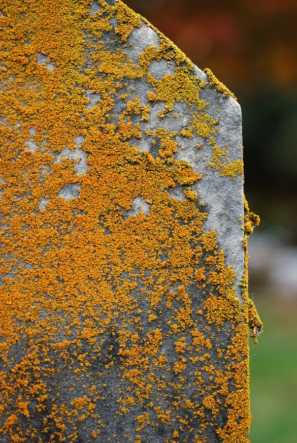
Gallowayella weberi can grow on diverse substrates including bark and rock, as well as on artificial ones such as concrete and tombstones. It is native to North America, and herbarium-based mapping indicates that it is widespread across the eastern and central United States, with its centre of distribution in the coastal plain of the southeastern United States. At the Delaware locality originally used to describe Xanthomendoza rosmarieae, the lichen was abundant on roadside plantings of Norway maple (Acer platanoides).
- Habitat atau konteks temuan
- Informasi belum tersedia pada sumber rilis ini.
- Substrat
- Informasi belum tersedia pada sumber rilis ini.
- Peran atau konteks
- Pembentuk liken
Reproduksi
The medulla, which is the inner tissue beneath the cortex, has a net-like structure made of short cells. Rhizines, which are root-like structures helping the lichen to anchor to its , are mostly present and are white in color, turning yellow when exposed. They can be either attached to the substrate or free. Soralia, the structures for asexual reproduction, are found at the lobe tips and often form small structures resembling bird nests, with the soredia (powdery reproductive propagules) being yellow in color. Apothecia (fruiting bodies) are rare in this species and range from 0.7 to 2.0 mm in width. The measure between 12.7 and 14.0 by 7.0 to 7.7 μm, with a septum thickness of 2.9 to 4.6 μm.
Catatan identitas dan cakupan
- Nama diterima mengikuti Catalogue of Life Base2026-09-11; uraian mempertahankan konsep dan nama sumber aslinya.
- Original geographic, specimen and life-cycle scope retained; source import awaits individual editorial review.
- Impor dan terjemahan otomatis belum diperiksa editorial per profil. Struktur reproduktif yang disebutkan tidak membuktikan seluruh siklus hidup diketahui.
- Pengukuran pada uraian mengikuti spesimen, kultur, dan metode sumber; angka tidak diperluas ke semua populasi.
Catatan distribusi dalam narasi mengikuti sumbernya. Dokumentasi spesimen atau foto mengikuti catatan sumber. Bukti wilayah Indonesia dan penilaian konservasi tidak disimpulkan dari gambar.
Melihat dengan konteks
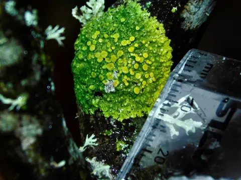
Dokumentasi Xanthomendoza weberi menurut asosiasi foto pada record takson iNaturalist. Kredit: Douglas Goldman. Identitas mengikuti sumber; bukan penentuan spesimen independen.
- Jenis
- Fotografi
- Kredit
- (c) Douglas Goldman, some rights reserved (CC BY-SA), uploaded by Douglas Goldman
- Lisensi
- CC BY-SA 4.0
- Sumber media
- Buka entri sumber
Dokumentasi tambahan
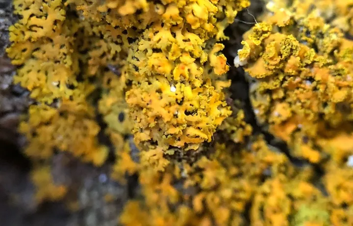
Foto yang diasosiasikan dengan Gallowayella weberi pada iNaturalist. Identifikasi mengikuti sumber; bukan penentuan spesimen independen.
Kredit dan penggunaan
(c) Nova Patch, some rights reserved (CC BY-SA), uploaded by Nova Patch
Ukuran dan format gambar disesuaikan tanpa pemotongan, retus, atau penambahan skala. Lisensi sumber dipertahankan.
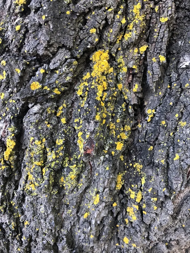
Foto yang diasosiasikan dengan Gallowayella weberi pada iNaturalist. Identifikasi mengikuti sumber; bukan penentuan spesimen independen.
Kredit dan penggunaan
(c) Nova Patch, some rights reserved (CC BY-SA), uploaded by Nova Patch
Ukuran dan format gambar disesuaikan tanpa pemotongan, retus, atau penambahan skala. Lisensi sumber dipertahankan.
Sumber profil
- [1] Gallowayella weberi — Wikipedia (en)
- [2] Catalogue of Life Base2026-09-11 — Gallowayella weberi
- [3] Dokumentasi visual — Gallowayella weberi
- [4] Dokumentasi visual — Gallowayella weberi
- [5] Dokumentasi visual — Gallowayella weberi
Rujukan halaman ini
Fungi. Gallowayella weberi. Rilis data fungi-2026-10-12-expansion-2. https://fungi-atlas.mgadib.chatgpt.site/jamur/gallowayella-weberi/.
Status: impor sumber; pemeriksaan editorial per profil belum selesai. Untuk klaim biologis, gunakan rujukan pada bagian terkait dan periksa konteks sumber aslinya.