01 / Morfologi

Ciri dan struktur

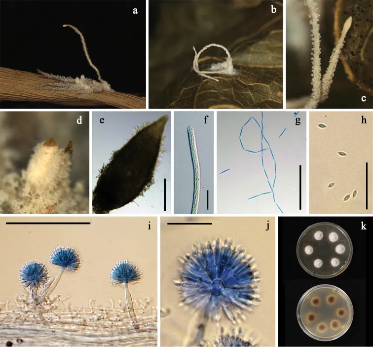
Spiders totally covered by the white to cream mycelial mat. A single synnema or synnemata in pairs cream to light brown, often darker than the mycelia covering the host, narrowing toward the apex and terminating in a swollen sterile tip with acute apex (Fig. 3a–c). Conidiophores arising laterally from the outer layer of synnemata, crowded, (23–)31–53(–83) × (4–)5.5–6.5(–7.5) μm, mostly verrucose, occasionally slightly roughed for very short conidiophores, abruptly narrowing to a slender apex and forming a globose to subglobose vesicle (Fig. 3c, i). Vesicles 6–7(–8) μm in diam, each bearing a number of metulae (Fig. 3j). Metulae obovoid to broadly obovoid, (7–)7.5–9(–10) × (4.5–)5–5.5(–6) μm (Fig. 3j). Phialides borne on metulae, narrowly obovoid, 7–8.5(–10) × 2–3 μm bearing fusiform to broadly fusiform conidia, (3.5–)4–5(–6) × 1.5–2(–2.5) μm (Fig. 3h, j). Conidial heads spherical, (31–)32–34.5(–37) μm in diam (Fig. 3j). Perithecia mostly appearing in pairs, ovoid, superficial with a loose covering of white to cream mycelia, reddish yellow, up to 1,000 μm in length, 320–350 μm in width (Fig. 3d–e). Asci 600–700 × 7–8 μm (Fig. 3f). Ascospores often disarticulating into part-spores. Part-spores fusiform, 12–15 × 2–3 μm (Fig. 3g). Granulomanus -like asexual morph absent. Colonies derived from conidia, on PDA slow-growing, attaining a diam of 1.1±0.03 cm in 20 days at 25 °C, white, velvety; reverse cream, becoming light brown with age toward center (Fig. 3k). Sporulation not observed.

Sebagian uraian ditampilkan dalam bahasa sumber; terjemahannya belum tersedia atau belum lolos pemeriksaan.

Rujukan: [1]

Pengukuran mengikuti objek, spesimen atau kultur, dan metode yang disebutkan sumber. Baca uraian asli untuk konteks lengkap.

02 / Kehidupan

Habitat dan peran ekologis

Thailand, Chiang Mai, Chiang Dao District, Ban Huathung; 19°420'N, 98°971'E; on Araneida attached to the underside of unidentified dicot leaf; 5 October 2012; K. Tasanathai, A. Khonsanit, W. Noisripoom, P. Srikitikulchai, R. Promharn (Holotype no. BBH 32918, ex-type culture no. BCC 56802, isolated from conidia) GenBank: ITS = MT477070, LSU = MT477063, TEF1 = MT503329, RPB1 = MT503322, RPB2 = MT503337. Colonies derived from conidia, on PDA slow-growing, attaining a diam of 1.1±0.03 cm in 20 days at 25 °C, white, velvety; reverse cream, becoming light brown with age toward center (Fig. 3k). Sporulation not observed.

Sebagian uraian ditampilkan dalam bahasa sumber; terjemahannya belum tersedia atau belum lolos pemeriksaan.

Rujukan: [1]

Habitat atau konteks temuan
kultur yang didokumentasikan
Substrat
Informasi belum tersedia pada sumber rilis ini.
Peran atau konteks
Belum dikategorikan pada rilis ini.
Temukan hubungan serupa
03 / Siklus hidup

Reproduksi

Spiders totally covered by the white to cream mycelial mat. A single synnema or synnemata in pairs cream to light brown, often darker than the mycelia covering the host, narrowing toward the apex and terminating in a swollen sterile tip with acute apex (Fig. 3a–c). Conidiophores arising laterally from the outer layer of synnemata, crowded, (23–)31–53(–83) × (4–)5.5–6.5(–7.5) μm, mostly verrucose, occasionally slightly roughed for very short conidiophores, abruptly narrowing to a slender apex and forming a globose to subglobose vesicle (Fig. 3c, i). Vesicles 6–7(–8) μm in diam, each bearing a number of metulae (Fig. 3j). Metulae obovoid to broadly obovoid, (7–)7.5–9(–10) × (4.5–)5–5.5(–6) μm (Fig. 3j).

Sebagian uraian ditampilkan dalam bahasa sumber; terjemahannya belum tersedia atau belum lolos pemeriksaan.

Rujukan: [1]

04 / Batas informasi

Catatan identitas dan cakupan

  • Nama diterima mengikuti Catalogue of Life Base2026-09-11; uraian mempertahankan konsep dan nama sumber aslinya.
  • Preserve original treatment localities, hosts and substrate scope. Materials examined describe documented specimens, not the full global range.
  • Impor dan terjemahan otomatis belum diperiksa editorial per profil. Struktur reproduktif yang disebutkan tidak membuktikan seluruh siklus hidup diketahui.
  • Pengukuran pada uraian mengikuti spesimen, kultur, dan metode sumber; angka tidak diperluas ke semua populasi.

Catatan distribusi dalam narasi mengikuti sumbernya. Dokumentasi spesimen atau foto mengikuti catatan sumber. Bukti wilayah Indonesia dan penilaian konservasi tidak disimpulkan dari gambar.

05 / Dokumentasi

Melihat dengan konteks

Gibellula fusiformispora a fungus on a spider (BBH 38838) b fungus on a spider (BBH 32918) c synnemata (BBH 32918) d, e perithecia (BBH 38838) f ascus (BBH 38838) g ascospores (BBH 38838) h conidia (BBH 32918) i conidiophores showing conidial heads (BBH 32918) j conidial head bearing conidia (BCC 32918) k colonies obverse and reverse on PDA at 25 °C after 20 days. Scale bars: 250 μm (e); 100 μm (i); 50 μm (g); 20 μm (f, h, j).

Jenis
Pelat ilmiah
Kredit
Wilawan Kuephadungphan, Kanoksri Tasanathai, Booppa Petcharad, Artit Khonsanit, Marc Stadler, J. Jennifer Luangsa-ard
Lisensi
CC BY 4.0
Sumber media
Buka entri sumber

Gambar menunjukkan objek menurut identifikasi sumber, bukan verifikasi independen spesimen oleh Fungi. Tidak ada skala baru yang ditambahkan.

06 / Rujukan

Sumber profil

  1. [1] Phylogeny- and morphology-based recognition of new species in the spider-parasitic genus Gibellula (Hypocreales, Cordycipitaceae) from Thailand — Gibellula fusiformisporaBagian sumber: PMC7481265_section_15
    Tanggal penghimpunan sumber: 2026-10-12
    Teks sumber: CC BY 4.0 · Kuephadungphan W, Tasanathai K, Petcharad B, Khonsanit A, Stadler M, Luangsa-Ard JJ.
  2. [2] Catalogue of Life Base2026-09-11 — Gibellula fusiformisporaBagian sumber: ColDP NameUsage.tsv ID 9Z7SC; accepted species; source 2073
    Tanggal penghimpunan sumber: 2026-10-12
  3. [3] Dokumentasi visual — Gibellula fusiformisporaBagian sumber: Individual media record; identification follows the source, not independent determination
    Tanggal penghimpunan sumber: 2026-10-12
    Media sumber: CC BY 4.0 · Wilawan Kuephadungphan, Kanoksri Tasanathai, Booppa Petcharad, Artit Khonsanit, Marc Stadler, J. Jennifer Luangsa-ard

Adaptasi teks: CC BY 4.0. Kuephadungphan W, Tasanathai K, Petcharad B, Khonsanit A, Stadler M, Luangsa-Ard JJ. — Phylogeny- and morphology-based recognition of new species in the spider-parasitic genus Gibellula (Hypocreales, Cordycipitaceae) from Thailand — Gibellula fusiformispora

Uraian asli dan riwayat sumber

Riwayat atau halaman sumber

Ringkasan sumber

Spiders totally covered by the white to cream mycelial mat. A single synnema or synnemata in pairs cream to light brown, often darker than the mycelia covering the host, narrowing toward the apex and terminating in a swollen sterile tip with acute apex (Fig. 3a–c). Conidiophores arising laterally from the outer layer of synnemata, crowded, (23–)31–53(–83) × (4–)5.5–6.5(–7.5) μm, mostly verrucose, occasionally slightly roughed for very short conidiophores, abruptly narrowing to a slender apex and forming a globose to subglobose vesicle (Fig. 3c, i).

Revisi sumber

Morfologi sumber

Spiders totally covered by the white to cream mycelial mat. A single synnema or synnemata in pairs cream to light brown, often darker than the mycelia covering the host, narrowing toward the apex and terminating in a swollen sterile tip with acute apex (Fig. 3a–c). Conidiophores arising laterally from the outer layer of synnemata, crowded, (23–)31–53(–83) × (4–)5.5–6.5(–7.5) μm, mostly verrucose, occasionally slightly roughed for very short conidiophores, abruptly narrowing to a slender apex and forming a globose to subglobose vesicle (Fig. 3c, i). Vesicles 6–7(–8) μm in diam, each bearing a number of metulae (Fig. 3j). Metulae obovoid to broadly obovoid, (7–)7.5–9(–10) × (4.5–)5–5.5(–6) μm (Fig. 3j). Phialides borne on metulae, narrowly obovoid, 7–8.5(–10) × 2–3 μm bearing fusiform to broadly fusiform conidia, (3.5–)4–5(–6) × 1.5–2(–2.5) μm (Fig. 3h, j). Conidial heads spherical, (31–)32–34.5(–37) μm in diam (Fig. 3j). Perithecia mostly appearing in pairs, ovoid, superficial with a loose covering of white to cream mycelia, reddish yellow, up to 1,000 μm in length, 320–350 μm in width (Fig. 3d–e). Asci 600–700 × 7–8 μm (Fig. 3f). Ascospores often disarticulating into part-spores. Part-spores fusiform, 12–15 × 2–3 μm (Fig. 3g). Granulomanus -like asexual morph absent. Colonies derived from conidia, on PDA slow-growing, attaining a diam of 1.1±0.03 cm in 20 days at 25 °C, white, velvety; reverse cream, becoming light brown with age toward center (Fig. 3k). Sporulation not observed.

Revisi sumber

Ekologi sumber

Thailand, Chiang Mai, Chiang Dao District, Ban Huathung; 19°420'N, 98°971'E; on Araneida attached to the underside of unidentified dicot leaf; 5 October 2012; K. Tasanathai, A. Khonsanit, W. Noisripoom, P. Srikitikulchai, R. Promharn (Holotype no. BBH 32918, ex-type culture no. BCC 56802, isolated from conidia) GenBank: ITS = MT477070, LSU = MT477063, TEF1 = MT503329, RPB1 = MT503322, RPB2 = MT503337. Colonies derived from conidia, on PDA slow-growing, attaining a diam of 1.1±0.03 cm in 20 days at 25 °C, white, velvety; reverse cream, becoming light brown with age toward center (Fig. 3k). Sporulation not observed.

Revisi sumber

Struktur reproduksi sumber

Spiders totally covered by the white to cream mycelial mat. A single synnema or synnemata in pairs cream to light brown, often darker than the mycelia covering the host, narrowing toward the apex and terminating in a swollen sterile tip with acute apex (Fig. 3a–c). Conidiophores arising laterally from the outer layer of synnemata, crowded, (23–)31–53(–83) × (4–)5.5–6.5(–7.5) μm, mostly verrucose, occasionally slightly roughed for very short conidiophores, abruptly narrowing to a slender apex and forming a globose to subglobose vesicle (Fig. 3c, i). Vesicles 6–7(–8) μm in diam, each bearing a number of metulae (Fig. 3j). Metulae obovoid to broadly obovoid, (7–)7.5–9(–10) × (4.5–)5–5.5(–6) μm (Fig. 3j).

Revisi sumber

Rujukan halaman ini

Fungi. Gibellula fusiformispora. Rilis data fungi-2026-10-12-expansion-2. https://fungi-atlas.mgadib.chatgpt.site/jamur/gibellula-fusiformispora/.

Status: impor sumber; pemeriksaan editorial per profil belum selesai. Untuk klaim biologis, gunakan rujukan pada bagian terkait dan periksa konteks sumber aslinya.