Ciri dan struktur
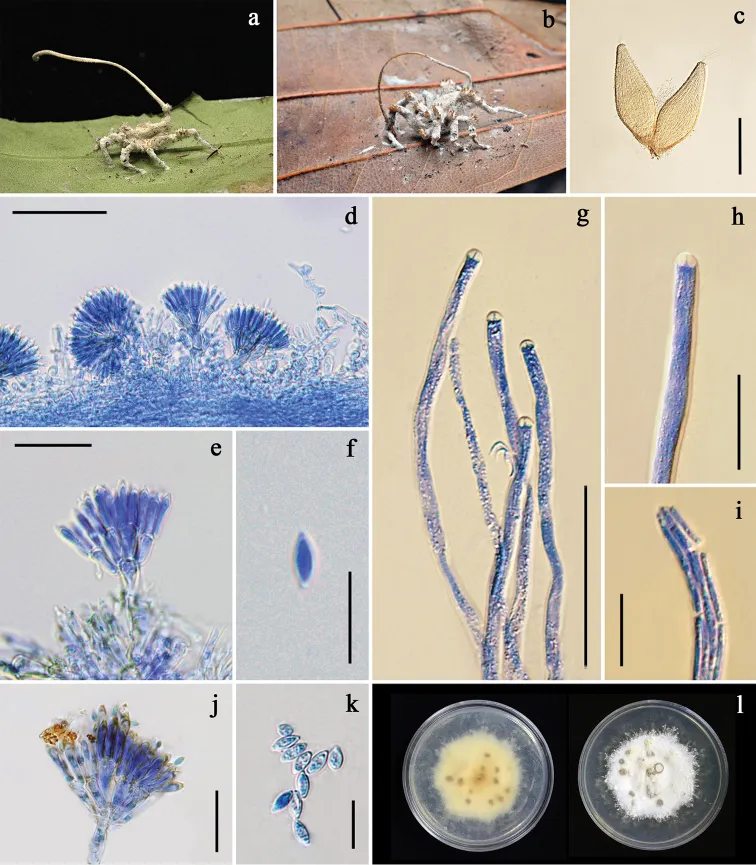
White to grayish- or brownish-white mycelial mat velvety, completely covering the spider host, firmly attaching the underside of living leaf by the mycelia covering its legs (Fig. 5a, b). Synnema solitary, arising from the posterior of the host abdomen, cylindrical, consisting of a compact bundle of parallel hyphae, 15–20 mm long with blunt tip. Conidiophores arising laterally from synnema, stout, smooth, mostly biverticillate, 20–29(–30) × 4 μm (Fig. 5d). Vesicles absent or hardly developed, bearing multiple metulae. Metulae obovoid, slightly broadening toward the base, (7–)9.5–12.5(–15) × (2–)3–5(–7) μm (Fig. 5e). A number of phialides borne on each metula, broadly cylindrical, abruptly tapering toward the apex, forming thickened distinct short neck, (9–)10–12.5(–14) × (2–)2.5–3.5(–4) μm, each bearing a conidium (Fig. 5e). Conidia fusiform, 5–7(–9) × (1.5–)2–3 μm (Fig. 5f). Sexual morph occasionally present. Perithecia occurring on the mycelial mat covering the host legs, occasionally on synnema particularly at base, superficial, mostly arranged in groups, ovoid, reddish yellow or light honey-brown, one-third immersed in the loose network of mycelia, 750–836(–870) × 310–361(–380) μm (Fig. 5c). Asci over 550 μm in length, (3–)4–5.5(–7) μm in width, ascus tip (4–)4.5–5 × 3–3.5(–4) μm (Fig. 5g, h). Ascospores often breaking into part-spores. Part-spores bacilliform, (9–)10–15(–22) × 1.5–2 μm (Fig. 5i). Granulomanus -like asexual morph absent. Colonies derived from conidia, on PDA slow-growing, attaining a diam of 1.5±0.1 cm in 4 weeks at 25 °C, floccose, forming irregular margin, white, reverse cream, darkening toward center with age (Fig. 5l).
Habitat dan peran ekologis
Thailand, Nakhon Ratchasima, Khao Yai National Park, Mo Sing To Nature Trail, 14°711'N, 101°421'E; on Portia sp. attached to the underside of unidentified dicot leaf; 1 June 2011; K. Tasanathai, P. Srikitikulchai, S. Mongkolsamrit, A. Khonsanit, K. Sansatchanon, W. Noisripoom (Holotype no. BBH 31439, ex-type culture no. BCC 47975, isolated from ascospores and BCC 47976, isolated from conidia) GenBank (BCC 47976): ITS = MT477078, LSU = MT477066, TEF1 = MT503335, RPB1 = MT503325, RPB2 = MT503339. Colonies derived from conidia, on PDA slow-growing, attaining a diam of 1.5±0.1 cm in 4 weeks at 25 °C, floccose, forming irregular margin, white, reverse cream, darkening toward center with age (Fig. 5l). Sporulation occurring after 3–4 months with the absence of synnema, forming a group of conidiophores, grey and scatter. Conidiophores biverticillate. Vesicles absent or hardly developed. Metulae obovoid, (10–)11–14.5(–16) × 3–5.5(–7) μm, each bearing cylindrical phialides, (10–)11.5–14(–16) × 3–4 μm. Conidia fusiform, 5–6(–7) × 3–3.5(–4) μm. Additional specimens examined. Thailand, Chumphon, Phato District, Phato Watershed Conservation and Management Unit; 9°784'N, 98°699'E; on Portia sp., underside of unidentified dicot leaf; 10 March 2011; K.
- Habitat atau konteks temuan
- kultur yang didokumentasikan
- Substrat
- Informasi belum tersedia pada sumber rilis ini.
- Peran atau konteks
- Belum dikategorikan pada rilis ini.
Reproduksi
White to grayish- or brownish-white mycelial mat velvety, completely covering the spider host, firmly attaching the underside of living leaf by the mycelia covering its legs (Fig. 5a, b). Synnema solitary, arising from the posterior of the host abdomen, cylindrical, consisting of a compact bundle of parallel hyphae, 15–20 mm long with blunt tip. Conidiophores arising laterally from synnema, stout, smooth, mostly biverticillate, 20–29(–30) × 4 μm (Fig. 5d). Vesicles absent or hardly developed, bearing multiple metulae. Metulae obovoid, slightly broadening toward the base, (7–)9.5–12.5(–15) × (2–)3–5(–7) μm (Fig. 5e). A number of phialides borne on each metula, broadly cylindrical, abruptly tapering toward the apex, forming thickened distinct short neck, (9–)10–12.5(–14) × (2–)2.5–3.5(–4) μm, each bearing a conidium (Fig. 5e).
Catatan identitas dan cakupan
- Nama diterima mengikuti Catalogue of Life Base2026-09-11; uraian mempertahankan konsep dan nama sumber aslinya.
- Preserve original treatment localities, hosts and substrate scope. Materials examined describe documented specimens, not the full global range.
- Impor dan terjemahan otomatis belum diperiksa editorial per profil. Struktur reproduktif yang disebutkan tidak membuktikan seluruh siklus hidup diketahui.
- Pengukuran pada uraian mengikuti spesimen, kultur, dan metode sumber; angka tidak diperluas ke semua populasi.
Catatan distribusi dalam narasi mengikuti sumbernya. Dokumentasi spesimen atau foto mengikuti catatan sumber. Bukti wilayah Indonesia dan penilaian konservasi tidak disimpulkan dari gambar.
Melihat dengan konteks
Gibellula scorpioides a fungus on a spider (BBH 29669) b fungus on a spider (BBH 31439) c perithecia (BBH 31439) d conidiophores arising on synnema (BBH 29669) e penicillate conidiophore (BBH 29669) f conidia (BBH 29669) g asci (BBH 31439) h ascus with apical apparatus (BBH 31439) i ascospores (BBH 31439) j penicillate conidiophore produced on PDA k conidia on PDA l colonies obverse and reverse on PDA at 25 °C after 4 months. Scale bars: 500 μm (c); 50 μm (d, g); 20 μm (e, h–j); 10 μm (f, k).
- Jenis
- Pelat ilmiah
- Kredit
- Wilawan Kuephadungphan, Kanoksri Tasanathai, Booppa Petcharad, Artit Khonsanit, Marc Stadler, J. Jennifer Luangsa-ard
- Lisensi
- CC BY 4.0
- Sumber media
- Buka entri sumber
Sumber profil
- [1] Phylogeny- and morphology-based recognition of new species in the spider-parasitic genus Gibellula (Hypocreales, Cordycipitaceae) from Thailand — Gibellula scorpioides
- [2] Catalogue of Life Base2026-09-11 — Gibellula scorpioides
- [3] Dokumentasi visual — Gibellula scorpioides
Rujukan halaman ini
Fungi. Gibellula scorpioides. Rilis data fungi-2026-10-12-expansion-2. https://fungi-atlas.mgadib.chatgpt.site/jamur/gibellula-scorpioides/.
Status: impor sumber; pemeriksaan editorial per profil belum selesai. Untuk klaim biologis, gunakan rujukan pada bagian terkait dan periksa konteks sumber aslinya.