Ciri dan struktur
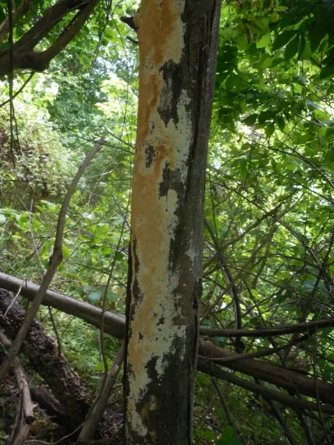
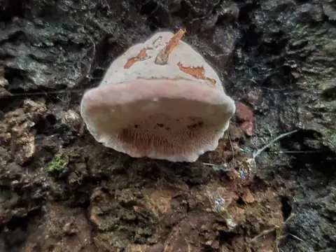
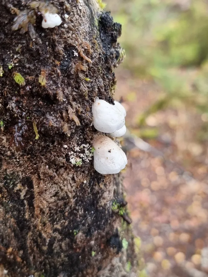
Hymenophore annual, solitary or growing caespitose from a common base, laterally attached by a short stem 3-4 mm. x 2-4 mm., sometimes growing from a stem-like elongation of the upper surface, occasionally dimidiate and attached by a broad base, firm but brittle, entirely white when fresh, drying cream. Pileus shell-shaped, spathulate, bell-shaped or occasionally applanate, 1-3.5 cm. x 1.5-2 cm. x 1-2 mm.; surface white, becoming cream, smooth, dull, sometimes obscurely sulcate, cuticle 30-40 µ thick, of compacted hyphae with ends upturned and arranged in a close palisade; margin thin, acute, interned, entire or when old crenate or toothed; hymenial surface plane or concave, even, white, drying cream, dissepiments not toothed. Context white, 0.25-1 mm. thick, densely woven, separated from the pores by a delicate wood-coloured woven layer; generative hyphae 6-10 µ thick, lumen 2-3 µ, freely branched, septate, staining, clamp connections present, small. Pores round or angular, 0.2-0.5 mm. deep, white in section, 100-150 µ diameter, or 6-7 per mm.; dissepiments 50-150 µ thick, mostly 100 µ, of woven mainly parallel hyphae, slightly tapering, apex delicately velutinate. Basidial type merulioid, basidia cylindrical, 6-8 x 2-3 µ, arranged in a dense persistent palisade. Spores allantoid, 3-3.5 x 1-1.5 µ, smooth. hyaline.
Habitat dan peran ekologis
Growing on bark and decorticated wood of dead branches, usually solitary, sometimes caespitose, occasionally dimidiate.
- Habitat atau konteks temuan
- Informasi belum tersedia pada sumber rilis ini.
- Substrat
- kulit kayu
- Peran atau konteks
- Belum dikategorikan pada rilis ini.
Reproduksi
Hymenophore annual, solitary or growing caespitose from a common base, laterally attached by a short stem 3-4 mm. x 2-4 mm., sometimes growing from a stem-like elongation of the upper surface, occasionally dimidiate and attached by a broad base, firm but brittle, entirely white when fresh, drying cream. Pileus shell-shaped, spathulate, bell-shaped or occasionally applanate, 1-3.5 cm. x 1.5-2 cm. x 1-2 mm.; surface white, becoming cream, smooth, dull, sometimes obscurely sulcate, cuticle 30-40 µ thick, of compacted hyphae with ends upturned and arranged in a close palisade; margin thin, acute, interned, entire or when old crenate or toothed; hymenial surface plane or concave, even, white, drying cream, dissepiments not toothed.
Catatan identitas dan cakupan
- Nama diterima mengikuti Catalogue of Life Base2026-09-11; uraian mempertahankan konsep dan nama sumber aslinya.
- Historical description as cited by BiotaNZ. Preserve explicit New Zealand/locality, host and specimen qualifiers. Distribution is the cited source scope, not a current global range.
- Impor dan terjemahan otomatis belum diperiksa editorial per profil. Struktur reproduktif yang disebutkan tidak membuktikan seluruh siklus hidup diketahui.
- Pengukuran pada uraian mengikuti spesimen, kultur, dan metode sumber; angka tidak diperluas ke semua populasi.
Catatan distribusi dalam narasi mengikuti sumbernya. Dokumentasi spesimen atau foto mengikuti catatan sumber. Bukti wilayah Indonesia dan penilaian konservasi tidak disimpulkan dari gambar.
Melihat dengan konteks
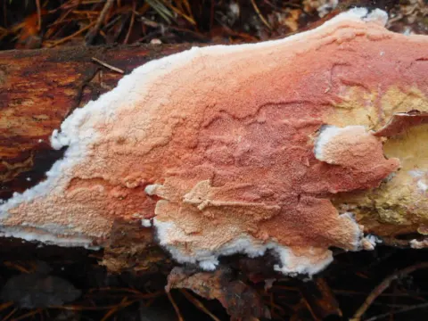
Dokumentasi Gloeoporus phlebophorus menurut asosiasi foto pada record takson iNaturalist. Kredit: Joss Simpson. Identitas mengikuti sumber; bukan penentuan spesimen independen.
- Jenis
- Fotografi
- Kredit
- (c) Joss Simpson, some rights reserved (CC BY)
- Lisensi
- CC BY 4.0
- Sumber media
- Buka entri sumber
Sumber profil
- [1] Cunningham, G.H. 1948: New Zealand Polyporaceae. 3. The genus Polyporus. New Zealand Department of Scientific and Industrial Research, Plant Diseases Division, Bulletin. 74. — Polyporus phlebophorus
- [2] Catalogue of Life Base2026-09-11 — Gloeoporus phlebophorus
- [3] Dokumentasi visual — Gloeoporus phlebophorus
Rujukan halaman ini
Fungi. Gloeoporus phlebophorus. Rilis data fungi-2026-10-12-expansion-2. https://fungi-atlas.mgadib.chatgpt.site/jamur/gloeoporus-phlebophorus/.
Status: impor sumber; pemeriksaan editorial per profil belum selesai. Untuk klaim biologis, gunakan rujukan pada bagian terkait dan periksa konteks sumber aslinya.