Ciri dan struktur
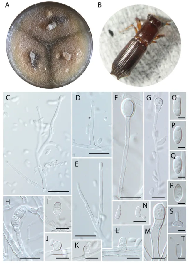
Diagnosis. Fungus associated with Euplatypus longius mycangium, inhabiting Zanthoxylum sp. (Rutaceae), exhibiting typical septate chlamydospores of 8–21 × 5–7 µm. Fungus producing typical terminal chlamydospore in culture. Description. Colonies initially cream, turning brown with age; reverse light orange on PDA. Fungi rapidly occupying the entire plate. Sexual morph was not observed. Asexual morph was composed of cylindrical, hyaline, regularly septate hyphae, ranging from 2–4 µm thick. Three types of conidiation were observed. The first type is sessile, micronematous, laterally forming globose to hemispherical conidia, sometimes leaving a scar after conidium release, formed solitarily and directly on the vegetative hyphae, 5–6.5 × 3.5–5 µm (Figure 2 J–L). The second type are terminally formed chlamydospores, hyaline, cylindrical, with 0–2 septa, developing a slightly verrucose wall with age (Figure 2 H), 8–21 × 5–7 µm (Figure 2 F–I,M). The third type formed on hyaline phialides, produced terminally or laterally on the main hypha, erect, occasionally irregular, cylindrical, sometimes tapering slightly towards the apex, (15–) 21 (–30) × 2.5 µm (Figure 2 C–E). Conidia hyaline, produced singly, aseptate, globose to elongate, sometimes curved, smooth-walled, commonly exhibiting germ tubes, (3.5–) 6 (–7) × 2.5–3 (–4) µm (Figure 2 S,T).
Habitat dan peran ekologis
Distribution. Thus far only known from Belize, the full distribution is unknown.
- Habitat atau konteks temuan
- Informasi belum tersedia pada sumber rilis ini.
- Substrat
- Informasi belum tersedia pada sumber rilis ini.
- Peran atau konteks
- Belum dikategorikan pada rilis ini.
Reproduksi
Description. Colonies initially cream, turning brown with age; reverse light orange on PDA. Fungi rapidly occupying the entire plate. Sexual morph was not observed. Asexual morph was composed of cylindrical, hyaline, regularly septate hyphae, ranging from 2–4 µm thick. Three types of conidiation were observed. The first type is sessile, micronematous, laterally forming globose to hemispherical conidia, sometimes leaving a scar after conidium release, formed solitarily and directly on the vegetative hyphae, 5–6.5 × 3.5–5 µm (Figure 2 J–L). The second type are terminally formed chlamydospores, hyaline, cylindrical, with 0–2 septa, developing a slightly verrucose wall with age (Figure 2 H), 8–21 × 5–7 µm (Figure 2 F–I,M).
Catatan identitas dan cakupan
- Nama diterima mengikuti Catalogue of Life Base2026-09-11; uraian mempertahankan konsep dan nama sumber aslinya.
- Preserve original treatment localities, hosts and substrate scope. Materials examined describe documented specimens, not the full global range.
- Impor dan terjemahan otomatis belum diperiksa editorial per profil. Struktur reproduktif yang disebutkan tidak membuktikan seluruh siklus hidup diketahui.
- Pengukuran pada uraian mengikuti spesimen, kultur, dan metode sumber; angka tidak diperluas ke semua populasi.
Catatan distribusi dalam narasi mengikuti sumbernya. Dokumentasi spesimen atau foto mengikuti catatan sumber. Bukti wilayah Indonesia dan penilaian konservasi tidak disimpulkan dari gambar.
Melihat dengan konteks
Harringtonia chlamydospora in pure culture on PDA. (A) Colony growth after 3 weeks at 25 °C. (B) Beetle vector, Euplatypus longius. (C) Terminal phialide. (D) Lateral phialide. (E) Terminal phialide. (F, G) Early stages of terminal chlamydospores. (H, I) Fully developed chlamydospore. (J – L) Micronematous conidiogenous cells formed laterally on the vegetative hypae. (M) Chlamydospore. (N) Germinating conidia. (O – R) Conidia. (S, T) Conidia exhibiting germ tube. Scale bars: (C – E) = 10 µm, (F, G) = 20 µm, (H) = 20 µm, (I) = 10 µm, (J – L) = 5 µm, (M) = 10 µm, (N) = 5 µm, (O – T) = 2 µm.
- Jenis
- Pelat ilmiah
- Kredit
- © 2022 by the authors.
- Lisensi
- CC BY 4.0
- Sumber media
- Buka entri sumber
Sumber profil
- [1] Four New Species of Harringtonia: Unravelling the Laurel Wilt Fungal Genus — Harringtonia chlamydospora
- [2] Catalogue of Life Base2026-09-11 — Harringtonia chlamydospora
- [3] Dokumentasi visual — Harringtonia chlamydospora
Rujukan halaman ini
Fungi. Harringtonia chlamydospora. Rilis data fungi-2026-10-12-expansion-2. https://fungi-atlas.mgadib.chatgpt.site/jamur/harringtonia-chlamydospora/.
Status: impor sumber; pemeriksaan editorial per profil belum selesai. Untuk klaim biologis, gunakan rujukan pada bagian terkait dan periksa konteks sumber aslinya.