01 / Morfologi

Ciri dan struktur

Sporodochia discoid; 0.1-1.25 mm. diam., immersed, surface alone showing, pulverulent, consisting of closely compacted tinted hyphae, 25-80 mmm. long, 2.5 mmm. thick. Conidia unicellular, globose, or shortly elliptical, 7-14 mmm. diam., epispore smooth, tinted dingy-violet or violet-brown, 1 mmm. thick.

Sebagian uraian ditampilkan dalam bahasa sumber; terjemahannya belum tersedia atau belum lolos pemeriksaan.

Rujukan: [1]

Pengukuran mengikuti objek, spesimen atau kultur, dan metode yang disebutkan sumber. Baca uraian asli untuk konteks lengkap.

02 / Kehidupan

Habitat dan peran ekologis

Habitat: Parasitic upon Aecidium otagense Linds. on Clematis Colensoi Hook. f. Miramar (Wellington), 20 m., J. W. Bird! 5 Nov., 1920. This fungus is conspicuous owing to the powdery nature of the spore-masses, and the purple colour of the spores and sporodochia. These are plano-discoid in shape, and are surrounded by the ruptured epidermis and partly disintegrated peridia of the aecidia; the conidiophores are closely packed together, and somewhat resemble the hymenium of Stereum or some similar Basidiomycete. On their apices are borne the spores, which, owing to the method of production, frequently occur in chains. In the specimens at hand the parasite is seen frequently to infect the aecidia before they appear on the surface - 4.e., before they dehisce - as when sections are examined aecidia in different stages of development may be seen in all stages of infection. But by far the greater number of parasitized aecidia appear to have been infected after they have expanded, since the sporidochia of the parasite are frequently seen to be partially surrounded by portions of the revolute margins, which give a very ragged appearance to the shoots of Clematis upon which the aecidia are located.

Sebagian uraian ditampilkan dalam bahasa sumber; terjemahannya belum tersedia atau belum lolos pemeriksaan.

Rujukan: [1]

Habitat atau konteks temuan
Informasi belum tersedia pada sumber rilis ini.
Substrat
Informasi belum tersedia pada sumber rilis ini.
Peran atau konteks
Belum dikategorikan pada rilis ini.
Temukan hubungan serupa
03 / Siklus hidup

Reproduksi

Sporodochia discoid; 0.1-1.25 mm. diam., immersed, surface alone showing, pulverulent, consisting of closely compacted tinted hyphae, 25-80 mmm. long, 2.5 mmm. thick. Conidia unicellular, globose, or shortly elliptical, 7-14 mmm. diam., epispore smooth, tinted dingy-violet or violet-brown, 1 mmm. thick.

Sebagian uraian ditampilkan dalam bahasa sumber; terjemahannya belum tersedia atau belum lolos pemeriksaan.

Rujukan: [1]

Sinonim menurut acuan taksonomi rilis: Cordalia persicina; Corticium sanguineum var. lilacinum; Helicobasidium brebissonii; Helicobasidium brebissonii f. barlae; Helicobasidium purpureum var. barlae; Helicobasidium purpureum var. orientale; Helicobasis purpureus; Hypochnus purpureus; Rhizoctonia allii; Rhizoctonia asparagi; Rhizoctonia crocorum; Rhizoctonia medicaginis; Rhizoctonia rubiae; Rhizoctonia violacea; Rhizoctonia violacea f. dauci; Sclerotium crocorum; Sclerotium medicaginis; Sclerotium sanguineum; Stypinella purpurea; Thanatophytum crocorum; Tuber parasiticum; Tubercularia persicina; Tuberculina persicina; Tuberculina sanguinea
04 / Batas informasi

Catatan identitas dan cakupan

  • Nama diterima mengikuti Catalogue of Life Base2026-09-11; uraian mempertahankan konsep dan nama sumber aslinya.
  • Historical description as cited by BiotaNZ. Preserve explicit New Zealand/locality, host and specimen qualifiers. Distribution is the cited source scope, not a current global range.
  • Impor dan terjemahan otomatis belum diperiksa editorial per profil. Struktur reproduktif yang disebutkan tidak membuktikan seluruh siklus hidup diketahui.
  • Pengukuran pada uraian mengikuti spesimen, kultur, dan metode sumber; angka tidak diperluas ke semua populasi.
  • Uraian sumber memuat penulisan satuan lama atau tidak standar. Angka dan satuannya dipertahankan apa adanya dan belum dinormalisasi.

Catatan distribusi dalam narasi mengikuti sumbernya. Dokumentasi spesimen atau foto mengikuti catatan sumber. Bukti wilayah Indonesia dan penilaian konservasi tidak disimpulkan dari gambar.

05 / Dokumentasi

Melihat dengan konteks

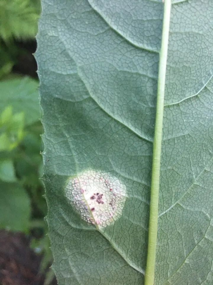
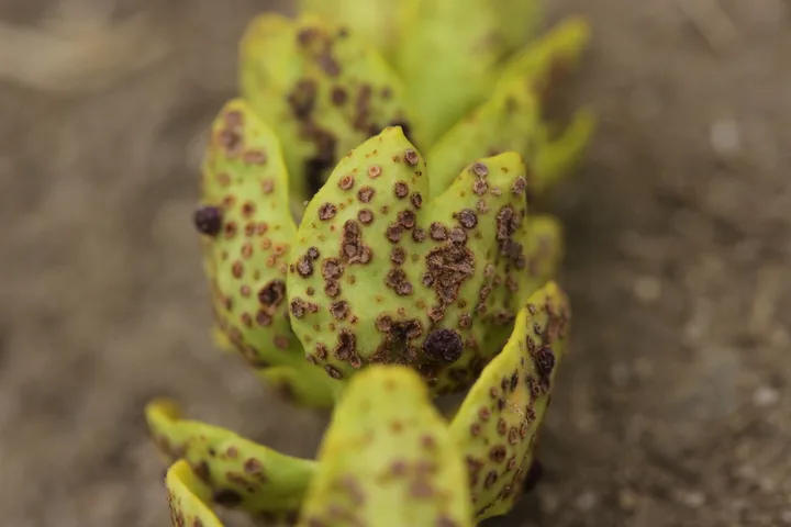
Foto yang diasosiasikan dengan Helicobasidium purpureum pada iNaturalist. Identifikasi mengikuti sumber; bukan penentuan spesimen independen.

Jenis
Fotografi
Kredit
(c) Björn Sothmann, some rights reserved (CC BY-SA), uploaded by Björn Sothmann
Sumber media
Buka entri sumber

Gambar menunjukkan objek menurut identifikasi sumber, bukan verifikasi independen spesimen oleh Fungi. Tidak ada skala baru yang ditambahkan.

Dokumentasi tambahan

06 / Rujukan

Sumber profil

  1. [1] Cunningham, G.H. 1924: The Uredinales, or rust-fungi, of New Zealand: supplement to Part 1; and Part 2. Transactions and Proceedings of the New Zealand Institute 55: 1-58. — Tuberculina persicinaBagian sumber: biota:1cb0ded1-36b9-11d5-9548-00d0592d548c:1cb1a86c-36b9-11d5-9548-00d0592d548c
    Tanggal penghimpunan sumber: 2026-10-12
    Teks sumber: CC BY 4.0 · Cunningham, G.H. 1924: The Uredinales, or rust-fungi, of New Zealand: supplement to Part 1; and Part 2. Transactions and Proceedings of the New Zealand Institute 55: 1-58.
  2. [2] Catalogue of Life Base2026-09-11 — Helicobasidium purpureumBagian sumber: ColDP NameUsage.tsv ID 3K7XX; accepted species; source 2073
    Tanggal penghimpunan sumber: 2026-10-12
  3. [3] Dokumentasi visual — Helicobasidium purpureumBagian sumber: Identitas mengikuti sumber dokumentasi; bukan penentuan spesimen independen.
    Tanggal penghimpunan sumber: 2026-10-17
    Media sumber: CC BY-SA 4.0 · (c) Björn Sothmann, some rights reserved (CC BY-SA), uploaded by Björn Sothmann
  4. [4] Dokumentasi visual — Helicobasidium purpureumBagian sumber: Identitas mengikuti sumber dokumentasi; bukan penentuan spesimen independen.
    Tanggal penghimpunan sumber: 2026-10-17
    Teks sumber: CC BY 4.0 · (c) Enrico Tomschke, some rights reserved (CC BY), uploaded by Enrico Tomschke

Adaptasi teks: CC BY 4.0. Cunningham, G.H. 1924: The Uredinales, or rust-fungi, of New Zealand: supplement to Part 1; and Part 2. Transactions and Proceedings of the New Zealand Institute 55: 1-58. — Cunningham, G.H. 1924: The Uredinales, or rust-fungi, of New Zealand: supplement to Part 1; and Part 2. Transactions and Proceedings of the New Zealand Institute 55: 1-58. — Tuberculina persicina

Uraian asli dan riwayat sumber

Riwayat atau halaman sumber

Ringkasan sumber

Sporodochia discoid; 0.1-1.25 mm. diam., immersed, surface alone showing, pulverulent, consisting of closely compacted tinted hyphae, 25-80 mmm. long, 2.5 mmm. thick. Conidia unicellular, globose, or shortly elliptical, 7-14 mmm. diam., epispore smooth, tinted dingy-violet or violet-brown, 1 mmm. thick.

Revisi sumber

Morfologi sumber

Sporodochia discoid; 0.1-1.25 mm. diam., immersed, surface alone showing, pulverulent, consisting of closely compacted tinted hyphae, 25-80 mmm. long, 2.5 mmm. thick. Conidia unicellular, globose, or shortly elliptical, 7-14 mmm. diam., epispore smooth, tinted dingy-violet or violet-brown, 1 mmm. thick.

Revisi sumber

Ekologi sumber

Habitat: Parasitic upon Aecidium otagense Linds. on Clematis Colensoi Hook. f. Miramar (Wellington), 20 m., J. W. Bird! 5 Nov., 1920. This fungus is conspicuous owing to the powdery nature of the spore-masses, and the purple colour of the spores and sporodochia. These are plano-discoid in shape, and are surrounded by the ruptured epidermis and partly disintegrated peridia of the aecidia; the conidiophores are closely packed together, and somewhat resemble the hymenium of Stereum or some similar Basidiomycete. On their apices are borne the spores, which, owing to the method of production, frequently occur in chains. In the specimens at hand the parasite is seen frequently to infect the aecidia before they appear on the surface - 4.e., before they dehisce - as when sections are examined aecidia in different stages of development may be seen in all stages of infection. But by far the greater number of parasitized aecidia appear to have been infected after they have expanded, since the sporidochia of the parasite are frequently seen to be partially surrounded by portions of the revolute margins, which give a very ragged appearance to the shoots of Clematis upon which the aecidia are located.

Revisi sumber

Struktur reproduksi sumber

Sporodochia discoid; 0.1-1.25 mm. diam., immersed, surface alone showing, pulverulent, consisting of closely compacted tinted hyphae, 25-80 mmm. long, 2.5 mmm. thick. Conidia unicellular, globose, or shortly elliptical, 7-14 mmm. diam., epispore smooth, tinted dingy-violet or violet-brown, 1 mmm. thick.

Revisi sumber

Rujukan halaman ini

Fungi. Helicobasidium purpureum. Rilis data fungi-2026-10-12-expansion-2. https://fungi-atlas.mgadib.chatgpt.site/jamur/helicobasidium-purpureum/.

Status: impor sumber; pemeriksaan editorial per profil belum selesai. Untuk klaim biologis, gunakan rujukan pada bagian terkait dan periksa konteks sumber aslinya.