Ciri dan struktur
The combination of a deeply rooting stipe, about 60 full length lamellae (from stipe to margin of pileus) and spores where almost every spore has a strongly loosening perispore forming a clear layer around the spore, separate this taxon from all other members of H. sect. Porphyrospora, as does the ITS-sequence. Basidiomes scattered. Pileus 37–64 mm wide, convex to broadly umbonate; surface dry or slightly viscid, without veil remnants on the pileus; cuticle color predominantly cream to pale buff (4A3, 4A4) in the center with paler margin, off-white to pale cream (4A2); pileus margin entire, hygrophanous. Lamellae adnate, moderately dense, thin, with approx. 60 full length lamellae and 2–3 lamellulae between the lamellae, off-white to cream when young, later pinkish or grayish red to purplish and eventually vinaceous to purple-brown following spore maturity; edges weakly fimbriate and white; the white edge remains when the basidiome is dried but the reddish brown color of the lamellae disappears with time. Stipe 160–194 mm long (including the ‘root’) and with central width 4–9 mm, cylindrical, distinctly and deeply rooting, white or alutaceous; surface dry, fibrillose, pruinose in the upper part, discoloring with handling and age. Flesh whitish, hardly discoloring where bruised. Smell fragrant; taste bitter. Spore deposit porphyry-brown (10E4). Exsiccata with no particular characteristics. Basidiospores based on n = 94 spores of the holotype, 5% to 95% percentile range 8.7–10.2 × 5.6–6.6 µm, with median 9.5 × 6.2 µm and av. 9.5 × 6.2 µm with S. D. length 0.47 µm and width 0.34 µm; Q value 5% to 95% percentile range 1.43–1.65, with median 1.53 and av. 1.54 with S.
Habitat dan peran ekologis
Only known from the type locality in Endau-Rompin (Johor) National Park, Malaysia. Scattered in lowland dipterocarp-oak woodland on the side of the path.
- Habitat atau konteks temuan
- hutan
- Substrat
- Informasi belum tersedia pada sumber rilis ini.
- Peran atau konteks
- Belum dikategorikan pada rilis ini.
Reproduksi
The combination of a deeply rooting stipe, about 60 full length lamellae (from stipe to margin of pileus) and spores where almost every spore has a strongly loosening perispore forming a clear layer around the spore, separate this taxon from all other members of H. sect. Porphyrospora, as does the ITS-sequence. Basidiomes scattered. Pileus 37–64 mm wide, convex to broadly umbonate; surface dry or slightly viscid, without veil remnants on the pileus; cuticle color predominantly cream to pale buff (4A3, 4A4) in the center with paler margin, off-white to pale cream (4A2); pileus margin entire, hygrophanous. Lamellae adnate, moderately dense, thin, with approx. 60 full length lamellae and 2–3 lamellulae between the lamellae, off-white to cream when young, later pinkish or grayish red to purplish and eventually vinaceous to purple-brown following spore maturity; edges weakly fimbriate and white; the white edge remains when the basidiome is dried but the reddish brown color of the lamellae disappears with time.
Catatan identitas dan cakupan
- Nama diterima mengikuti Catalogue of Life Base2026-09-11; uraian mempertahankan konsep dan nama sumber aslinya.
- Preserve original treatment localities, hosts and substrate scope. Materials examined describe documented specimens, not the full global range.
- Impor dan terjemahan otomatis belum diperiksa editorial per profil. Struktur reproduktif yang disebutkan tidak membuktikan seluruh siklus hidup diketahui.
- Pengukuran pada uraian mengikuti spesimen, kultur, dan metode sumber; angka tidak diperluas ke semua populasi.
Catatan distribusi dalam narasi mengikuti sumbernya. Dokumentasi spesimen atau foto mengikuti catatan sumber. Bukti wilayah Indonesia dan penilaian konservasi tidak disimpulkan dari gambar.
Melihat dengan konteks
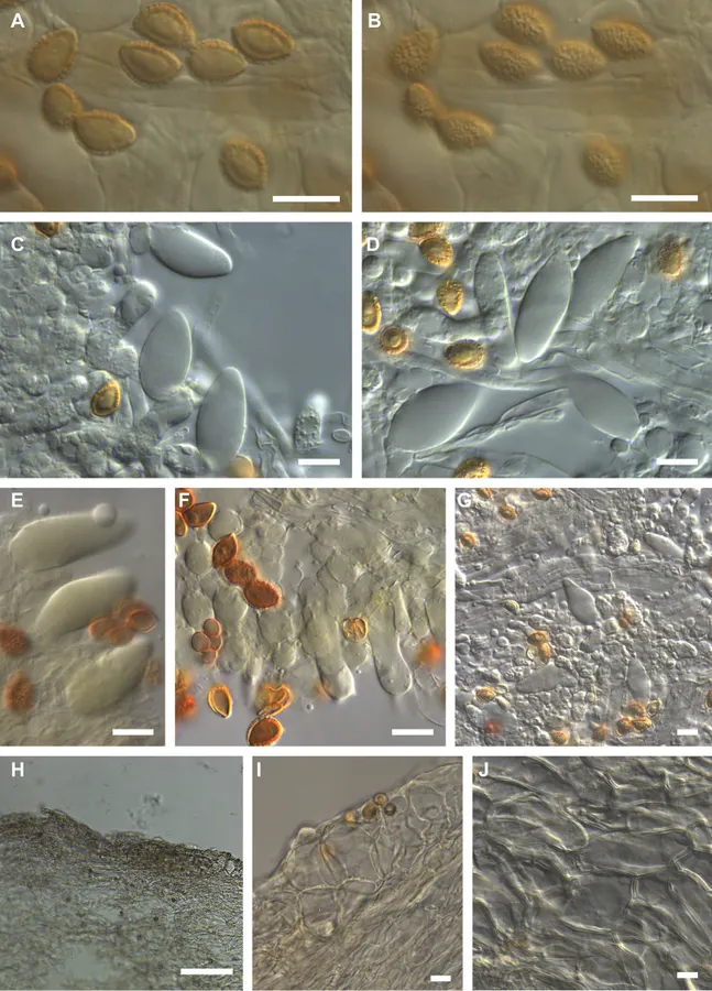
Microscopic features of Hebeloma radicans holotype (E. Horak 13265) A spores in KOH ×1600 B spore ornamentation in KOH ×1600 C cheilocystidia and basidium in KOH ×1000 D cheilocystidia and basidium in KOH ×1000 E pleurocystidia in Melzer’s reagent ×1000 F basidia in KOH ×1000 G pleurocystidia in KOH ×500 H sectional view of ixocutis showing thin gelatinous epicutis in KOH ×125 I sectional view of subcutis and trama below subcutis in KOH ×500 J sectional view of trama below subcutis in KOH ×500. Scale bars: 10 µm, 100 µm (H). Photographs H.J. Beker.
- Jenis
- Pelat ilmiah
- Kredit
- Ursula Eberhardt, Nicole Schütz, Henry J. Beker, Su See Lee, Egon Horak
- Lisensi
- CC BY 4.0
- Sumber media
- Buka entri sumber
Dokumentasi tambahan
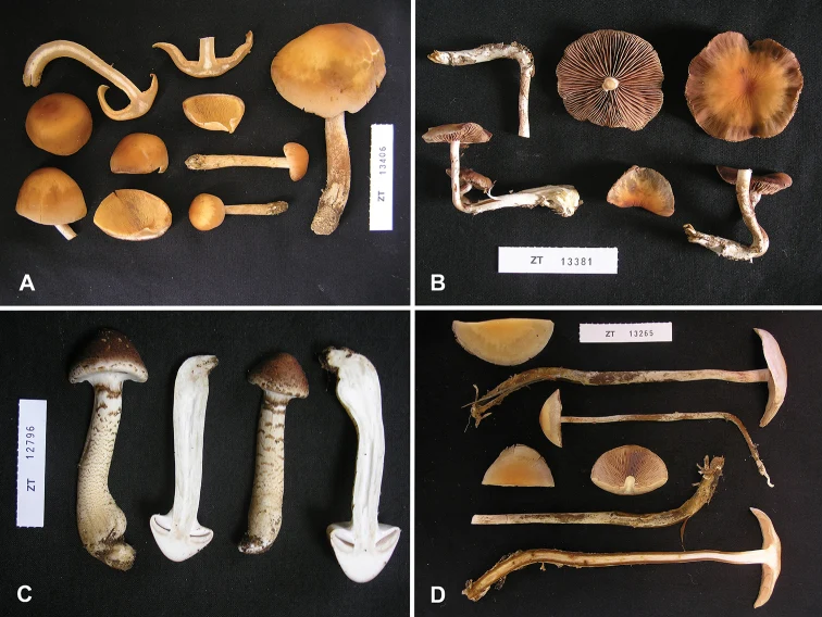
Pelat perbandingan tubuh buah. Hebeloma radicans ditampilkan pada panel D; panel lainnya memuat spesies pembanding. Pembagian panel mengikuti keterangan sumber.
Kredit dan penggunaan
Ursula Eberhardt, Nicole Schütz, Henry J. Beker, Su See Lee, Egon Horak
Ukuran dan format gambar disesuaikan tanpa pemotongan, retus, atau penambahan skala. Lisensi sumber dipertahankan.
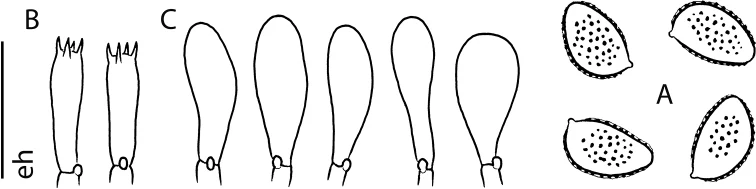
Microscopic features of Hebeloma radicans holotype (E. Horak 13265) A spores ×2000 B basidia ×1000 C cheilocystidia ×1000. Scale bar: 10 µm ×2000, 20 µm ×1000 and 40 µm ×500. Drawing E. Horak.
Kredit dan penggunaan
Ursula Eberhardt, Nicole Schütz, Henry J. Beker, Su See Lee, Egon Horak
Ukuran dan format gambar disesuaikan tanpa pemotongan, retus, atau penambahan skala. Lisensi sumber dipertahankan.
Sumber profil
- [1] Hebeloma in the Malay Peninsula: Masquerading within Psathyrella — Hebeloma radicans
- [2] Catalogue of Life Base2026-09-11 — Hebeloma radicans
- [3] Dokumentasi visual — Hebeloma radicans
- [4] Dokumentasi visual — Hebeloma radicans
- [5] Dokumentasi visual — Hebeloma radicans
Rujukan halaman ini
Fungi. Hebeloma radicans. Rilis data fungi-2026-10-12-expansion-2. https://fungi-atlas.mgadib.chatgpt.site/jamur/hebeloma-radicans/.
Status: impor sumber; pemeriksaan editorial per profil belum selesai. Untuk klaim biologis, gunakan rujukan pada bagian terkait dan periksa konteks sumber aslinya.