Ciri dan struktur
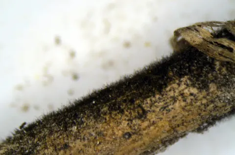
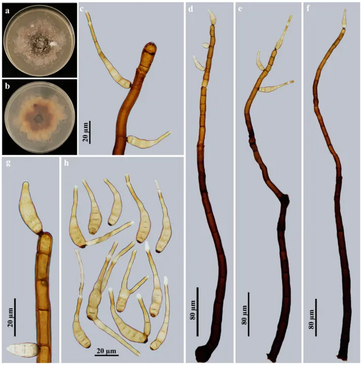
Description: Saprobic on decaying wood in terrestrial habitats. Anamorph hyphomycetous. Colonies on natural substrate effuse, scattered, hairy, brown to dark brown, velvety. Mycelium superficial and immersed in the substratum, composed of branched, septate, smooth, pale brown to brown, thick-walled hyphae. Conidiophores macronematous, mononematous, solitary or in groups of 2–4, simple, erect, straight or flexuous, cylindrical, smooth, 18–29-septate, blackish brown, paler towards the apex, with several cylindrical, enteroblastic percurrent extensions with well-defined small pores at the apex and laterally beneath the upper 1–5 septa, 544–712.5 × 12.5–17 μm (x ¯ = 622 × 15.2 μm, n = 8). Conidiogenous cells polytretic, integrated, terminal and intercalary, cylindrical, brown, smooth, with noncicatrized, distinct pores. Conidial secession schizolytic. Conidia acropleurogenous, solitary, dry, obclavate, rostrate, straight or curved, 6–13-distoseptate, pale brown, smooth, (20.7–)41.5–82.8 × 6.9–10.4 µm (x ¯ = 64 × 8 µm, n = 29), tapering to 1.7–3.5 µm near the apex, 3–7 μm wide at the base. Basal cell or apical portion sometimes with branches that developed a rostrate. Culture characteristics: Colony on PDA reaching 60–70 mm diam. after 4 weeks in an incubator under dark conditions at 25 °C, irregular circular, surface gray-brown with blackish brown in the center and gray mat of aerial hyphae at the margin; reverse rosy-brown with dark brown center and pale brown periphery.
Habitat dan peran ekologis
Description: Saprobic on decaying wood in terrestrial habitats. Anamorph hyphomycetous. Colonies on natural substrate effuse, scattered, hairy, brown to dark brown, velvety. Mycelium superficial and immersed in the substratum, composed of branched, septate, smooth, pale brown to brown, thick-walled hyphae. Conidiophores macronematous, mononematous, solitary or in groups of 2–4, simple, erect, straight or flexuous, cylindrical, smooth, 18–29-septate, blackish brown, paler towards the apex, with several cylindrical, enteroblastic percurrent extensions with well-defined small pores at the apex and laterally beneath the upper 1–5 septa, 544–712.5 × 12.5–17 μm (x ¯ = 622 × 15.2 μm, n = 8). Conidiogenous cells polytretic, integrated, terminal and intercalary, cylindrical, brown, smooth, with noncicatrized, distinct pores. Conidial secession schizolytic. Conidia acropleurogenous, solitary, dry, obclavate, rostrate, straight or curved, 6–13-distoseptate, pale brown, smooth, (20.7–)41.5–82.8 × 6.9–10.4 µm (x ¯ = 64 × 8 µm, n = 29), tapering to 1.7–3.5 µm near the apex, 3–7 μm wide at the base. Basal cell or apical portion sometimes with branches that developed a rostrate. Culture characteristics: Colony on PDA reaching 60–70 mm diam. after 4 weeks in an incubator under dark conditions at 25 °C, irregular circular, surface gray-brown with blackish brown in the center and gray mat of aerial hyphae at the margin; reverse rosy-brown with dark brown center and pale brown periphery.
- Habitat atau konteks temuan
- kultur yang didokumentasikan
- Substrat
- kayu
- Peran atau konteks
- Pengurai
Reproduksi
Description: Saprobic on decaying wood in terrestrial habitats. Anamorph hyphomycetous. Colonies on natural substrate effuse, scattered, hairy, brown to dark brown, velvety. Mycelium superficial and immersed in the substratum, composed of branched, septate, smooth, pale brown to brown, thick-walled hyphae. Conidiophores macronematous, mononematous, solitary or in groups of 2–4, simple, erect, straight or flexuous, cylindrical, smooth, 18–29-septate, blackish brown, paler towards the apex, with several cylindrical, enteroblastic percurrent extensions with well-defined small pores at the apex and laterally beneath the upper 1–5 septa, 544–712.5 × 12.5–17 μm (x ¯ = 622 × 15.2 μm, n = 8). Conidiogenous cells polytretic, integrated, terminal and intercalary, cylindrical, brown, smooth, with noncicatrized, distinct pores.
Catatan identitas dan cakupan
- Nama diterima mengikuti Catalogue of Life Base2026-09-11; uraian mempertahankan konsep dan nama sumber aslinya.
- Preserve original treatment localities, hosts and substrate scope. Materials examined describe documented specimens, not the full global range.
- Impor dan terjemahan otomatis belum diperiksa editorial per profil. Struktur reproduktif yang disebutkan tidak membuktikan seluruh siklus hidup diketahui.
- Pengukuran pada uraian mengikuti spesimen, kultur, dan metode sumber; angka tidak diperluas ke semua populasi.
Catatan distribusi dalam narasi mengikuti sumbernya. Dokumentasi spesimen atau foto mengikuti catatan sumber. Bukti wilayah Indonesia dan penilaian konservasi tidak disimpulkan dari gambar.
Melihat dengan konteks
Helminthosporium meilingense (HJAUP M1076, holotype): (a) surface of colony after 4 weeks on PDA; (b) reverse of colony after 4 weeks on PDA; (c,g) conidiogenous cells and conidia; (d – f) conidiophores, conidiogenous cells and conidia; (h) conidia.
- Jenis
- Pelat ilmiah
- Kredit
- © 2023 by the authors.
- Lisensi
- CC BY 4.0
- Sumber media
- Buka entri sumber
Sumber profil
- [1] Morphology and Multigene Phylogeny Revealed Three New Species of Helminthosporium (Massarinaceae, Pleosporales) from China — Helminthosporium meilingense
- [2] Catalogue of Life Base2026-09-11 — Helminthosporium meilingense
- [3] Dokumentasi visual — Helminthosporium meilingense
Rujukan halaman ini
Fungi. Helminthosporium meilingense. Rilis data fungi-2026-10-12-expansion-2. https://fungi-atlas.mgadib.chatgpt.site/jamur/helminthosporium-meilingense/.
Status: impor sumber; pemeriksaan editorial per profil belum selesai. Untuk klaim biologis, gunakan rujukan pada bagian terkait dan periksa konteks sumber aslinya.