01 / Morfologi

Ciri dan struktur

(after Bohn 1993): Colonies on PDA and MEA 30–33 mm after 10 d (52–62 mm after 20 d), appearing leathery, at first whitish to greyish, later becoming greyish-orange, particularly on MEA; margin superficial, entire on MEA but fimbriate to lobate on PDA; exudate and diffusible pigment absent; reverse greyish-orange, especially at the margin; brownish, thick-walled, chlamydospore-like swollen portions 6–18 µm diam. present. Conidiomata appearing after ca. 14 d as dark green pustules of various sizes, irregularly scattered over the colony surface, but sometimes arranged in concentric rings, particularly in old cultures, initially covered by mycelium but becoming almost black and shiny at later stages due to the mass of conidia; conidiomata sporodochial (acervular?), irregular, dark green, up to 2 mm diam., scattered, gregarious or coalescent, composed of a 50–70 µm high stroma of textura intricata and conidiophores. Marginal hyphae and setae absent. Conidiophores arising from the stroma, branched, septate, yellowish to brownish, ca. 40–75 µm × 2–4 µm. Conidiogenous cells cylindrical to subulate, 15–25 × 2–3 µm, arranged in verticils of 2–4 at the top of the conidiophore, sometimes also intercalary, provided with conspicuous, pigmented collarettes and producing conidia by percurrent growth. Conidia black and shiny in mass, olivaceous to brownish under the microscope, straight, cylindrical with rounded ends, sometimes slightly narrowing towards the base or apiculate, (9–)10–12(–15) × (5–)6(–7) µm, with smooth wall. Teleomorph not formed after 3 months incubation. Culture (own observations): Colony on MEA circular, first hyaline, turning and long remaining whitish, with age forming narrow concentric zones with tooth-like margins and turning pale brownish.

Sebagian uraian ditampilkan dalam bahasa sumber; terjemahannya belum tersedia atau belum lolos pemeriksaan.

Rujukan: [1]

Pengukuran mengikuti objek, spesimen atau kultur, dan metode yang disebutkan sumber. Baca uraian asli untuk konteks lengkap.

02 / Kehidupan

Habitat dan peran ekologis

Melanconis groenlandica is known from North America (Greenland, USA) and Japan from Betula maximowicziana, B. nana and B. papyrifera.

Sebagian uraian ditampilkan dalam bahasa sumber; terjemahannya belum tersedia atau belum lolos pemeriksaan.

Rujukan: [1]

Habitat atau konteks temuan
Informasi belum tersedia pada sumber rilis ini.
Substrat
Informasi belum tersedia pada sumber rilis ini.
Peran atau konteks
Belum dikategorikan pada rilis ini.
Temukan hubungan serupa
03 / Siklus hidup

Reproduksi

(after Bohn 1993): Colonies on PDA and MEA 30–33 mm after 10 d (52–62 mm after 20 d), appearing leathery, at first whitish to greyish, later becoming greyish-orange, particularly on MEA; margin superficial, entire on MEA but fimbriate to lobate on PDA; exudate and diffusible pigment absent; reverse greyish-orange, especially at the margin; brownish, thick-walled, chlamydospore-like swollen portions 6–18 µm diam. present. Conidiomata appearing after ca. 14 d as dark green pustules of various sizes, irregularly scattered over the colony surface, but sometimes arranged in concentric rings, particularly in old cultures, initially covered by mycelium but becoming almost black and shiny at later stages due to the mass of conidia; conidiomata sporodochial (acervular?), irregular, dark green, up to 2 mm diam., scattered, gregarious or coalescent, composed of a 50–70 µm high stroma of textura intricata and conidiophores.

Sebagian uraian ditampilkan dalam bahasa sumber; terjemahannya belum tersedia atau belum lolos pemeriksaan.

Rujukan: [1]

Sinonim menurut acuan taksonomi rilis: Myrothecium groenlandicum
04 / Batas informasi

Catatan identitas dan cakupan

  • Nama diterima mengikuti Catalogue of Life Base2026-09-11; uraian mempertahankan konsep dan nama sumber aslinya.
  • Preserve original treatment localities, hosts and substrate scope. Materials examined describe documented specimens, not the full global range.
  • Impor dan terjemahan otomatis belum diperiksa editorial per profil. Struktur reproduktif yang disebutkan tidak membuktikan seluruh siklus hidup diketahui.
  • Pengukuran pada uraian mengikuti spesimen, kultur, dan metode sumber; angka tidak diperluas ke semua populasi.

Catatan distribusi dalam narasi mengikuti sumbernya. Dokumentasi spesimen atau foto mengikuti catatan sumber. Bukti wilayah Indonesia dan penilaian konservasi tidak disimpulkan dari gambar.

05 / Dokumentasi

Melihat dengan konteks

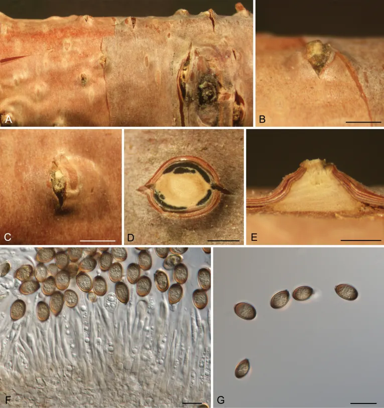
Morphology of Melanconis groenlandica from Betula tianchanica. A–C. Habit of conidiomata on branch; D. Transverse section through conidiomata; E. Longitudinal section through conidiomata; F. Conidiophores and conidiogenous cells; G. Alpha conidia. Scale bars: 500 μm (B–E); 10 μm (F, G).

Jenis
Pelat ilmiah
Kredit
Caixia Wang, Ning Jiang, Chuli Liu, ZiYan Xu, Hailong Lu, Rong Ma
Lisensi
CC BY 4.0
Sumber media
Buka entri sumber

Gambar menunjukkan objek menurut identifikasi sumber, bukan verifikasi independen spesimen oleh Fungi. Tidak ada skala baru yang ditambahkan.

06 / Rujukan

Sumber profil

  1. [1] The genus Melanconis (Diaporthales) — Melanconis groenlandicaBagian sumber: PMC7062851_section_23
    Tanggal penghimpunan sumber: 2026-10-12
    Teks sumber: CC BY 4.0 · Jaklitsch WM, Voglmayr H.
  2. [2] Catalogue of Life Base2026-09-11 — Melanconis groenlandicaBagian sumber: ColDP NameUsage.tsv ID 739DD; accepted species; source 2073
    Tanggal penghimpunan sumber: 2026-10-12
  3. [3] Dokumentasi visual — Melanconis groenlandicaBagian sumber: Individual media record; identification follows the source, not independent determination
    Tanggal penghimpunan sumber: 2026-10-12
    Media sumber: CC BY 4.0 · Caixia Wang, Ning Jiang, Chuli Liu, ZiYan Xu, Hailong Lu, Rong Ma

Adaptasi teks: CC BY 4.0. Jaklitsch WM, Voglmayr H. — The genus Melanconis (Diaporthales) — Melanconis groenlandica

Uraian asli dan riwayat sumber

Riwayat atau halaman sumber

Ringkasan sumber

(after Bohn 1993): Colonies on PDA and MEA 30–33 mm after 10 d (52–62 mm after 20 d), appearing leathery, at first whitish to greyish, later becoming greyish-orange, particularly on MEA; margin superficial, entire on MEA but fimbriate to lobate on PDA; exudate and diffusible pigment absent; reverse greyish-orange, especially at the margin; brownish, thick-walled, chlamydospore-like swollen portions 6–18 µm diam. present. Conidiomata appearing after ca. 14 d as dark green pustules of various sizes, irregularly scattered over the colony surface, but sometimes arranged in concentric rings, particularly in old cultures, initially covered by mycelium but becoming almost black and shiny at later stages due to the mass of conidia; conidiomata sporodochial (acervular?), irregular, dark green, up to 2 mm diam., scattered, gregarious or coalescent, composed of a 50–70 µm high stroma of textura intricata and conidiophores.

Revisi sumber

Morfologi sumber

(after Bohn 1993): Colonies on PDA and MEA 30–33 mm after 10 d (52–62 mm after 20 d), appearing leathery, at first whitish to greyish, later becoming greyish-orange, particularly on MEA; margin superficial, entire on MEA but fimbriate to lobate on PDA; exudate and diffusible pigment absent; reverse greyish-orange, especially at the margin; brownish, thick-walled, chlamydospore-like swollen portions 6–18 µm diam. present. Conidiomata appearing after ca. 14 d as dark green pustules of various sizes, irregularly scattered over the colony surface, but sometimes arranged in concentric rings, particularly in old cultures, initially covered by mycelium but becoming almost black and shiny at later stages due to the mass of conidia; conidiomata sporodochial (acervular?), irregular, dark green, up to 2 mm diam., scattered, gregarious or coalescent, composed of a 50–70 µm high stroma of textura intricata and conidiophores. Marginal hyphae and setae absent. Conidiophores arising from the stroma, branched, septate, yellowish to brownish, ca. 40–75 µm × 2–4 µm. Conidiogenous cells cylindrical to subulate, 15–25 × 2–3 µm, arranged in verticils of 2–4 at the top of the conidiophore, sometimes also intercalary, provided with conspicuous, pigmented collarettes and producing conidia by percurrent growth. Conidia black and shiny in mass, olivaceous to brownish under the microscope, straight, cylindrical with rounded ends, sometimes slightly narrowing towards the base or apiculate, (9–)10–12(–15) × (5–)6(–7) µm, with smooth wall. Teleomorph not formed after 3 months incubation. Culture (own observations): Colony on MEA circular, first hyaline, turning and long remaining whitish, with age forming narrow concentric zones with tooth-like margins and turning pale brownish.

Revisi sumber

Ekologi sumber

Melanconis groenlandica is known from North America (Greenland, USA) and Japan from Betula maximowicziana, B. nana and B. papyrifera.

Revisi sumber

Struktur reproduksi sumber

(after Bohn 1993): Colonies on PDA and MEA 30–33 mm after 10 d (52–62 mm after 20 d), appearing leathery, at first whitish to greyish, later becoming greyish-orange, particularly on MEA; margin superficial, entire on MEA but fimbriate to lobate on PDA; exudate and diffusible pigment absent; reverse greyish-orange, especially at the margin; brownish, thick-walled, chlamydospore-like swollen portions 6–18 µm diam. present. Conidiomata appearing after ca. 14 d as dark green pustules of various sizes, irregularly scattered over the colony surface, but sometimes arranged in concentric rings, particularly in old cultures, initially covered by mycelium but becoming almost black and shiny at later stages due to the mass of conidia; conidiomata sporodochial (acervular?), irregular, dark green, up to 2 mm diam., scattered, gregarious or coalescent, composed of a 50–70 µm high stroma of textura intricata and conidiophores.

Revisi sumber

Rujukan halaman ini

Fungi. Melanconis groenlandica. Rilis data fungi-2026-10-12-expansion-2. https://fungi-atlas.mgadib.chatgpt.site/jamur/melanconis-groenlandica/.

Status: impor sumber; pemeriksaan editorial per profil belum selesai. Untuk klaim biologis, gunakan rujukan pada bagian terkait dan periksa konteks sumber aslinya.