Saprobic on Raksida sawit yang membusuk (Phoenix paludosa). Morph seksual tidak ditentukan. Aseksual morph koloni di substratum superficial, effuse, tufted, hitam. Mycelium immersed, terdiri dari bercabang, bersepta, halus, cokelat pucat hifa. Conidiophores 125–170 μm long (\(\bar{x} \) = 147.7 μm, n = 20), 3.5–6.5 μm wide (\(\bar{x} \) = 5.5 μm, n = 20), macronematous, 3–5 in loose flared fascicle or rarely single, unbranched, multi-septate, thick-walled, smooth, slightly geniculate near the apex, straight or flexuous, with percurrent proliferation that is often nodose, brown below, becoming pale brown above to subhyaline at the tip.
Sebagian uraian ditampilkan dalam bahasa sumber; terjemahannya belum tersedia atau belum lolos pemeriksaan.
Profil ini diimpor dari uraian spesies pada sumber berlisensi dan sebagian teks diterjemahkan otomatis. Pemeriksaan editorial per profil belum selesai. Bagian yang belum diterjemahkan atau belum lolos pemeriksaan terjemahan tetap ditampilkan dalam bahasa sumber. Angka dan nama ilmiah dipertahankan.
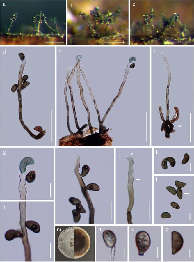
Melanographium phoenicis (MFLU18-1587, holotype). a – c Colonies on natural substratum. d, e Conidiophores with conidiogenous cells bearing condia, note (e) conidiophores in loose flared fascicles. f Conidiophores in loosely fascicles. g – i Attached conidia at the proliferation of conidiophores that often at nodose. j Conidiogenous cells with conidiogenous scars. k, l Conidia. m Colony on PDA. n – p Conidia producing in culture. Scale bars: a – c = 100 μm, d – f = 50 μm, g, h, k, l = 10 μm, i, j = 20 μm, n – p = 5 μm Penulis dan sumber artikel: Hyde, Kevin D.; Dong, Yang; Phookamsak, Rungtiwa; Jeewon, Rajesh; Bhat, D. Jayarama; Jones, E. B. Gareth; Liu, Ning-Guo; A… (kredit lengkap di bawah) · CC BY 4.0 · Ukuran dan format gambar disesuaikan; lisensi asli dipertahankan.
01 / Morfologi
Ciri dan struktur
Saprobic on decaying rachides of palm (Phoenix paludosa). Sexual morph Undetermined. Asexual morph Colonies on the substratum superficial, effuse, tufted, black. Mycelium immersed, composed of branched, septate, smooth, pale brown hyphae. Conidiophores 125–170 μm long (\(\bar{x} \) = 147.7 μm, n = 20), 3.5–6.5 μm wide (\(\bar{x} \) = 5.5 μm, n = 20), macronematous, 3–5 in loose flared fascicle or rarely single, unbranched, multi-septate, thick-walled, smooth, slightly geniculate near the apex, straight or flexuous, with percurrent proliferation that is often nodose, brown below, becoming pale brown above to subhyaline at the tip. Conidiogenous cells polyblastic, integrated, terminal and intercalary, cylindrical, cicatrized, intercalary conidiogenous loci often at the nodose proliferations, with conidiogenous scars. Conidia 11–18 μm long (\(\bar{x} \) = 14.4 μm, n = 30), 6–9 μm wide (\(\bar{x} \) = 7.9 μm, n = 50), holoblastic, solitary, aseptate, olivaceous to dark brown, reniform or obovoid, with a subhyaline longitudinal germ-slit on the convex side, guttulate, verruculose, smooth-walled. Conidial secession schizolytic. Culture characteristics: Conidia germinated on PDA within 24 h and been transferred to MEA. Colonies growing on MEA, velvety, with flat surface, firstly whitish, and becoming olive to grey-green or light grey-green. Mycelium superficial and partially immersed, branched, septate, hyaline to pale brown, smooth. Conidia produced in culture smaller than that found in substrate, 8.2–14.5 μm long (\(\bar{x} \) = 11.7 μm, n = 10), 5.9–8.7 μm wide (\(\bar{x} \) = 6.9 μm, n = 10).
Sebagian uraian ditampilkan dalam bahasa sumber; terjemahannya belum tersedia atau belum lolos pemeriksaan.
Pengukuran mengikuti objek, spesimen atau kultur, dan metode yang disebutkan sumber. Baca uraian asli untuk konteks lengkap.
02 / Kehidupan
Habitat dan peran ekologis
Saprobic on decaying rachides of palm (Phoenix paludosa). Sexual morph Undetermined. Asexual morph Colonies on the substratum superficial, effuse, tufted, black. Mycelium immersed, composed of branched, septate, smooth, pale brown hyphae. Conidiophores 125–170 μm long (\(\bar{x} \) = 147.7 μm, n = 20), 3.5–6.5 μm wide (\(\bar{x} \) = 5.5 μm, n = 20), macronematous, 3–5 in loose flared fascicle or rarely single, unbranched, multi-septate, thick-walled, smooth, slightly geniculate near the apex, straight or flexuous, with percurrent proliferation that is often nodose, brown below, becoming pale brown above to subhyaline at the tip. Conidiogenous cells polyblastic, integrated, terminal and intercalary, cylindrical, cicatrized, intercalary conidiogenous loci often at the nodose proliferations, with conidiogenous scars. Conidia 11–18 μm long (\(\bar{x} \) = 14.4 μm, n = 30), 6–9 μm wide (\(\bar{x} \) = 7.9 μm, n = 50), holoblastic, solitary, aseptate, olivaceous to dark brown, reniform or obovoid, with a subhyaline longitudinal germ-slit on the convex side, guttulate, verruculose, smooth-walled. Conidial secession schizolytic. Culture characteristics: Conidia germinated on PDA within 24 h and been transferred to MEA.
Sebagian uraian ditampilkan dalam bahasa sumber; terjemahannya belum tersedia atau belum lolos pemeriksaan.
Saprobic on decaying rachides of palm (Phoenix paludosa). Sexual morph Undetermined. Asexual morph Colonies on the substratum superficial, effuse, tufted, black. Mycelium immersed, composed of branched, septate, smooth, pale brown hyphae. Conidiophores 125–170 μm long (\(\bar{x} \) = 147.7 μm, n = 20), 3.5–6.5 μm wide (\(\bar{x} \) = 5.5 μm, n = 20), macronematous, 3–5 in loose flared fascicle or rarely single, unbranched, multi-septate, thick-walled, smooth, slightly geniculate near the apex, straight or flexuous, with percurrent proliferation that is often nodose, brown below, becoming pale brown above to subhyaline at the tip. Conidiogenous cells polyblastic, integrated, terminal and intercalary, cylindrical, cicatrized, intercalary conidiogenous loci often at the nodose proliferations, with conidiogenous scars.
Sebagian uraian ditampilkan dalam bahasa sumber; terjemahannya belum tersedia atau belum lolos pemeriksaan.
Nama diterima mengikuti Catalogue of Life Base2026-09-11; uraian mempertahankan konsep dan nama sumber aslinya.
Preserve original monograph treatment localities, hosts and specimen/substrate scope. Materials examined document specimens, not the full global range.
Impor dan terjemahan otomatis belum diperiksa editorial per profil. Struktur reproduktif yang disebutkan tidak membuktikan seluruh siklus hidup diketahui.
Pengukuran pada uraian mengikuti spesimen, kultur, dan metode sumber; angka tidak diperluas ke semua populasi.
Catatan distribusi dalam narasi mengikuti sumbernya. Dokumentasi spesimen atau foto mengikuti catatan sumber. Bukti wilayah Indonesia dan penilaian konservasi tidak disimpulkan dari gambar.
05 / Dokumentasi
Melihat dengan konteks
Melanographium phoenicis (MFLU18-1587, holotype). a – c Colonies on natural substratum. d, e Conidiophores with conidiogenous cells bearing condia, note (e) conidiophores in loose flared fascicles. f Conidiophores in loosely fascicles. g – i Attached conidia at the proliferation of conidiophores that often at nodose. j Conidiogenous cells with conidiogenous scars. k, l Conidia. m Colony on PDA. n – p Conidia producing in culture. Scale bars: a – c = 100 μm, d – f = 50 μm, g, h, k, l = 10 μm, i, j = 20 μm, n – p = 5 μm
Jenis
Pelat ilmiah
Kredit
Penulis dan sumber artikel: Hyde, Kevin D.; Dong, Yang; Phookamsak, Rungtiwa; Jeewon, Rajesh; Bhat, D. Jayarama; Jones, E. B. Gareth; Liu, Ning-Guo; Abeywickrama, Pranami D.; Mapook, Ausana; Wei, Deping; Perera, Rekhani H.; Manawasinghe, Ishara S.; Pem, Dhandevi; Bundhun, Digvijayini; Karunarathna, Anuruddha; Ekanayaka, Anusha H.; Bao, Dan-Feng; Li, Junfu; Samarakoon, Milan C.; Chaiwan, Napalai; Lin, Chuan-Gen; Phutthacharoen, Kunthida; Zhang, Sheng-Nan; Senanayake, Indunil C.; Goonasekara, Ishani D.; Thambugala, Kasun M.; Phukhamsakda, Chayanard; Tennakoon, Danushka S.; Jiang, Hong-Bo; Yang, Jing; Zeng, Ming; Huanraluek, Naruemon; Liu, Jian-Kui (Jack); Wijesinghe, Subodini N.; Tian, Qing; Tibpromma, Saowaluck; Brahmanage, Rashika S.; Boonmee, Saranyaphat; Huang, Shi-Ke; Thiyagaraja, Vinodhini; Lu, Yong-Zhong; Jayawardena, Ruvishika S.; Dong, Wei; Yang, Er-Fu; Singh, Sanjay K.; Singh, Shiv Mohan; Rana, Shiwali; Lad, Sneha S.; Anand, Garima; Devadatha, Bandarupalli; Niranjan, M.; Sarma, V. Venkateswara; Liimatainen, Kare; Aguirre-Hudson, Begoña; Niskanen, Tuula; Overall, Andy; Alvarenga, Renato Lúcio Mendes; Gibertoni, Tatiana Baptista; Pfliegler, Walter P.; Horváth, Enikő; Imre, Alexandra; Alves, Amanda Lucia; da Silva Santos, Ana Carla; Tiago, Patricia Vieira; Bulgakov, Timur S.; Wanasinghe, Dhanushaka N.; Bahkali, Ali H.; Doilom, Mingkwan; Elgorban, Abdallah M.; Maharachchikumbura, Sajeewa S. N.; Rajeshkumar, Kunhiraman C.; Haelewaters, Danny; Mortimer, Peter E.; Zhao, Qi; Lumyong, Saisamorn; Xu, Jianchu; Sheng, Jun
Saprobic on decaying rachides of palm (Phoenix paludosa).
Sexual morph Undetermined.
Asexual morph Colonies on the substratum superficial, effuse, tufted, black.
Mycelium immersed, composed of branched, septate, smooth, pale brown hyphae.
Conidiophores 125–170 μm long (\(\bar{x} \) = 147.7 μm, n = 20), 3.5–6.5 μm wide (\(\bar{x} \) = 5.5 μm, n = 20), macronematous, 3–5 in loose flared fascicle or rarely single, unbranched, multi-septate, thick-walled, smooth, slightly geniculate near the apex, straight or flexuous, with percurrent proliferation that is often nodose, brown below, becoming pale brown above to subhyaline at the tip.
Saprobic on decaying rachides of palm (Phoenix paludosa).
Sexual morph Undetermined.
Asexual morph Colonies on the substratum superficial, effuse, tufted, black.
Mycelium immersed, composed of branched, septate, smooth, pale brown hyphae.
Conidiophores 125–170 μm long (\(\bar{x} \) = 147.7 μm, n = 20), 3.5–6.5 μm wide (\(\bar{x} \) = 5.5 μm, n = 20), macronematous, 3–5 in loose flared fascicle or rarely single, unbranched, multi-septate, thick-walled, smooth, slightly geniculate near the apex, straight or flexuous, with percurrent proliferation that is often nodose, brown below, becoming pale brown above to subhyaline at the tip.
Conidiogenous cells polyblastic, integrated, terminal and intercalary, cylindrical, cicatrized, intercalary conidiogenous loci often at the nodose proliferations, with conidiogenous scars.
Conidia 11–18 μm long (\(\bar{x} \) = 14.4 μm, n = 30), 6–9 μm wide (\(\bar{x} \) = 7.9 μm, n = 50), holoblastic, solitary, aseptate, olivaceous to dark brown, reniform or obovoid, with a subhyaline longitudinal germ-slit on the convex side, guttulate, verruculose, smooth-walled.
Conidial secession schizolytic.
Culture characteristics: Conidia germinated on PDA within 24 h and been transferred to MEA.
Colonies growing on MEA, velvety, with flat surface, firstly whitish, and becoming olive to grey-green or light grey-green.
Mycelium superficial and partially immersed, branched, septate, hyaline to pale brown, smooth.
Conidia produced in culture smaller than that found in substrate, 8.2–14.5 μm long (\(\bar{x} \) = 11.7 μm, n = 10), 5.9–8.7 μm wide (\(\bar{x} \) = 6.9 μm, n = 10).
Saprobic on decaying rachides of palm (Phoenix paludosa).
Sexual morph Undetermined.
Asexual morph Colonies on the substratum superficial, effuse, tufted, black.
Mycelium immersed, composed of branched, septate, smooth, pale brown hyphae.
Conidiophores 125–170 μm long (\(\bar{x} \) = 147.7 μm, n = 20), 3.5–6.5 μm wide (\(\bar{x} \) = 5.5 μm, n = 20), macronematous, 3–5 in loose flared fascicle or rarely single, unbranched, multi-septate, thick-walled, smooth, slightly geniculate near the apex, straight or flexuous, with percurrent proliferation that is often nodose, brown below, becoming pale brown above to subhyaline at the tip.
Conidiogenous cells polyblastic, integrated, terminal and intercalary, cylindrical, cicatrized, intercalary conidiogenous loci often at the nodose proliferations, with conidiogenous scars.
Conidia 11–18 μm long (\(\bar{x} \) = 14.4 μm, n = 30), 6–9 μm wide (\(\bar{x} \) = 7.9 μm, n = 50), holoblastic, solitary, aseptate, olivaceous to dark brown, reniform or obovoid, with a subhyaline longitudinal germ-slit on the convex side, guttulate, verruculose, smooth-walled.
Conidial secession schizolytic.
Culture characteristics: Conidia germinated on PDA within 24 h and been transferred to MEA.
Saprobic on decaying rachides of palm (Phoenix paludosa).
Sexual morph Undetermined.
Asexual morph Colonies on the substratum superficial, effuse, tufted, black.
Mycelium immersed, composed of branched, septate, smooth, pale brown hyphae.
Conidiophores 125–170 μm long (\(\bar{x} \) = 147.7 μm, n = 20), 3.5–6.5 μm wide (\(\bar{x} \) = 5.5 μm, n = 20), macronematous, 3–5 in loose flared fascicle or rarely single, unbranched, multi-septate, thick-walled, smooth, slightly geniculate near the apex, straight or flexuous, with percurrent proliferation that is often nodose, brown below, becoming pale brown above to subhyaline at the tip.
Conidiogenous cells polyblastic, integrated, terminal and intercalary, cylindrical, cicatrized, intercalary conidiogenous loci often at the nodose proliferations, with conidiogenous scars.
Status: impor sumber; pemeriksaan editorial per profil belum selesai. Untuk klaim biologis, gunakan rujukan pada bagian terkait dan periksa konteks sumber aslinya.
Saprobic on a dry palm branch. Morph seksual: Ascomata 440–680 µm lebar, 470–540 µm tinggi, tenggelam, kesepian atau gregarious, muncul sebagai bintik hitam kecil, di pembagian silang globosa ke subglobosa dengan dasar datar. Ostioles cent…