01 / Morfologi

Ciri dan struktur

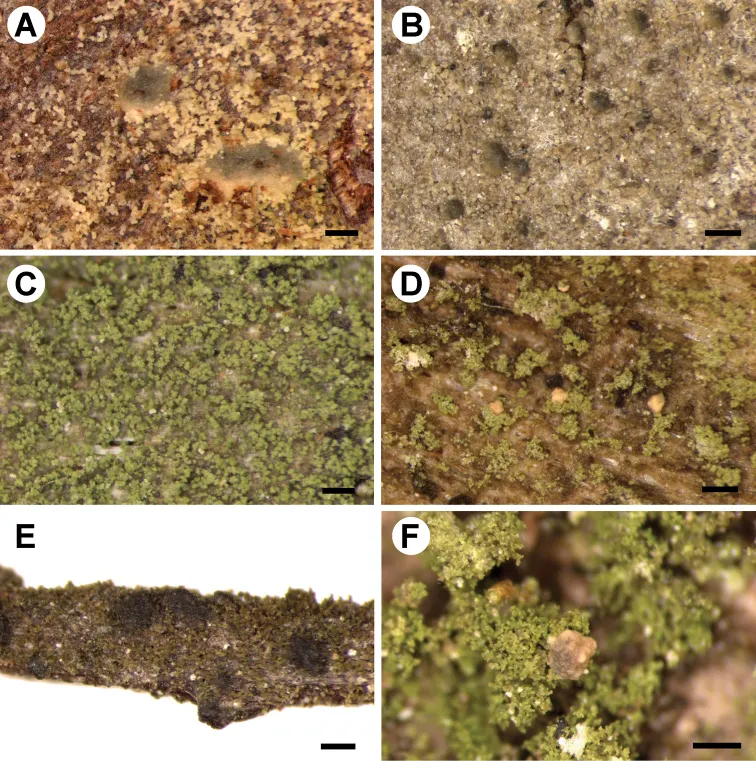
Species morphologically similar to Micarea viridileprosa, characterised by sorediate thallus, delimited or diffuse and confluent soralia with green or locally bluish soredia produced from the thallus areoles, white and immarginate when mature apothecia, 0.2–0.3 mm in diam., cylindrical to ellipsoidal (0–)1-septate ascospores measuring 9.5–13 × 2.8–3.5 µm and the presence of methoxymicareic acid. Thallus diffuse, up to 10 cm wide, consisting of finely granular soredia, often with a powdery appearance, vivid green or green, sometimes with bluish tinge; prothallus not seen; areoles up to 25 µm in diam., green, soon bursting to produce soredia; soralia at first delimited, produced from small, convex areoles, soon fused and confluent, sometimes forming a sorediate continuous layer; soredia simple, up to 20 µm in diam., sometimes slightly elongated or in more or less rounded consoredia up 35 µm in diam. apothecia rarely present, adnate, first with indistinct margin, then immarginate, 0.2–0.3 mm in diam., white or slightly brownish; excipulum in young apothecia present, 15–25 µm wide, of thin irregular hyphae; hymenium ca. 30‒42 µm tall; epihymenium and hypothecium hyaline; paraphyses thick (in K), branched and anastomosing, ca. 1.2‒1.5 µm wide; asci 29–35 × 7–10 µm, 8-spored; ascospores cylindrical to ellipsoidal, 9.5–13 × 2.8–3.5 µm, (0–)1-septate; micropycnidia present in some specimens, ca. 60 µm in diam., with dark brown tops (K–); microconidia narrow fusiform to bacilliform, 7 × 0.8 µm; mesopycnidia, mesoconidia 3.8 × 1.4 µm; crystalline granules (studied in polarised light) visible in hymenium and in thallus, soluble in K.

Sebagian uraian ditampilkan dalam bahasa sumber; terjemahannya belum tersedia atau belum lolos pemeriksaan.

Rujukan: [1]

Pengukuran mengikuti objek, spesimen atau kultur, dan metode yang disebutkan sumber. Baca uraian asli untuk konteks lengkap.

02 / Kehidupan

Habitat dan peran ekologis

The new species occurs on acidic bark of various trees such as Alnus, Betula, Fagus and Quercus, usually in humid forests, also on decaying wood (logs and stumps) and rarely on terrestrial decaying mosses in, for example, steep slopes in heath and dunes. It is a very common species in the south of the Netherlands and some areas in Poland and is mostly found on microhabitats where only few other lichens species co-occur. On several occasions, Normandina pulchella (Borrer) Nyl. and squamules of Cladonia spp. are the only accompanying lichens. To date, the species has been found in Belgium, Germany, the Netherlands, Poland and Portugal. One specimen of Micarea microsorediata was invaded by Nectriopsis micareae Diederich, van den Boom & Ernst (see below additional specimens examined).

Sebagian uraian ditampilkan dalam bahasa sumber; terjemahannya belum tersedia atau belum lolos pemeriksaan.

Rujukan: [1]

Habitat atau konteks temuan
hutan
Substrat
kayu
Peran atau konteks
Pembentuk liken
Temukan hubungan serupa
03 / Siklus hidup

Reproduksi

Species morphologically similar to Micarea viridileprosa, characterised by sorediate thallus, delimited or diffuse and confluent soralia with green or locally bluish soredia produced from the thallus areoles, white and immarginate when mature apothecia, 0.2–0.3 mm in diam., cylindrical to ellipsoidal (0–)1-septate ascospores measuring 9.5–13 × 2.8–3.5 µm and the presence of methoxymicareic acid. Thallus diffuse, up to 10 cm wide, consisting of finely granular soredia, often with a powdery appearance, vivid green or green, sometimes with bluish tinge; prothallus not seen; areoles up to 25 µm in diam., green, soon bursting to produce soredia; soralia at first delimited, produced from small, convex areoles, soon fused and confluent, sometimes forming a sorediate continuous layer; soredia simple, up to 20 µm in diam., sometimes slightly elongated or in more or less rounded consoredia up 35 µm in diam. apothecia rarely present, adnate, first with indistinct margin, then immarginate, 0.2–0.3 mm in diam., white or slightly brownish; excipulum in young apothecia present, 15–25 µm wide, of thin irregular hyphae; hymenium ca. 30‒42 µm tall; epihymenium and hypothecium hyaline; paraphyses thick (in K), branched and anastomosing, ca. 1.2‒1.5 µm wide; asci 29–35 × 7–10 µm, 8-spored; ascospores cylindrical to ellipsoidal, 9.5–13 × 2.8–3.5 µm, (0–)1-septate; micropycnidia present in some specimens, ca. 60 µm in diam., with dark brown tops (K–); microconidia narrow fusiform to bacilliform, 7 × 0.8 µm; mesopycnidia, mesoconidia 3.8 × 1.4 µm; crystalline granules (studied in polarised light) visible in hymenium and in thallus, soluble in K.

Sebagian uraian ditampilkan dalam bahasa sumber; terjemahannya belum tersedia atau belum lolos pemeriksaan.

Rujukan: [1]

04 / Batas informasi

Catatan identitas dan cakupan

  • Nama diterima mengikuti Catalogue of Life Base2026-09-11; uraian mempertahankan konsep dan nama sumber aslinya.
  • Preserve original treatment localities, hosts and substrate scope. Materials examined describe documented specimens, not the full global range.
  • Impor dan terjemahan otomatis belum diperiksa editorial per profil. Struktur reproduktif yang disebutkan tidak membuktikan seluruh siklus hidup diketahui.
  • Pengukuran pada uraian mengikuti spesimen, kultur, dan metode sumber; angka tidak diperluas ke semua populasi.

Catatan distribusi dalam narasi mengikuti sumbernya. Dokumentasi spesimen atau foto mengikuti catatan sumber. Bukti wilayah Indonesia dan penilaian konservasi tidak disimpulkan dari gambar.

05 / Dokumentasi

Melihat dengan konteks

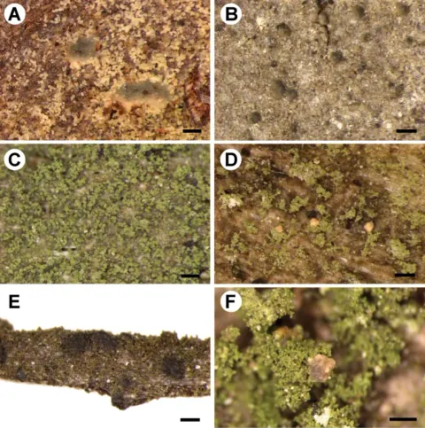
Morphology of newly described Micarea species A M. aeruginoprasina (holotype) B M. azorica (holotype) C M. isidioprasina (holotype) D M. microsorediata (holotype) E M. nigra (holotype) F M. pauli (holotype). Scale bars: 200 µm (A−C, E); 300 µm (D, F).

Jenis
Pelat ilmiah
Kredit
Beata Guzow-Krzemińska, Emmanuël Sérusiaux, Pieter P. G. van den Boom, A. Maarten Brand, Annina Launis, Anna Łubek, Martin Kukwa
Lisensi
CC BY 4.0
Sumber media
Buka entri sumber

Gambar menunjukkan objek menurut identifikasi sumber, bukan verifikasi independen spesimen oleh Fungi. Tidak ada skala baru yang ditambahkan.

06 / Rujukan

Sumber profil

  1. [1] Understanding the evolution of phenotypical characters in the Micarea prasina group (Pilocarpaceae) and descriptions of six new species within the group — Micarea microsorediataBagian sumber: PMC6685933_section_37
    Tanggal penghimpunan sumber: 2026-10-12
    Teks sumber: CC BY 4.0 · Guzow-Krzemińska B, Sérusiaux E, van den Boom PPG, Brand AM, Launis A, Łubek A, Kukwa M.
  2. [2] Catalogue of Life Base2026-09-11 — Micarea microsorediataBagian sumber: ColDP NameUsage.tsv ID VJHMK; accepted species; source 2073
    Tanggal penghimpunan sumber: 2026-10-12
  3. [3] Dokumentasi visual — Micarea microsorediataBagian sumber: Individual media record; identification follows the source, not independent determination
    Tanggal penghimpunan sumber: 2026-10-12
    Media sumber: CC BY 4.0 · Beata Guzow-Krzemińska, Emmanuël Sérusiaux, Pieter P. G. van den Boom, A. Maarten Brand, Annina Launis, Anna Łubek, Martin Kukwa

Adaptasi teks: CC BY 4.0. Guzow-Krzemińska B, Sérusiaux E, van den Boom PPG, Brand AM, Launis A, Łubek A, Kukwa M. — Understanding the evolution of phenotypical characters in the Micarea prasina group (Pilocarpaceae) and descriptions of six new species within the group — Micarea microsorediata

Uraian asli dan riwayat sumber

Riwayat atau halaman sumber

Ringkasan sumber

Species morphologically similar to Micarea viridileprosa, characterised by sorediate thallus, delimited or diffuse and confluent soralia with green or locally bluish soredia produced from the thallus areoles, white and immarginate when mature apothecia, 0.2–0.3 mm in diam., cylindrical to ellipsoidal (0–)1-septate ascospores measuring 9.5–13 × 2.8–3.5 µm and the presence of methoxymicareic acid. Thallus diffuse, up to 10 cm wide, consisting of finely granular soredia, often with a powdery appearance, vivid green or green, sometimes with bluish tinge; prothallus not seen; areoles up to 25 µm in diam., green, soon bursting to produce soredia; soralia at first delimited, produced from small, convex areoles, soon fused and confluent, sometimes forming a sorediate continuous layer; soredia simple, up to 20 µm in diam., sometimes slightly elongated or in more or less rounded consoredia up 35 µm in diam. apothecia rarely present, adnate, first with indistinct margin, then immarginate, 0.2–0.3 mm in diam., white or slightly brownish; excipulum in young apothecia present, 15–25 µm wide, of thin irregular hyphae; hymenium ca. 30‒42 µm tall; epihymenium and hypothecium hyaline; paraphyses thick (in K), branched and anastomosing, ca. 1.2‒1.5 µm wide; asci 29–35 × 7–10 µm, 8-spored; ascospores cylindrical to ellipsoidal, 9.5–13 × 2.8–3.5 µm, (0–)1-septate; micropycnidia present in some specimens, ca. 60 µm in diam., with dark brown tops (K–); microconidia narrow fusiform to bacilliform, 7 × 0.8 µm; mesopycnidia, mesoconidia 3.8 × 1.4 µm; crystalline granules (studied in polarised light) visible in hymenium and in thallus, soluble in K.

Revisi sumber

Morfologi sumber

Species morphologically similar to Micarea viridileprosa, characterised by sorediate thallus, delimited or diffuse and confluent soralia with green or locally bluish soredia produced from the thallus areoles, white and immarginate when mature apothecia, 0.2–0.3 mm in diam., cylindrical to ellipsoidal (0–)1-septate ascospores measuring 9.5–13 × 2.8–3.5 µm and the presence of methoxymicareic acid. Thallus diffuse, up to 10 cm wide, consisting of finely granular soredia, often with a powdery appearance, vivid green or green, sometimes with bluish tinge; prothallus not seen; areoles up to 25 µm in diam., green, soon bursting to produce soredia; soralia at first delimited, produced from small, convex areoles, soon fused and confluent, sometimes forming a sorediate continuous layer; soredia simple, up to 20 µm in diam., sometimes slightly elongated or in more or less rounded consoredia up 35 µm in diam. apothecia rarely present, adnate, first with indistinct margin, then immarginate, 0.2–0.3 mm in diam., white or slightly brownish; excipulum in young apothecia present, 15–25 µm wide, of thin irregular hyphae; hymenium ca. 30‒42 µm tall; epihymenium and hypothecium hyaline; paraphyses thick (in K), branched and anastomosing, ca. 1.2‒1.5 µm wide; asci 29–35 × 7–10 µm, 8-spored; ascospores cylindrical to ellipsoidal, 9.5–13 × 2.8–3.5 µm, (0–)1-septate; micropycnidia present in some specimens, ca. 60 µm in diam., with dark brown tops (K–); microconidia narrow fusiform to bacilliform, 7 × 0.8 µm; mesopycnidia, mesoconidia 3.8 × 1.4 µm; crystalline granules (studied in polarised light) visible in hymenium and in thallus, soluble in K.

Revisi sumber

Ekologi sumber

The new species occurs on acidic bark of various trees such as Alnus, Betula, Fagus and Quercus, usually in humid forests, also on decaying wood (logs and stumps) and rarely on terrestrial decaying mosses in, for example, steep slopes in heath and dunes. It is a very common species in the south of the Netherlands and some areas in Poland and is mostly found on microhabitats where only few other lichens species co-occur. On several occasions, Normandina pulchella (Borrer) Nyl. and squamules of Cladonia spp. are the only accompanying lichens. To date, the species has been found in Belgium, Germany, the Netherlands, Poland and Portugal. One specimen of Micarea microsorediata was invaded by Nectriopsis micareae Diederich, van den Boom & Ernst (see below additional specimens examined).

Revisi sumber

Struktur reproduksi sumber

Species morphologically similar to Micarea viridileprosa, characterised by sorediate thallus, delimited or diffuse and confluent soralia with green or locally bluish soredia produced from the thallus areoles, white and immarginate when mature apothecia, 0.2–0.3 mm in diam., cylindrical to ellipsoidal (0–)1-septate ascospores measuring 9.5–13 × 2.8–3.5 µm and the presence of methoxymicareic acid. Thallus diffuse, up to 10 cm wide, consisting of finely granular soredia, often with a powdery appearance, vivid green or green, sometimes with bluish tinge; prothallus not seen; areoles up to 25 µm in diam., green, soon bursting to produce soredia; soralia at first delimited, produced from small, convex areoles, soon fused and confluent, sometimes forming a sorediate continuous layer; soredia simple, up to 20 µm in diam., sometimes slightly elongated or in more or less rounded consoredia up 35 µm in diam. apothecia rarely present, adnate, first with indistinct margin, then immarginate, 0.2–0.3 mm in diam., white or slightly brownish; excipulum in young apothecia present, 15–25 µm wide, of thin irregular hyphae; hymenium ca. 30‒42 µm tall; epihymenium and hypothecium hyaline; paraphyses thick (in K), branched and anastomosing, ca. 1.2‒1.5 µm wide; asci 29–35 × 7–10 µm, 8-spored; ascospores cylindrical to ellipsoidal, 9.5–13 × 2.8–3.5 µm, (0–)1-septate; micropycnidia present in some specimens, ca. 60 µm in diam., with dark brown tops (K–); microconidia narrow fusiform to bacilliform, 7 × 0.8 µm; mesopycnidia, mesoconidia 3.8 × 1.4 µm; crystalline granules (studied in polarised light) visible in hymenium and in thallus, soluble in K.

Revisi sumber

Rujukan halaman ini

Fungi. Micarea microsorediata. Rilis data fungi-2026-10-12-expansion-2. https://fungi-atlas.mgadib.chatgpt.site/jamur/micarea-microsorediata/.

Status: impor sumber; pemeriksaan editorial per profil belum selesai. Untuk klaim biologis, gunakan rujukan pada bagian terkait dan periksa konteks sumber aslinya.