Ciri dan struktur
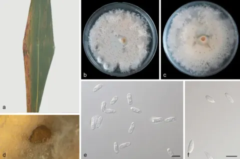
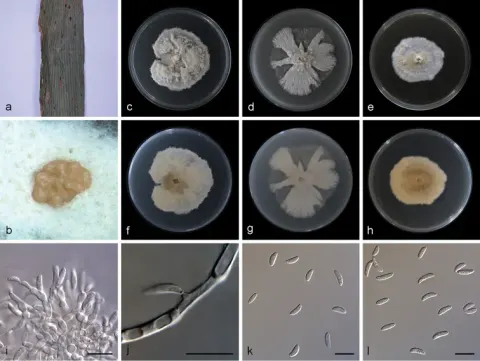
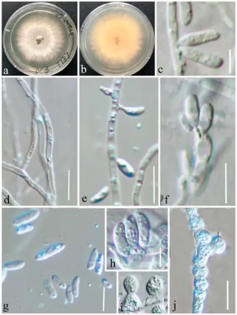
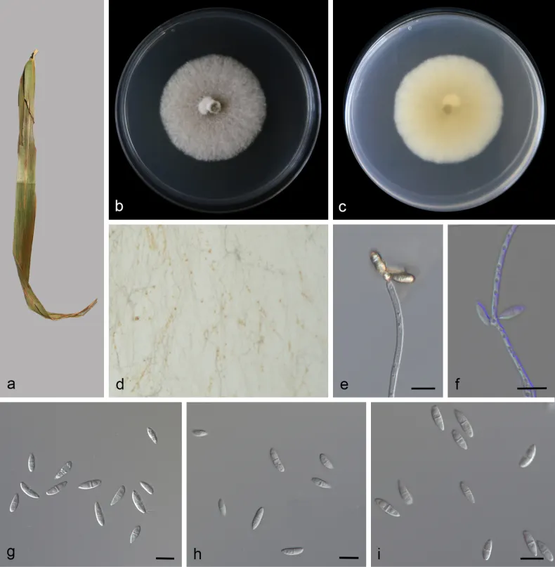
On leaves of Phragmites australis. Asexual morph: Mycelium superficial, abundant, hyphae hyaline, branched, septate, thin-walled, 1.99–3.04 µm wide. Conidiophores solitary, simple, rarely branched, straight, rarely bent, cylindrical, 49.70 × 2.96 µm, often not clearly differentiated from the mycelium, hyaline. Conidiogenous cells terminal or lateral, monoblastic or polyblastic, cylindrical, 2.21–3.44 × 2.21–2.87 μm. Conidia dry, fusiform, straight, smooth with thin walls, hyaline, (0–1)-septate, 9.21–14.07 × 2.96–4.39 μm (av. = 11.54 ± 1.31 × 3.50 ± 0.48 μm, n = 26), sometimes borne directly from hyphae. Chlamydospores not observed. Sexual morphs unknown, see Fig. 10. Colonies on PDA. 48.45–50.35 mm in diameter after 7 d at 25 °C in darkness, with a growth rate of 6.92–7.19 mm/day, entire at edge, white in obverse and pale yellow reverse.
Habitat dan peran ekologis
Associated with diseased leaves of Licuala grandis, Miscanthus sinensis, Phragmites australis, Phyllostachys nigra, and Pseudosasa japonica.
- Habitat atau konteks temuan
- Informasi belum tersedia pada sumber rilis ini.
- Substrat
- Informasi belum tersedia pada sumber rilis ini.
- Peran atau konteks
- Belum dikategorikan pada rilis ini.
Reproduksi
On leaves of Phragmites australis. Asexual morph: Mycelium superficial, abundant, hyphae hyaline, branched, septate, thin-walled, 1.99–3.04 µm wide. Conidiophores solitary, simple, rarely branched, straight, rarely bent, cylindrical, 49.70 × 2.96 µm, often not clearly differentiated from the mycelium, hyaline. Conidiogenous cells terminal or lateral, monoblastic or polyblastic, cylindrical, 2.21–3.44 × 2.21–2.87 μm. Conidia dry, fusiform, straight, smooth with thin walls, hyaline, (0–1)-septate, 9.21–14.07 × 2.96–4.39 μm (av. = 11.54 ± 1.31 × 3.50 ± 0.48 μm, n = 26), sometimes borne directly from hyphae. Chlamydospores not observed. Sexual morphs unknown, see Fig. 10.
Catatan identitas dan cakupan
- Nama diterima mengikuti Catalogue of Life Base2026-09-11; uraian mempertahankan konsep dan nama sumber aslinya.
- Preserve original treatment localities, hosts and substrate scope. Materials examined describe documented specimens, not the full global range.
- Impor dan terjemahan otomatis belum diperiksa editorial per profil. Struktur reproduktif yang disebutkan tidak membuktikan seluruh siklus hidup diketahui.
- Pengukuran pada uraian mengikuti spesimen, kultur, dan metode sumber; angka tidak diperluas ke semua populasi.
Catatan distribusi dalam narasi mengikuti sumbernya. Dokumentasi spesimen atau foto mengikuti catatan sumber. Bukti wilayah Indonesia dan penilaian konservasi tidak disimpulkan dari gambar.
Melihat dengan konteks
Morphology of Microdochium miscanthi (SAUCC 7610-2). a on leaf of Phragmites australis b, c colonies (b-above, c-reverse) after 7 d on PDA d colony on PDA producing conidia masses e conidiophores with conidiogenous cells and conidia f hyaline, conidiogenous cells and conidia g–i conidia. Scale bars: 10 μm (e–i).
- Jenis
- Pelat ilmiah
- Kredit
- Qiyun Liu, Yuxin Shang, Jinyang Yuan, Zhaoxue Zhang, Congcong Ai, Jiwen Xia, Xiuguo Zhang, Zhuang Li
- Lisensi
- CC BY 4.0
- Sumber media
- Buka entri sumber
Sumber profil
- [1] Multilocus phylogeny, morphology and taxonomy of Microdochium (Microdochiaceae): insights into evolutionary divergence times and historical biogeography — Microdochium miscanthi
- [2] Catalogue of Life Base2026-09-11 — Microdochium miscanthi
- [3] Dokumentasi visual — Microdochium miscanthi
Rujukan halaman ini
Fungi. Microdochium miscanthi. Rilis data fungi-2026-10-12-expansion-2. https://fungi-atlas.mgadib.chatgpt.site/jamur/microdochium-miscanthi/.
Status: impor sumber; pemeriksaan editorial per profil belum selesai. Untuk klaim biologis, gunakan rujukan pada bagian terkait dan periksa konteks sumber aslinya.