Ciri dan struktur
Sexual morph not observed. Asexual morph on SNA. Vegetative hyphae hyaline, smooth-walled, septate, branched, 1–3 μm wide, lacking chlamydospores. Sporulation abundant, conidia formed directly on hyphal cells, in conidiomata and by microcyclic conidiation. Conidiophores on hyphae reduced to conidiogenous cells, conidiogenous loci formed terminally. Conidiogenous cells enteroblastic, hyaline, smooth-walled, mostly reduced to mere openings with collarettes formed directly on hyphal cells, discrete phialides rare, navicular, constricted at the base, 5.5–14.5 × 1.5–2.5 μm; collarettes rarely visible or flaring, < 0.5–3 μm long, opening 0.5–1.5 μm, periclinal thickening sometimes visible. Conidia aggregated in masses around the hyphae, hyaline, smooth-walled, aseptate, oblong to ellipsoidal, mostly straight, sometimes slightly curved, with both ends rounded, sometimes with a prominent scar on one end, (2.5–)3–5(–6) × 1–1.5(–2) μm, mean ± SD = 3.9 ± 0.9 × 1.4 ± 0.2 μm, L/W ratio= 2.8. Conidiomata produced on OA in 2–4 wk; solitary or aggregated, globose to subglobose, unilocular, immersed to superficial, 50–340 μm wide, olivaceous to black, mostly glabrous, sometimes with a few hairs, opening with an irregular rupture. Conidiophores reduced to conidiogenous cells. Conidiogenus cells enteroblastic, hyaline, smooth-walled, conidiogenous loci formed terminally, discrete phialides, globose to ampulliform or navicular, 3.5–7.5 × 2–3.5 μm, opening 0.5–1 μm, periclinal thickening sometimes visible. Conidia hyaline, smooth-walled, cylindrical to ellipsoidal, sometimes slightly curved, with both ends rounded, (2.5–)3–4.5(–6) × (1–)1.5–2(–3) μm, mean ± SD = 3.8 ± 0.8 × 1.7 ± 0.4 μm, L/W ratio = 2.2.
Habitat dan peran ekologis
Two strains of Minutiella pruni-avium were isolated from wood of Prunus avium. The LSU sequences of these strains differ in three and one nucleotides from those of M. tardicola and Minutiella sp., respectively. The ITS region shows 11 differences to M. tardicola and 9 differences to Minutiella sp. The TUB sequence of M. tardicola and Minutiella sp. differ in one nucleotide, however, in 35 and 33 nucleotides compared to M. pruni-avium. Minutiella pruni-avium differs from Minutiella tardicola and the strains of Minutiella sp. by forming larger conidiomata, longer discrete phialides and flaring collarettes of up to 3 μm. The closest match in a blastn search with the ITS sequence of strain GLMC 1624 is the type strain of Minutiella tardicola CBS 121757 with 97.9% identity (NR132006, Damm et al. 2010).
- Habitat atau konteks temuan
- Informasi belum tersedia pada sumber rilis ini.
- Substrat
- Informasi belum tersedia pada sumber rilis ini.
- Peran atau konteks
- Belum dikategorikan pada rilis ini.
Reproduksi
Sexual morph not observed. Asexual morph on SNA. Vegetative hyphae hyaline, smooth-walled, septate, branched, 1–3 μm wide, lacking chlamydospores. Sporulation abundant, conidia formed directly on hyphal cells, in conidiomata and by microcyclic conidiation. Conidiophores on hyphae reduced to conidiogenous cells, conidiogenous loci formed terminally. Conidiogenous cells enteroblastic, hyaline, smooth-walled, mostly reduced to mere openings with collarettes formed directly on hyphal cells, discrete phialides rare, navicular, constricted at the base, 5.5–14.5 × 1.5–2.5 μm; collarettes rarely visible or flaring, < 0.5–3 μm long, opening 0.5–1.5 μm, periclinal thickening sometimes visible. Conidia aggregated in masses around the hyphae, hyaline, smooth-walled, aseptate, oblong to ellipsoidal, mostly straight, sometimes slightly curved, with both ends rounded, sometimes with a prominent scar on one end, (2.5–)3–5(–6) × 1–1.5(–2) μm, mean ± SD = 3.9 ± 0.9 × 1.4 ± 0.2 μm, L/W ratio= 2.8.
Catatan identitas dan cakupan
- Nama diterima mengikuti Catalogue of Life Base2026-09-11; uraian mempertahankan konsep dan nama sumber aslinya.
- Preserve original treatment localities, hosts and substrate scope. Materials examined describe documented specimens, not the full global range.
- Impor dan terjemahan otomatis belum diperiksa editorial per profil. Struktur reproduktif yang disebutkan tidak membuktikan seluruh siklus hidup diketahui.
- Pengukuran pada uraian mengikuti spesimen, kultur, dan metode sumber; angka tidak diperluas ke semua populasi.
Catatan distribusi dalam narasi mengikuti sumbernya. Dokumentasi spesimen atau foto mengikuti catatan sumber. Bukti wilayah Indonesia dan penilaian konservasi tidak disimpulkan dari gambar.
Melihat dengan konteks
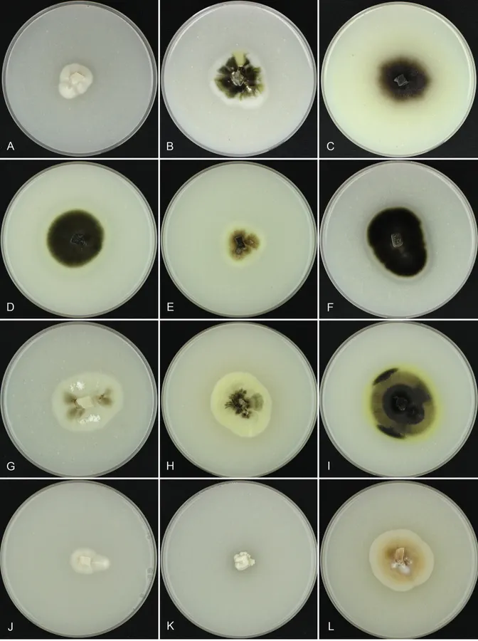
Colony surface of analysed strains on OA medium. A Arboricolonus simplex GLMC 459 T B Cadophora africana CBS 120890 T C C. bubakii CBS 198.30 T D C. luteo-olivacea GLMC 1264 E C. novi-eboraci GLMC 1472 F C. obscura CBS 269.33 G C. prunicola CBS 120891 T H C. prunicola GLMC 1633 I C. ramosa GLMC 377 T J Minutiella pruni-avium GLMC 1624 T K Proliferodiscus ingens GLMC 1751 T L Proliferodiscus sp. GLMC 460. Cultures A, J–L after 4 wk. Cultures B–I after 2 wk. Strains with a superscript T are ex-type cultures.
- Jenis
- Pelat ilmiah
- Kredit
- Steffen Bien, Ulrike Damm
- Lisensi
- CC BY 4.0
- Sumber media
- Buka entri sumber
Dokumentasi tambahan
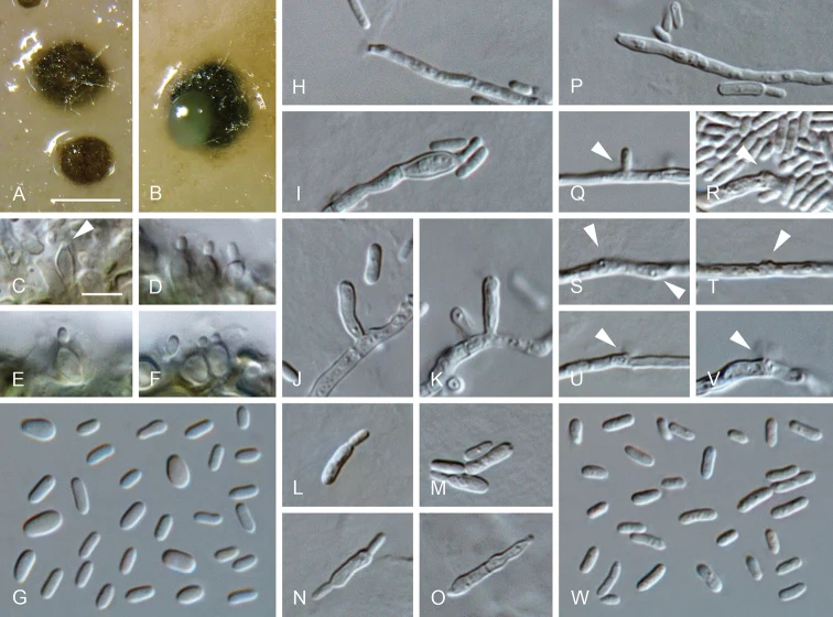
Minutiella pruni-avium sp. nov. A, B conidiomata C–F conidiogenous cells lining the inner wall of a conidioma G conidia formed in conidiomata H–K, P–V conidiogenous cells formed on hyphal cells (arrows indicate conidiogenous openings) L–O mother cells W conidia formed on hyphal cells A–G from OA H–W from SNA A, B SM, C–W LM. Scale bars: 200 μm (A applies to B), 5 μm (C applies to D–W).
Kredit dan penggunaan
Steffen Bien, Ulrike Damm
Ukuran dan format gambar disesuaikan tanpa pemotongan, retus, atau penambahan skala. Lisensi sumber dipertahankan.
Sumber profil
- [1] Arboricolonus simplex gen. et sp. nov. and novelties in Cadophora, Minutiella and Proliferodiscus from Prunus wood in Germany — Minutiella pruni-avium
- [2] Catalogue of Life Base2026-09-11 — Minutiella pruni-avium
- [3] Dokumentasi visual — Minutiella pruni-avium
- [4] Dokumentasi visual — Minutiella pruni-avium
Rujukan halaman ini
Fungi. Minutiella pruni-avium. Rilis data fungi-2026-10-12-expansion-2. https://fungi-atlas.mgadib.chatgpt.site/jamur/minutiella-pruni-avium/.
Status: impor sumber; pemeriksaan editorial per profil belum selesai. Untuk klaim biologis, gunakan rujukan pada bagian terkait dan periksa konteks sumber aslinya.