Ciri dan struktur
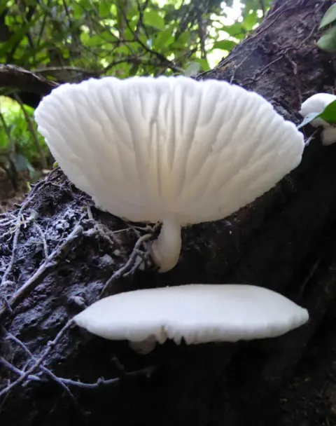
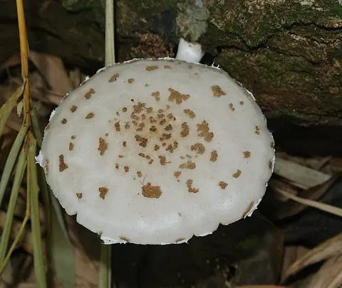
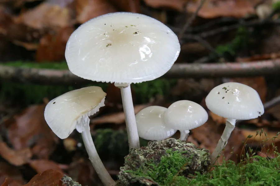
Caps are 2–8 cm across, strongly convex at first, but mildly flattening over time. They are pale greyish when young, becoming whiter and covered with a semi-translucent and slimy membrane, often with an ochraceous flush at the centre. The surface layer resembles spore-bearing tissue, with erect club-shaped cells, but lacks functional basidia (i.e. cap cuticle is hymeniform). The slender stems are 30–100 mm tall and 3–10 mm wide, white striate above a substantial membranous ring and slightly scaly and greyish below. Flesh is thin and white and the lamellae are adnate, broad and very distant. Cystidia are thin-walled cylindric or utriform. Spore print is white, they are smooth and subglobose in shape and very thickwalled at 13–18×12–15 μm. When porcelain mushrooms grow from the underside of the tree, the stems will curl in a way so the caps are all held horizontally, with the gills facing down. O. mucida are seldom confused with other fungi.
Habitat dan peran ekologis
Porcelain fungus is native to Europe, and specific to beech wood, where it appears in autumn on dead tree trunks and fallen branches, occasionally on dead branches high up in living trees. It is saprobic or weakly parasitic to living beech trees. While it has a strong tie to beech, it has been found growing on oak on rare occasions. O. mucida occurs throughout northern and central Europe, where beech is found and in its habitat this fungus is a common species. When O. mucida is found on a beech tree, it usually outcompetes other fungi locally by means of a powerful anti-fungal agent called strobilurin.
- Habitat atau konteks temuan
- Informasi belum tersedia pada sumber rilis ini.
- Substrat
- kayu
- Peran atau konteks
- Pengurai
Reproduksi
Caps are 2–8 cm across, strongly convex at first, but mildly flattening over time. They are pale greyish when young, becoming whiter and covered with a semi-translucent and slimy membrane, often with an ochraceous flush at the centre. The surface layer resembles spore-bearing tissue, with erect club-shaped cells, but lacks functional basidia (i.e. cap cuticle is hymeniform). Cystidia are thin-walled cylindric or utriform. Spore print is white, they are smooth and subglobose in shape and very thickwalled at 13–18×12–15 μm.
Catatan identitas dan cakupan
- Nama diterima mengikuti Catalogue of Life Base2026-09-11; uraian mempertahankan konsep dan nama sumber aslinya.
- Original geographic, specimen and life-cycle scope retained; source import awaits individual editorial review.
- Impor dan terjemahan otomatis belum diperiksa editorial per profil. Struktur reproduktif yang disebutkan tidak membuktikan seluruh siklus hidup diketahui.
- Pengukuran pada uraian mengikuti spesimen, kultur, dan metode sumber; angka tidak diperluas ke semua populasi.
Catatan distribusi dalam narasi mengikuti sumbernya. Dokumentasi spesimen atau foto mengikuti catatan sumber. Bukti wilayah Indonesia dan penilaian konservasi tidak disimpulkan dari gambar.
Melihat dengan konteks
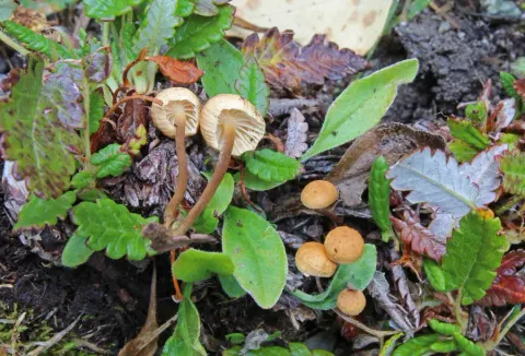
Dokumentasi Oudemansiella mucida menurut asosiasi foto pada record takson iNaturalist. Kredit: Alexandre Dulaunoy. Identitas mengikuti sumber; bukan penentuan spesimen independen.
- Jenis
- Fotografi
- Kredit
- (c) Alexandre Dulaunoy, some rights reserved (CC BY-SA)
- Lisensi
- CC BY-SA 4.0
- Sumber media
- Buka entri sumber
Sumber profil
- [1] Oudemansiella mucida — Wikipedia (en)
- [2] Catalogue of Life Base2026-09-11 — Mucidula mucida
- [3] Dokumentasi visual — Oudemansiella mucida
Rujukan halaman ini
Fungi. Mucidula mucida. Rilis data fungi-2026-10-12-expansion-2. https://fungi-atlas.mgadib.chatgpt.site/jamur/mucidula-mucida/.
Status: impor sumber; pemeriksaan editorial per profil belum selesai. Untuk klaim biologis, gunakan rujukan pada bagian terkait dan periksa konteks sumber aslinya.