01 / Morfologi

Ciri dan struktur

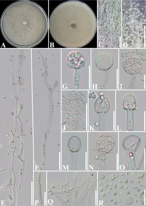
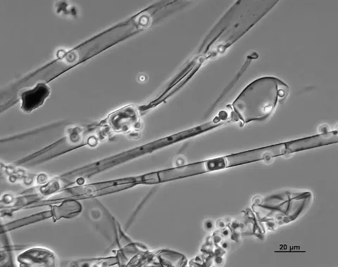
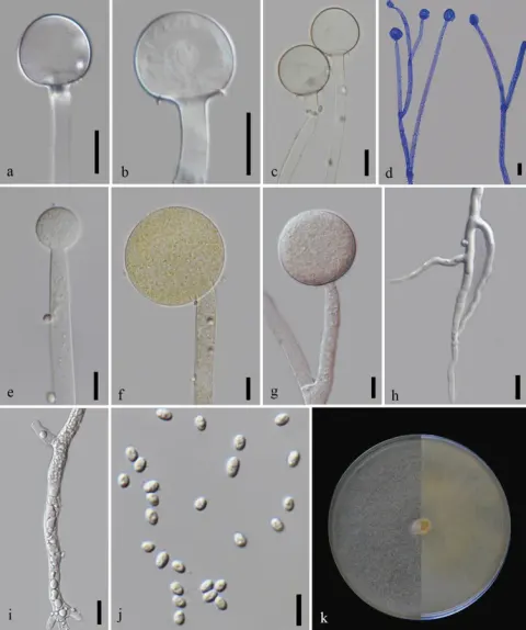
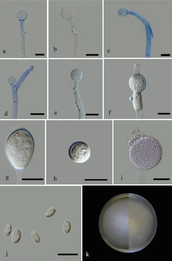
Asexual morph (based on cultures grown in MEA at 25 °C): Sporangiophores hyaline, up to 7.5 µm (x – = 5, n = 30) in diameter, undulate, occasionally curved, irregular septate near the base (1–2), unbranched, sympodial branches formed. Sporangia 20–47.5 × 20–45.5 µm (x – = 34 × 35.5 µm, n = 28), globose, smooth-walled, thick-walled, wall echinulate, deliquescent in mature sporangia, hyaline to pale brown. Columellae 12.5–23.5 × 12–22.5 µm ((x – = 16 × 15.5 µm, n = 30), mostly globose to subglobose, and sometimes oblong, obovoid, and rarely ellipsoid, without or with short collar, hyaline to pale brown, smooth-walled. Sporangiospores 3.5–6.5 × 2–4 µm (x – = 5 × 2.5 µm, n = 30) µm, mostly ellipsoidal, sometimes flattened on one side, oval or cylindrical, smooth-walled, hyaline with one or more granules. Chlamydospores abundant, intercalary, terminal, variable in shape and size. Rhizoids absent. Sexual morph not observed. Colonies on MEA reaching a diameter of 60 mm after 3 days of incubation at 25 °C. At temperatures 20 and 30 °C, the colony diameter is 46 mm and 45.5 mm respectively after 3 days. Colony growth is observed at 8 to 30 °C with optimal growth at 25 °C. Unbranched or sympodial branching of sporangiophore are prominent at 30 °C. Colonies on PDA, reaching 51 mm after 3 days of incubation at 25 °C. In both culture media, colony from above and reverse: white or very pale yellow, and cottony. No growth was observed at 37 °C. At 8 °C, the colony reached a diameter of 21 mm after 9 days but with no sporulation in MEA.

Sebagian uraian ditampilkan dalam bahasa sumber; terjemahannya belum tersedia atau belum lolos pemeriksaan.

Rujukan: [1]

Pengukuran mengikuti objek, spesimen atau kultur, dan metode yang disebutkan sumber. Baca uraian asli untuk konteks lengkap.

02 / Kehidupan

Habitat dan peran ekologis

Colonies on MEA reaching a diameter of 60 mm after 3 days of incubation at 25 °C. At temperatures 20 and 30 °C, the colony diameter is 46 mm and 45.5 mm respectively after 3 days. Colony growth is observed at 8 to 30 °C with optimal growth at 25 °C. Unbranched or sympodial branching of sporangiophore are prominent at 30 °C. Colonies on PDA, reaching 51 mm after 3 days of incubation at 25 °C. In both culture media, colony from above and reverse: white or very pale yellow, and cottony. No growth was observed at 37 °C. At 8 °C, the colony reached a diameter of 21 mm after 9 days but with no sporulation in MEA.

Sebagian uraian ditampilkan dalam bahasa sumber; terjemahannya belum tersedia atau belum lolos pemeriksaan.

Rujukan: [1]

Habitat atau konteks temuan
kultur yang didokumentasikan
Substrat
Informasi belum tersedia pada sumber rilis ini.
Peran atau konteks
Belum dikategorikan pada rilis ini.
Temukan hubungan serupa
03 / Siklus hidup

Reproduksi

Asexual morph (based on cultures grown in MEA at 25 °C): Sporangiophores hyaline, up to 7.5 µm (x – = 5, n = 30) in diameter, undulate, occasionally curved, irregular septate near the base (1–2), unbranched, sympodial branches formed. Sporangia 20–47.5 × 20–45.5 µm (x – = 34 × 35.5 µm, n = 28), globose, smooth-walled, thick-walled, wall echinulate, deliquescent in mature sporangia, hyaline to pale brown. Columellae 12.5–23.5 × 12–22.5 µm ((x – = 16 × 15.5 µm, n = 30), mostly globose to subglobose, and sometimes oblong, obovoid, and rarely ellipsoid, without or with short collar, hyaline to pale brown, smooth-walled. Sporangiospores 3.5–6.5 × 2–4 µm (x – = 5 × 2.5 µm, n = 30) µm, mostly ellipsoidal, sometimes flattened on one side, oval or cylindrical, smooth-walled, hyaline with one or more granules.

Sebagian uraian ditampilkan dalam bahasa sumber; terjemahannya belum tersedia atau belum lolos pemeriksaan.

Rujukan: [1]

Sinonim menurut acuan taksonomi rilis: Mucor subtilissimus
04 / Batas informasi

Catatan identitas dan cakupan

  • Nama diterima mengikuti Catalogue of Life Base2026-09-11; uraian mempertahankan konsep dan nama sumber aslinya.
  • Preserve original treatment localities, hosts and substrate scope. Materials examined describe documented specimens, not the full global range.
  • Impor dan terjemahan otomatis belum diperiksa editorial per profil. Struktur reproduktif yang disebutkan tidak membuktikan seluruh siklus hidup diketahui.
  • Pengukuran pada uraian mengikuti spesimen, kultur, dan metode sumber; angka tidak diperluas ke semua populasi.

Catatan distribusi dalam narasi mengikuti sumbernya. Dokumentasi spesimen atau foto mengikuti catatan sumber. Bukti wilayah Indonesia dan penilaian konservasi tidak disimpulkan dari gambar.

05 / Dokumentasi

Melihat dengan konteks

Mucor nederlandicus (MFLU 21–007) a-c columella with highly reduced collar d sterile sporangium e columella with visible collar f-h chlamydospores i mature sporangia j sporangiospores k front and reverse of the colony in MEA. Scale bars: 20 µm (a–e, i); 10 µm (f–h).

Jenis
Pelat ilmiah
Kredit
Vedprakash G. Hurdeal, Eleni Gentekaki, Kevin D. Hyde, Thuong T. T. Nguyen, Hyang Burm Lee
Lisensi
CC BY 4.0
Sumber media
Buka entri sumber

Gambar menunjukkan objek menurut identifikasi sumber, bukan verifikasi independen spesimen oleh Fungi. Tidak ada skala baru yang ditambahkan.

06 / Rujukan

Sumber profil

  1. [1] Novel Mucor species (Mucoromycetes, Mucoraceae) from northern Thailand — Mucor nederlandicusBagian sumber: PMC8575866_section_26
    Tanggal penghimpunan sumber: 2026-10-12
    Teks sumber: CC BY 4.0 · Hurdeal VG, Gentekaki E, Hyde KD, Nguyen TTT, Lee HB.
  2. [2] Catalogue of Life Base2026-09-11 — Mucor nederlandicusBagian sumber: ColDP NameUsage.tsv ID 6S37C; accepted species; source 2073
    Tanggal penghimpunan sumber: 2026-10-12
  3. [3] Dokumentasi visual — Mucor nederlandicusBagian sumber: Individual media record; identification follows the source, not independent determination
    Tanggal penghimpunan sumber: 2026-10-12
    Media sumber: CC BY 4.0 · Vedprakash G. Hurdeal, Eleni Gentekaki, Kevin D. Hyde, Thuong T. T. Nguyen, Hyang Burm Lee

Adaptasi teks: CC BY 4.0. Hurdeal VG, Gentekaki E, Hyde KD, Nguyen TTT, Lee HB. — Novel Mucor species (Mucoromycetes, Mucoraceae) from northern Thailand — Mucor nederlandicus

Uraian asli dan riwayat sumber

Riwayat atau halaman sumber

Ringkasan sumber

Asexual morph (based on cultures grown in MEA at 25 °C): Sporangiophores hyaline, up to 7.5 µm (x – = 5, n = 30) in diameter, undulate, occasionally curved, irregular septate near the base (1–2), unbranched, sympodial branches formed. Sporangia 20–47.5 × 20–45.5 µm (x – = 34 × 35.5 µm, n = 28), globose, smooth-walled, thick-walled, wall echinulate, deliquescent in mature sporangia, hyaline to pale brown. Columellae 12.5–23.5 × 12–22.5 µm ((x – = 16 × 15.5 µm, n = 30), mostly globose to subglobose, and sometimes oblong, obovoid, and rarely ellipsoid, without or with short collar, hyaline to pale brown, smooth-walled.

Revisi sumber

Morfologi sumber

Asexual morph (based on cultures grown in MEA at 25 °C): Sporangiophores hyaline, up to 7.5 µm (x – = 5, n = 30) in diameter, undulate, occasionally curved, irregular septate near the base (1–2), unbranched, sympodial branches formed. Sporangia 20–47.5 × 20–45.5 µm (x – = 34 × 35.5 µm, n = 28), globose, smooth-walled, thick-walled, wall echinulate, deliquescent in mature sporangia, hyaline to pale brown. Columellae 12.5–23.5 × 12–22.5 µm ((x – = 16 × 15.5 µm, n = 30), mostly globose to subglobose, and sometimes oblong, obovoid, and rarely ellipsoid, without or with short collar, hyaline to pale brown, smooth-walled. Sporangiospores 3.5–6.5 × 2–4 µm (x – = 5 × 2.5 µm, n = 30) µm, mostly ellipsoidal, sometimes flattened on one side, oval or cylindrical, smooth-walled, hyaline with one or more granules. Chlamydospores abundant, intercalary, terminal, variable in shape and size. Rhizoids absent. Sexual morph not observed. Colonies on MEA reaching a diameter of 60 mm after 3 days of incubation at 25 °C. At temperatures 20 and 30 °C, the colony diameter is 46 mm and 45.5 mm respectively after 3 days. Colony growth is observed at 8 to 30 °C with optimal growth at 25 °C. Unbranched or sympodial branching of sporangiophore are prominent at 30 °C. Colonies on PDA, reaching 51 mm after 3 days of incubation at 25 °C. In both culture media, colony from above and reverse: white or very pale yellow, and cottony. No growth was observed at 37 °C. At 8 °C, the colony reached a diameter of 21 mm after 9 days but with no sporulation in MEA.

Revisi sumber

Ekologi sumber

Colonies on MEA reaching a diameter of 60 mm after 3 days of incubation at 25 °C. At temperatures 20 and 30 °C, the colony diameter is 46 mm and 45.5 mm respectively after 3 days. Colony growth is observed at 8 to 30 °C with optimal growth at 25 °C. Unbranched or sympodial branching of sporangiophore are prominent at 30 °C. Colonies on PDA, reaching 51 mm after 3 days of incubation at 25 °C. In both culture media, colony from above and reverse: white or very pale yellow, and cottony. No growth was observed at 37 °C. At 8 °C, the colony reached a diameter of 21 mm after 9 days but with no sporulation in MEA.

Revisi sumber

Struktur reproduksi sumber

Asexual morph (based on cultures grown in MEA at 25 °C): Sporangiophores hyaline, up to 7.5 µm (x – = 5, n = 30) in diameter, undulate, occasionally curved, irregular septate near the base (1–2), unbranched, sympodial branches formed. Sporangia 20–47.5 × 20–45.5 µm (x – = 34 × 35.5 µm, n = 28), globose, smooth-walled, thick-walled, wall echinulate, deliquescent in mature sporangia, hyaline to pale brown. Columellae 12.5–23.5 × 12–22.5 µm ((x – = 16 × 15.5 µm, n = 30), mostly globose to subglobose, and sometimes oblong, obovoid, and rarely ellipsoid, without or with short collar, hyaline to pale brown, smooth-walled. Sporangiospores 3.5–6.5 × 2–4 µm (x – = 5 × 2.5 µm, n = 30) µm, mostly ellipsoidal, sometimes flattened on one side, oval or cylindrical, smooth-walled, hyaline with one or more granules.

Revisi sumber

Rujukan halaman ini

Fungi. Mucor nederlandicus. Rilis data fungi-2026-10-12-expansion-2. https://fungi-atlas.mgadib.chatgpt.site/jamur/mucor-nederlandicus/.

Status: impor sumber; pemeriksaan editorial per profil belum selesai. Untuk klaim biologis, gunakan rujukan pada bagian terkait dan periksa konteks sumber aslinya.