Ciri dan struktur
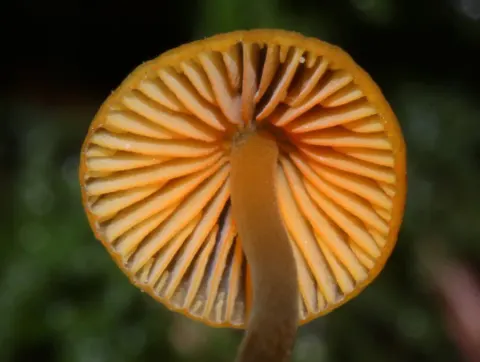
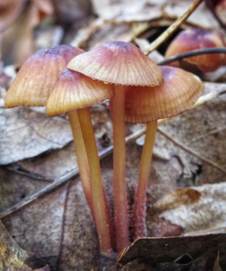
The cap is 1 – 30 mm broad, initially obtuse to convex, later becoming broadly umbonate. Eventually the cap flattens, sometimes with the disc (the central region of the cap) slightly depressed. The cap margin initially touches the stem; as it expands it becomes somewhat wavy. The extreme margin is marked by small lines, grooves or ridges when the cap is moist. The cap is initially covered with white powdery granules, but this later sloughs off to leave a smooth surface. The disc is brownish when young, but soon develops reddish tones; older specimens are bay to Isabella, with lighter-colored margins. The flesh is buff-colored, and oozes an orange-yellow juice when cut. The gills have an adnate attachment to the stem. They have a close to moderately crowded spacing, with between 23 and 26 gills reaching the stem. The gills are narrow to moderately broad, chamoisee when young, somewhat darker in age, with edges that are maroon, and either crenulate (finely scalloped) or even. The stem is 2 – 4 cm long, 2–3 mm thick, and more or less equal in width throughout. The stem base is rooted among the leaves and debris, and the base is covered with short stiff hairs pressed flat against the surface. Its surface is covered sparsely with minute purplish brown fibrils. The stem is dull reddish brown overall, but the color fades near the top. When mushroom tissue is cut or injured, it oozes a dull reddish-brown juice or, in old specimens, a dull orange juice. M. atkinsoniana mushrooms have no distinctive taste or odor. The spores are narrowly to broadly ellipsoid, amyloid (staining black to blue-black in Melzer's reagent), and measure 7–9 by 4–5 μm.
Habitat dan peran ekologis
The fungus is saprobic, and so derives nutrients from breaking down dead plant material, such as leaves, bark, needles, and twigs, that has fallen to the ground. Fruit bodies are usually found growing in groups or scattered on leaf litter in beech and beech-hemlock forests during the summer and autumn, although it has been recorded growing on lawns. It is also often found under eastern North American oaks. The distribution includes the US states Connecticut, New York, Ohio, Michigan, and Vermont. It has also been collected in Quebec, Canada.
- Habitat atau konteks temuan
- hutan
- Substrat
- Informasi belum tersedia pada sumber rilis ini.
- Peran atau konteks
- Pengurai
Reproduksi
The spores are narrowly to broadly ellipsoid, amyloid (staining black to blue-black in Melzer's reagent), and measure 7–9 by 4–5 μm. The basidia (spore-bearing cells) are four-spored and measure 28–30 by 6–7 μm. Cheilocystidia are plentiful, and arranged so as to form a sterile band on the edge of the gill. Measuring, they are narrowly fusoid (tapered at each end), smooth, and have dark reddish contents. The pleurocystidia (cystidia on the gill face) similar in appearance to the cheilocystidia but far less abundant. Gill tissue is yellowish to very faintly vinaceous-brown when stained with iodine. The tissue of the cap consists of several layers. The outer surface comprises a thin cuticle made of narrow hyphae filled with dark-reddish material.
Catatan identitas dan cakupan
- Nama diterima mengikuti Catalogue of Life Base2026-09-11; uraian mempertahankan konsep dan nama sumber aslinya.
- Original geographic, specimen and life-cycle scope retained; source import awaits individual editorial review.
- Impor dan terjemahan otomatis belum diperiksa editorial per profil. Struktur reproduktif yang disebutkan tidak membuktikan seluruh siklus hidup diketahui.
- Pengukuran pada uraian mengikuti spesimen, kultur, dan metode sumber; angka tidak diperluas ke semua populasi.
Catatan distribusi dalam narasi mengikuti sumbernya. Dokumentasi spesimen atau foto mengikuti catatan sumber. Bukti wilayah Indonesia dan penilaian konservasi tidak disimpulkan dari gambar.
Melihat dengan konteks
Dokumentasi Mycena atkinsoniana menurut asosiasi foto pada record takson iNaturalist. Kredit: Deana Tempest Thomas. Identitas mengikuti sumber; bukan penentuan spesimen independen.
- Jenis
- Fotografi
- Kredit
- (c) Deana Tempest Thomas, some rights reserved (CC BY), uploaded by Deana Tempest Thomas
- Lisensi
- CC BY 4.0
- Sumber media
- Buka entri sumber
Dokumentasi tambahan
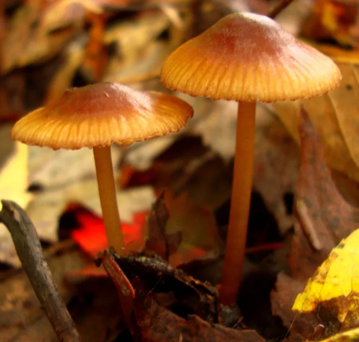
Foto yang diasosiasikan dengan Mycena atkinsoniana pada iNaturalist. Identifikasi mengikuti sumber; bukan penentuan spesimen independen.
Kredit dan penggunaan
(c) martin_livezey, some rights reserved (CC BY)
Ukuran dan format gambar disesuaikan tanpa pemotongan, retus, atau penambahan skala. Lisensi sumber dipertahankan.
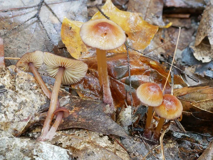
Foto yang diasosiasikan dengan Mycena atkinsoniana pada iNaturalist. Identifikasi mengikuti sumber; bukan penentuan spesimen independen.
Kredit dan penggunaan
no rights reserved, uploaded by Garrett Taylor
Ukuran dan format gambar disesuaikan tanpa pemotongan, retus, atau penambahan skala. Lisensi sumber dipertahankan.
Sumber profil
- [1] Mycena atkinsoniana — Wikipedia (en)
- [2] Catalogue of Life Base2026-09-11 — Mycena atkinsoniana
- [3] Dokumentasi visual — Mycena atkinsoniana
- [4] Dokumentasi visual — Mycena atkinsoniana
- [5] Dokumentasi visual — Mycena atkinsoniana
Rujukan halaman ini
Fungi. Mycena atkinsoniana. Rilis data fungi-2026-10-12-expansion-2. https://fungi-atlas.mgadib.chatgpt.site/jamur/mycena-atkinsoniana/.
Status: impor sumber; pemeriksaan editorial per profil belum selesai. Untuk klaim biologis, gunakan rujukan pada bagian terkait dan periksa konteks sumber aslinya.