Ciri dan struktur
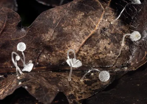
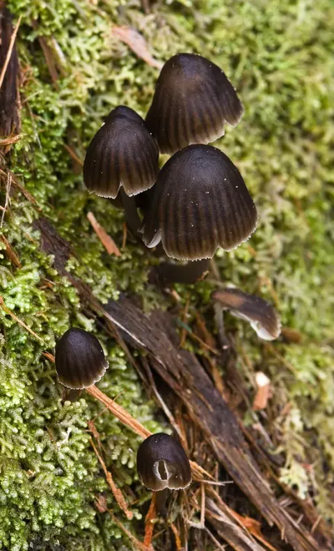
Growing on dead stem of Miscanthus. Pileus sparsely pruinose. Basidiospores cylindric. Cherocytes absent. Acanthocysts forming two types. Caulocystidia sphaero-pedunculate covered with spines. Clamps present. Pileus 3.5–7.8 mm in diam., hemispherical, broadly conical to convex, occasionally ± centrally depressed when young, sulcate, translucent-striate, pure white, pubescent to inconspicuously puberulous, margin nearly plane, undulate. Context white, thin, very fragile, about 1.0 mm thick at centre. Lamellae narrowly adnate or adnexed, off-white, concolorous with the sides. Stipe 26–38 × 0.5–1.0 mm, pure white, central, terete, hollow, equal, surface covered with slight white pubescent, base swollen but not discoid, pruinose. Odour and taste not distinctive. Basidiospores (6.2-)6.7–8.6(-9.1) × (3.1)3.3–4.2(4.5) μm, Q=1.8–2.3, Qav=2.07, cylindric to narrow-ellipsoid, hyaline, guttulate, thin walled, amyloid. Basidia 18–24 × 6–9 μm, clavate, hyaline, 4-spored. Cheilocystidia 13–26 × 9–14 μm, abundant, lageniform, utriform or sphaero-pedunculate, with short and conical spines. Pleurocystidia absent. Pileipellis hyphae 3–8 μm wide, strongly dextrinoid; cherocytes absent; universal veil composed of acanthocysts, forming two types, pyriform, vesicular or clavate, 12–32 × 10–17 μm, inamyloid. Hyphae of the stipitipellis 2–8 μm wide, with coarse excrescences, 0.9–2.8 × 0.5–0.9 μm, strongly dextrinoid; caulocystidia abundant, elliptic, utriform, sphaero-pedunculate, 15–37 × 7–15 μm, with conical or cylindrical spines inamyloid. Clamps present in all tissues.
Habitat dan peran ekologis
Growing on dead stem of Miscanthus. Pileus sparsely pruinose. Basidiospores cylindric. Cherocytes absent. Acanthocysts forming two types. Caulocystidia sphaero-pedunculate covered with spines. Clamps present. Solitary to scattered on dead stem of Miscanthus. The distinctive features of Mycena miscanthi include a white, granulose pileus, a pubescent stipe without forming a basal disc, narrow-ellipsoid spores, two types of acanthocysts and growth on dead stems of Miscanthus species. In combination, these features support the placement of M. miscanthi in sect. Amparoina stirps Alphitophora. Similar to M. miscanthi, M. alphitophora and M. depilata produce pure white basidiomata, cylindric spores and sphaero-pedunculate and spinulose cheilocystidia (Desjardin 1995; Aravindakshan and Manimohan 2015). However, the two types of acanthocysts and longer caulocystidia can be used to distinguish M. alphitophora and M. depilata from M. miscanthi (Desjardin 1995). Mycena hemitrichialis is closely allied to M. miscanthi, but differs in producing caulocystidia up to 400 μm in length that lack spinulae or with a few spinulae in the upper half (Singer 1989). Mycena distincta, which was originally described as M. alphitophora var. distincta, was elevated to species level by Manimohan and Leelavathy (1989).
- Habitat atau konteks temuan
- Informasi belum tersedia pada sumber rilis ini.
- Substrat
- Informasi belum tersedia pada sumber rilis ini.
- Peran atau konteks
- Belum dikategorikan pada rilis ini.
Reproduksi
Growing on dead stem of Miscanthus. Pileus sparsely pruinose. Basidiospores cylindric. Cherocytes absent. Acanthocysts forming two types. Caulocystidia sphaero-pedunculate covered with spines. Clamps present. Pileus 3.5–7.8 mm in diam., hemispherical, broadly conical to convex, occasionally ± centrally depressed when young, sulcate, translucent-striate, pure white, pubescent to inconspicuously puberulous, margin nearly plane, undulate. Context white, thin, very fragile, about 1.0 mm thick at centre. Lamellae narrowly adnate or adnexed, off-white, concolorous with the sides. Stipe 26–38 × 0.5–1.0 mm, pure white, central, terete, hollow, equal, surface covered with slight white pubescent, base swollen but not discoid, pruinose.
Catatan identitas dan cakupan
- Nama diterima mengikuti Catalogue of Life Base2026-09-11; uraian mempertahankan konsep dan nama sumber aslinya.
- Preserve original treatment localities, hosts and substrate scope. Materials examined describe documented specimens, not the full global range.
- Impor dan terjemahan otomatis belum diperiksa editorial per profil. Struktur reproduktif yang disebutkan tidak membuktikan seluruh siklus hidup diketahui.
- Pengukuran pada uraian mengikuti spesimen, kultur, dan metode sumber; angka tidak diperluas ke semua populasi.
Catatan distribusi dalam narasi mengikuti sumbernya. Dokumentasi spesimen atau foto mengikuti catatan sumber. Bukti wilayah Indonesia dan penilaian konservasi tidak disimpulkan dari gambar.
Melihat dengan konteks
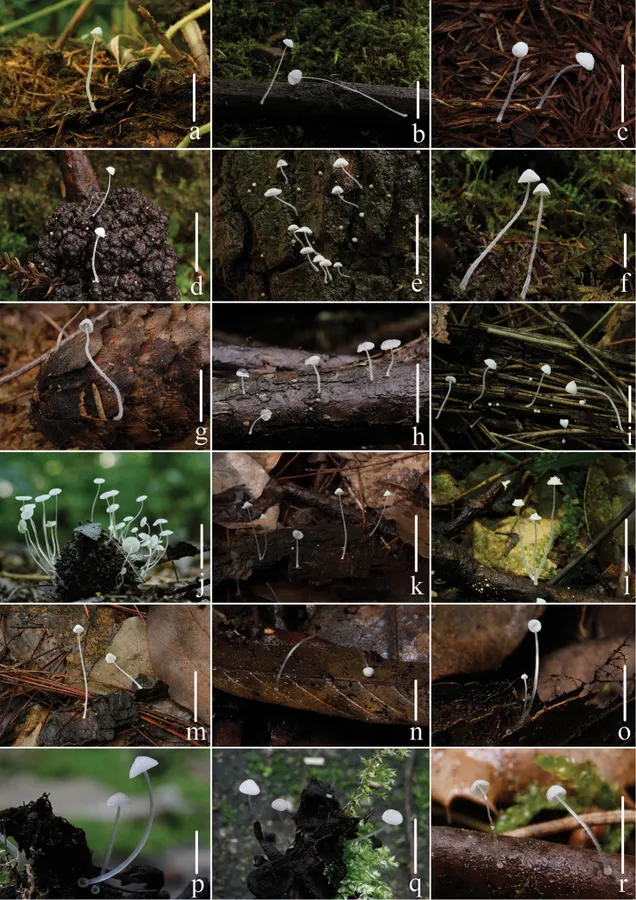
Basidiomata of sect. Amparoina species. stirps Alphitophora: a–b Mycena alphitophora (Berk.) Sacc. c–d Mycena bicystidiata T.Bau & Q.Na e Mycena corynephora Maas Geest. f–g Mycena griseotincta T.Bau & Q.Na h Mycena hygroporoides T.Bau & Q.Na i Mycena miscanthi T.Bau & Q.Na; stirps Amparoina: j Mycena castaneicola T.Bau & Q.Na k–m Mycena heteracantha (Singer) Desjardin. Basidiomata of sect. Saccariferae species n–o Mycena hyalinostipitata T.Bau & Q.Na p–q Mycena substylobates T.Bau & Q.Na r Mycena tenerrima (Berk.) Quél. (= Mycena adscendens Maas Geest.) Scale bars: 10 mm (a–g, i–m, r), 5 mm (h, n–q). Photographs a–r by Qin Na.
- Jenis
- Pelat ilmiah
- Kredit
- Qin Na, Tolgor Bau
- Lisensi
- CC BY 4.0
- Sumber media
- Buka entri sumber
Dokumentasi tambahan
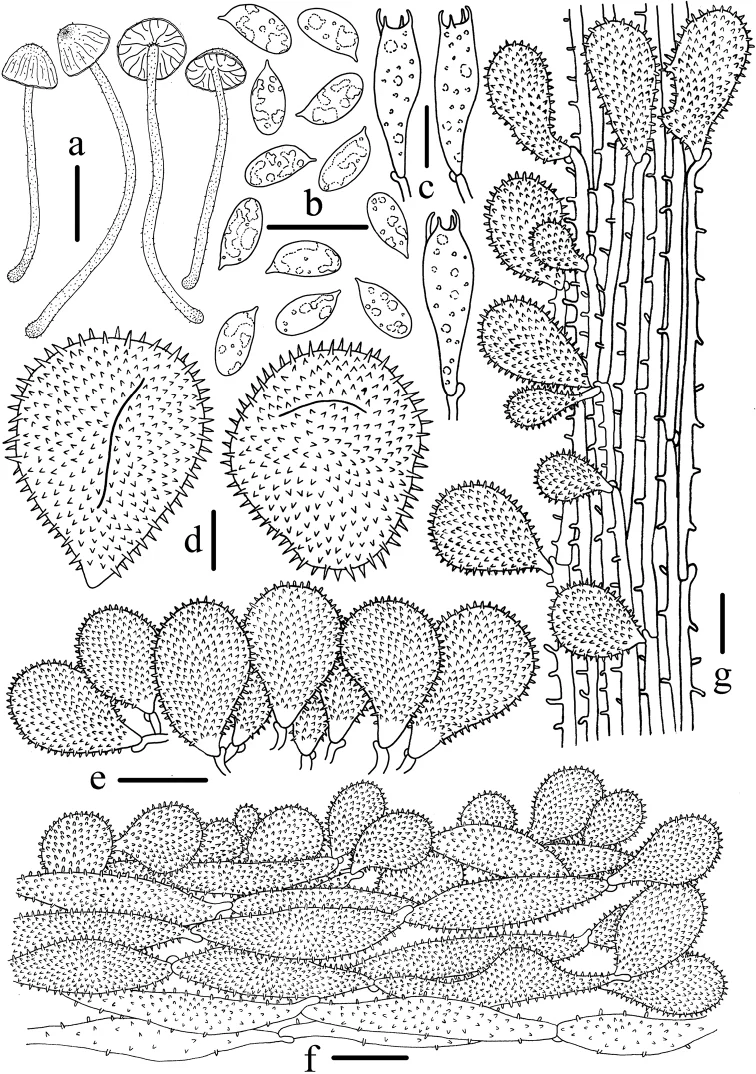
Microscopic features of Mycena miscanthi (HMJAU 43584, holotype) a Basidiomata b Basidiospores c Basidia d Universal veil acanthocysts e Cheilocystidia f Pileipellis g Caulocystidia. Scale bars: 10 mm (a); 10 μm (b–g). Drawing by Qin Na.
Kredit dan penggunaan
Qin Na, Tolgor Bau
Ukuran dan format gambar disesuaikan tanpa pemotongan, retus, atau penambahan skala. Lisensi sumber dipertahankan.
Sumber profil
- [1] Recognition of Mycena sect. Amparoina sect. nov. (Mycenaceae, Agaricales), including four new species and revision of the limits of sect. Sacchariferae — Mycena miscanthi
- [2] Catalogue of Life Base2026-09-11 — Mycena miscanthi
- [3] Dokumentasi visual — Mycena miscanthi
- [4] Dokumentasi visual — Mycena miscanthi
Rujukan halaman ini
Fungi. Mycena miscanthi. Rilis data fungi-2026-10-12-expansion-2. https://fungi-atlas.mgadib.chatgpt.site/jamur/mycena-miscanthi/.
Status: impor sumber; pemeriksaan editorial per profil belum selesai. Untuk klaim biologis, gunakan rujukan pada bagian terkait dan periksa konteks sumber aslinya.