Ciri dan struktur
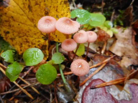
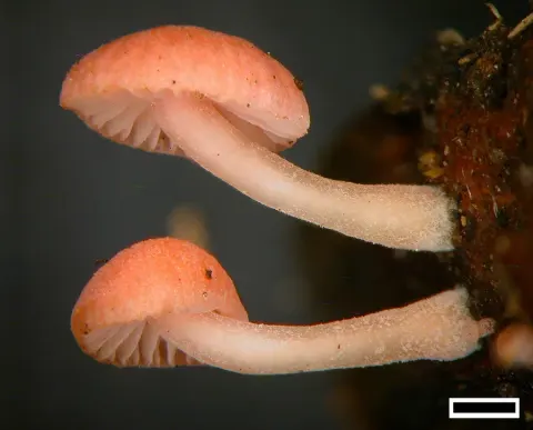
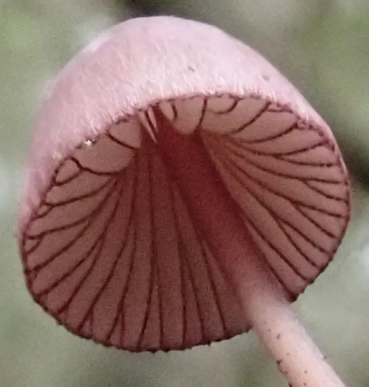
The cap is conical to bell-shaped, flattening in age, and reaches a diameter of 0.5–2.5 cm. The cap margin is usually bent inwards initially. The cap surface is initially covered with tiny white hairs, but later becomes smooth. It is slightly hygrophanous, and when moist, is slightly translucent, so that the outline of the gills underneath are apparent. Its color is dark purple in the center, fading to pale lilac at the margins; older specimens are purplish-gray. The flesh is thin and pliant, with a texture similar to cartilage. It is initially purplish-gray, becoming pale lilac to white in age. The odor is mild and the taste is indistinctive. The narrow gills have an ascending attachment to the stipe and are narrowly adnate. They are somewhat closely spaced, with pallid to grayish face color and dark grayish purple edges that are sometimes fringed. The tubular stipe measures 3–10 cm long by 1–3 mm thick. It is tough and cartilaginous, and its base it covered with white hairs. Overall, its color is that of the cap or paler, and often paler near the top. The spores are broadly ellipsoid in shape, amyloid, and have dimensions of either 8–10 by 6–7 μm or 10–14 by 6.7–8.5 μm depending on whether they originated from four-or two-spored basidia (spore-bearing cells), respectively. There are abundant cheilocystidia on the gill edges. They measure 30–50 by 7–12 μm, and are fusoid-ventricose, with tips that are broadly rounded. They are filled with a purplish sap and have granular contents. The cap tissue comprises a well-differentiated cuticle, a distinct hypoderm, and a filamentous tramal body.
Habitat dan peran ekologis
The fruit bodies grow singly or in clusters on the decaying wood of conifers, In the UK, the fungus is commonly found in Caledonian pine woods, and it is considered an indicator species for that habitat type.
- Habitat atau konteks temuan
- Informasi belum tersedia pada sumber rilis ini.
- Substrat
- Informasi belum tersedia pada sumber rilis ini.
- Peran atau konteks
- Belum dikategorikan pada rilis ini.
Reproduksi
The spores are broadly ellipsoid in shape, amyloid, and have dimensions of either 8–10 by 6–7 μm or 10–14 by 6.7–8.5 μm depending on whether they originated from four-or two-spored basidia (spore-bearing cells), respectively. There are abundant cheilocystidia on the gill edges. They measure 30–50 by 7–12 μm, and are fusoid-ventricose, with tips that are broadly rounded. They are filled with a purplish sap and have granular contents. The cap tissue comprises a well-differentiated cuticle, a distinct hypoderm, and a filamentous tramal body. Clamp connections in the hyphae are rare or absent.
Catatan identitas dan cakupan
- Nama diterima mengikuti Catalogue of Life Base2026-09-11; uraian mempertahankan konsep dan nama sumber aslinya.
- Original geographic, specimen and life-cycle scope retained; source import awaits individual editorial review.
- Impor dan terjemahan otomatis belum diperiksa editorial per profil. Struktur reproduktif yang disebutkan tidak membuktikan seluruh siklus hidup diketahui.
- Pengukuran pada uraian mengikuti spesimen, kultur, dan metode sumber; angka tidak diperluas ke semua populasi.
Catatan distribusi dalam narasi mengikuti sumbernya. Dokumentasi spesimen atau foto mengikuti catatan sumber. Bukti wilayah Indonesia dan penilaian konservasi tidak disimpulkan dari gambar.
Melihat dengan konteks
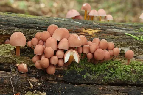
Dokumentasi Mycena purpureofusca menurut asosiasi foto pada record takson iNaturalist. Kredit: debk. Identitas mengikuti sumber; bukan penentuan spesimen independen.
- Jenis
- Fotografi
- Kredit
- (c) debk, some rights reserved (CC BY-SA)
- Lisensi
- CC BY-SA 4.0
- Sumber media
- Buka entri sumber
Sumber profil
- [1] Mycena purpureofusca — Wikipedia (en)
- [2] Catalogue of Life Base2026-09-11 — Mycena purpureofusca
- [3] Dokumentasi visual — Mycena purpureofusca
Rujukan halaman ini
Fungi. Mycena purpureofusca. Rilis data fungi-2026-10-12-expansion-2. https://fungi-atlas.mgadib.chatgpt.site/jamur/mycena-purpureofusca/.
Status: impor sumber; pemeriksaan editorial per profil belum selesai. Untuk klaim biologis, gunakan rujukan pada bagian terkait dan periksa konteks sumber aslinya.