Ciri dan struktur
Resembles N. acutata, but distinguishable by more sparsely formed conidiophores, longer asci, and longer ascospores with rounded extremes (vs. more acute in N. acutata). Apothecia developed from scuta. Scuta superficial, scattered to gregarious, flat discoid, approximately 0.2 mm diam., blackish brown (C80M100Y80–100K60), textura epidermoidea, consisting of thick-walled cells. Apothecia 0.2–0.4 mm high, with dark brown (C80M80Y80–100K60) receptacle; disc 0.5–1.6 mm diam., white to pale gray (K10) when fresh, shrunk to 0.4–1.3 mm diam., pale yellow (Y10) when dried. Ectal excipulum 37–50 µm thick at base, 25–35 µm thick at the upper flank to margin; cortical cells hemispherical to short clavate, 12–17 × 9–10(–12) µm at base, becoming slender and closely packed toward the upper flank to margin. Medullary excipulum 37–87 µm thick. Asci (63–)78–98(–105) × 5–8 µm, arising from croziers, with MLZ + apical pore. Ascospores (12–)17–26(–27.5) × 2.5–3.5 µm, long ellipsoid to fusiform with rounded extremes, (1–)3-septate, containing abundant guttules. Paraphyses (62–)74–90(–100) × 2.5–3 µm, simple, with long apical cell. Subiculum sparsely developed, covering the surface of substrates in patches, shiny brown; subicular hyphae straight to curved, usually constricted at septum, fascicular, 3–5 µm width with 0.5–1 µm thick-walls, septate every 15–25(–50) µm, branched at right angle, covered by gelatinous substance. Colony of NBRC 115371 on PDA flat to slightly winkled, entire to undulate, floccose to woolly, grayish brown (C20–30M40Y40K60) from the surface, forming indistinct section observed clearer from the reverse, without soluble pigment and crystals; aerial mycelium moderately abundant at the center, sparse at the edge, pale gray (K10–30) to white.
Habitat dan peran ekologis
TNS -F-86402, Sugadaira Research Station, Mountain Science Center, Ueda City, Nagano Pref., 5 June 2021, on decaying culms of Sasa kurilensis, ex-holotype culture NBRC 115371. Apothecia developed from scuta. Scuta superficial, scattered to gregarious, flat discoid, approximately 0.2 mm diam., blackish brown (C80M100Y80–100K60), textura epidermoidea, consisting of thick-walled cells. Apothecia 0.2–0.4 mm high, with dark brown (C80M80Y80–100K60) receptacle; disc 0.5–1.6 mm diam., white to pale gray (K10) when fresh, shrunk to 0.4–1.3 mm diam., pale yellow (Y10) when dried. Ectal excipulum 37–50 µm thick at base, 25–35 µm thick at the upper flank to margin; cortical cells hemispherical to short clavate, 12–17 × 9–10(–12) µm at base, becoming slender and closely packed toward the upper flank to margin. Medullary excipulum 37–87 µm thick. Asci (63–)78–98(–105) × 5–8 µm, arising from croziers, with MLZ + apical pore. Ascospores (12–)17–26(–27.5) × 2.5–3.5 µm, long ellipsoid to fusiform with rounded extremes, (1–)3-septate, containing abundant guttules. Paraphyses (62–)74–90(–100) × 2.5–3 µm, simple, with long apical cell. Subiculum sparsely developed, covering the surface of substrates in patches, shiny brown; subicular hyphae straight to curved, usually constricted at septum, fascicular, 3–5 µm width with 0.5–1 µm thick-walls, septate every 15–25(–50) µm, branched at right angle, covered by gelatinous substance.
- Habitat atau konteks temuan
- kultur yang didokumentasikan
- Substrat
- Informasi belum tersedia pada sumber rilis ini.
- Peran atau konteks
- Belum dikategorikan pada rilis ini.
Reproduksi
Resembles N. acutata, but distinguishable by more sparsely formed conidiophores, longer asci, and longer ascospores with rounded extremes (vs. more acute in N. acutata). Apothecia developed from scuta. Scuta superficial, scattered to gregarious, flat discoid, approximately 0.2 mm diam., blackish brown (C80M100Y80–100K60), textura epidermoidea, consisting of thick-walled cells. Apothecia 0.2–0.4 mm high, with dark brown (C80M80Y80–100K60) receptacle; disc 0.5–1.6 mm diam., white to pale gray (K10) when fresh, shrunk to 0.4–1.3 mm diam., pale yellow (Y10) when dried. Ectal excipulum 37–50 µm thick at base, 25–35 µm thick at the upper flank to margin; cortical cells hemispherical to short clavate, 12–17 × 9–10(–12) µm at base, becoming slender and closely packed toward the upper flank to margin.
Catatan identitas dan cakupan
- Nama diterima mengikuti Catalogue of Life Base2026-09-11; uraian mempertahankan konsep dan nama sumber aslinya.
- Preserve original treatment localities, hosts and substrate scope. Materials examined describe documented specimens, not the full global range.
- Impor dan terjemahan otomatis belum diperiksa editorial per profil. Struktur reproduktif yang disebutkan tidak membuktikan seluruh siklus hidup diketahui.
- Pengukuran pada uraian mengikuti spesimen, kultur, dan metode sumber; angka tidak diperluas ke semua populasi.
Catatan distribusi dalam narasi mengikuti sumbernya. Dokumentasi spesimen atau foto mengikuti catatan sumber. Bukti wilayah Indonesia dan penilaian konservasi tidak disimpulkan dari gambar.
Melihat dengan konteks
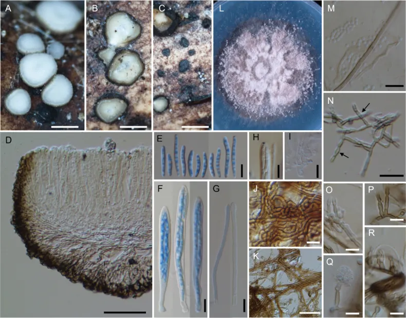
Neobelonopsis multiguttata (TNS -F-86402, holotype) A fresh apothecia on decaying culm of Sasa kurilensis B dried apothecia C stromata with sparse subiculum D vertical section of apothecium (in LA) E ascospores (in CB/ LA) F asci with ascospores (in CB/ LA) G paraphyses (in CB/ LA) H blue-stained apical pore of ascus (in Melzer’s solution after 3% KOH pretreatment) I croziers at the base of asci (in CB/ LA) J texture of stroma (in LA) K subicular hyphae (in lactic acid) L three months old colony on PDA M conidia (in CB/ LA) N–R conidiophores, arrows in N indicated conidiogenous cells (in CB/ LA). Scale bars: 0.5 mm (A–C); 50 μm (D, K); 20 μm (N), 10 μm (E–J, M, O–R).
- Jenis
- Pelat ilmiah
- Kredit
- Hiyori Itagaki, Tsuyoshi Hosoya
- Lisensi
- CC BY 4.0
- Sumber media
- Buka entri sumber
Dokumentasi tambahan
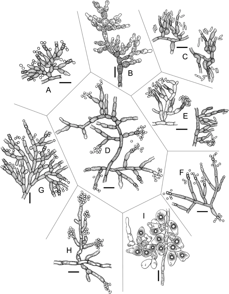
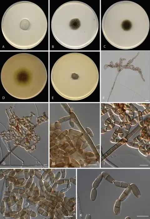
Asexual morph and hyphal structures of the Neobelonopsis and Trichobelonium species A conidiophores of N. acutata B monilioid hyphae of N. bicolor C conidiophores with conidia of N. cinnabarina D conidiophores with conidia of N. didymospora E conidiophores with conidia of N. microspora F conidiophores with conidia of N. multiguttata G conidiophores with conidia of N. obtusa H conidiophores with conidia of T. albobarbatum I hyphal mass with melanized appressorium-like structure of T. miscanthi. Scale bars: 10 μm.
Kredit dan penggunaan
Hiyori Itagaki, Tsuyoshi Hosoya
Ukuran dan format gambar disesuaikan tanpa pemotongan, retus, atau penambahan skala. Lisensi sumber dipertahankan.
Sumber profil
- [1] A new genus Neobelonopsis and two new species of Trichobelonium (Helotiales, Ascomycota) discovered mainly from poaceous grasses native to Asia in Japan — Neobelonopsis multiguttata
- [2] Catalogue of Life Base2026-09-11 — Neobelonopsis multiguttata
- [3] Dokumentasi visual — Neobelonopsis multiguttata
- [4] Dokumentasi visual — Neobelonopsis multiguttata
Rujukan halaman ini
Fungi. Neobelonopsis multiguttata. Rilis data fungi-2026-10-12-expansion-2. https://fungi-atlas.mgadib.chatgpt.site/jamur/neobelonopsis-multiguttata/.
Status: impor sumber; pemeriksaan editorial per profil belum selesai. Untuk klaim biologis, gunakan rujukan pada bagian terkait dan periksa konteks sumber aslinya.