01 / Morfologi

Ciri dan struktur

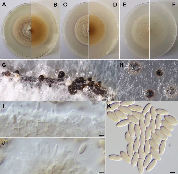
Phylogenetic analysis based on the ITS, ITS and tub2 and ITS and tef1-α dataset considered in the present study clustered the retrieved strains in a monophyletic lineage in the family Didymosphaeriaceae. Therefore, a new genus Neptunomyces gen. nov., and a new species Neptunomyces aureus sp. nov. are here proposed. Mycelium smooth, white, 2–3 μm wide hyphae. Hyphae thick-walled, smooth, hyaline and rarely with nucleus. Conidiomata aggregated or solitary, globose to subglobose, dark brown, immersed or rarely superficial. Conidiomata wall pseudoparenchymatous. Conidiophores reduced to ampulliform to subcylindrical, hyaline, smooth conidiogenous cells (mean ± S.D. = 5.2 ± 0.3 × 2.0 ± 0.6 μm, n = 20). Conidia solitary, subcylindrical with rounded apices, aseptate, initially hyaline, smooth, becoming golden yellow (mean ± S.D. = 7.0 ± 0.6 × 2.7 ± 0.2 μm, n = 100). Sexual morph unknown. On 2 weeks old PDA and OA plates, at 25 °C, colonies growing to 50 mm in diameter, regular and above and a little immersed into agar. PDA obverse white near the center getting flesh orange towards the borders; reverse buff orange in the center and lighter in periphery. OA obverse skimmed milk white; reverse snow white. On 2 weeks old MEA plates, at 25 °C, colonies growing to 44 mm in diameter, regular and above and a little immersed into agar. Obverse orange-colored white; reverse reddish orange in the center and ochre yellow in periphery. At 35 °C, there was no growth in any media tested.

Sebagian uraian ditampilkan dalam bahasa sumber; terjemahannya belum tersedia atau belum lolos pemeriksaan.

Rujukan: [1]

Pengukuran mengikuti objek, spesimen atau kultur, dan metode yang disebutkan sumber. Baca uraian asli untuk konteks lengkap.

02 / Kehidupan

Habitat dan peran ekologis

Portugal, Ria de Aveiro (40°40'38"N, 8°42'21"W), isolated from Gracilaria gracilis, 26 th September 2018, M. Gonçalves, (holotype: a dried culture sporulating on pine needles AVE-F-1; ex-type living culture, MUM 19.38 = CMG 10A). Portugal, Ria de Aveiro (Table 1), isolated from Ulva sp., Enteromorpha intestinalis and Enteromorpha sp. (Supp. material 1: Table S1). M. Gonçalves, living cultures CMG 11, CMG 12, CMG 13 and CMG 14.

Sebagian uraian ditampilkan dalam bahasa sumber; terjemahannya belum tersedia atau belum lolos pemeriksaan.

Rujukan: [1]

Habitat atau konteks temuan
kultur yang didokumentasikan
Substrat
Informasi belum tersedia pada sumber rilis ini.
Peran atau konteks
Belum dikategorikan pada rilis ini.
Temukan hubungan serupa
03 / Siklus hidup

Reproduksi

Mycelium smooth, white, 2–3 μm wide hyphae. Hyphae thick-walled, smooth, hyaline and rarely with nucleus. Conidiomata aggregated or solitary, globose to subglobose, dark brown, immersed or rarely superficial. Conidiomata wall pseudoparenchymatous. Conidiophores reduced to ampulliform to subcylindrical, hyaline, smooth conidiogenous cells (mean ± S.D. = 5.2 ± 0.3 × 2.0 ± 0.6 μm, n = 20). Conidia solitary, subcylindrical with rounded apices, aseptate, initially hyaline, smooth, becoming golden yellow (mean ± S.D. = 7.0 ± 0.6 × 2.7 ± 0.2 μm, n = 100). Sexual morph unknown.

Sebagian uraian ditampilkan dalam bahasa sumber; terjemahannya belum tersedia atau belum lolos pemeriksaan.

Rujukan: [1]

04 / Batas informasi

Catatan identitas dan cakupan

  • Nama diterima mengikuti Catalogue of Life Base2026-09-11; uraian mempertahankan konsep dan nama sumber aslinya.
  • Preserve original treatment localities, hosts and substrate scope. Materials examined describe documented specimens, not the full global range.
  • Impor dan terjemahan otomatis belum diperiksa editorial per profil. Struktur reproduktif yang disebutkan tidak membuktikan seluruh siklus hidup diketahui.
  • Pengukuran pada uraian mengikuti spesimen, kultur, dan metode sumber; angka tidak diperluas ke semua populasi.

Catatan distribusi dalam narasi mengikuti sumbernya. Dokumentasi spesimen atau foto mengikuti catatan sumber. Bukti wilayah Indonesia dan penilaian konservasi tidak disimpulkan dari gambar.

05 / Dokumentasi

Melihat dengan konteks

Neptunomyces aureus (MUM 19.38). A, B Colony after 2 weeks at 25 °C on PDA (obverse and reverse) C, D colony after 2 weeks at 25 °C on MEA (obverse and reverse) E, F colony after 2 weeks at 25 °C on OA (obverse and reverse) G, H conidiomata after 1 month at 25 °C on pine needles and PDA. I, J conidiogenous cells K conidia. Scale bars: 2.5 μm.

Jenis
Pelat ilmiah
Kredit
Micael F. M. Gonçalves, Tânia F. L. Vicente, Ana C. Esteves, Artur Alves
Lisensi
CC BY 4.0
Sumber media
Buka entri sumber

Gambar menunjukkan objek menurut identifikasi sumber, bukan verifikasi independen spesimen oleh Fungi. Tidak ada skala baru yang ditambahkan.

06 / Rujukan

Sumber profil

  1. [1] Neptunomyces aureus gen. et sp. nov. (Didymosphaeriaceae, Pleosporales) isolated from algae in Ria de Aveiro, Portugal — Neptunomyces aureusBagian sumber: PMC6838220_section_13
    Tanggal penghimpunan sumber: 2026-10-12
    Teks sumber: CC BY 4.0 · Gonçalves MFM, Vicente TFL, Esteves AC, Alves A.
  2. [2] Catalogue of Life Base2026-09-11 — Neptunomyces aureusBagian sumber: ColDP NameUsage.tsv ID 47474; accepted species; source 2073
    Tanggal penghimpunan sumber: 2026-10-12
  3. [3] Dokumentasi visual — Neptunomyces aureusBagian sumber: Individual media record; identification follows the source, not independent determination
    Tanggal penghimpunan sumber: 2026-10-12
    Media sumber: CC BY 4.0 · Micael F. M. Gonçalves, Tânia F. L. Vicente, Ana C. Esteves, Artur Alves

Adaptasi teks: CC BY 4.0. Gonçalves MFM, Vicente TFL, Esteves AC, Alves A. — Neptunomyces aureus gen. et sp. nov. (Didymosphaeriaceae, Pleosporales) isolated from algae in Ria de Aveiro, Portugal — Neptunomyces aureus

Uraian asli dan riwayat sumber

Riwayat atau halaman sumber

Ringkasan sumber

Phylogenetic analysis based on the ITS, ITS and tub2 and ITS and tef1-α dataset considered in the present study clustered the retrieved strains in a monophyletic lineage in the family Didymosphaeriaceae. Therefore, a new genus Neptunomyces gen. nov., and a new species Neptunomyces aureus sp. nov. are here proposed. Mycelium smooth, white, 2–3 μm wide hyphae. Hyphae thick-walled, smooth, hyaline and rarely with nucleus. Conidiomata aggregated or solitary, globose to subglobose, dark brown, immersed or rarely superficial.

Revisi sumber

Morfologi sumber

Phylogenetic analysis based on the ITS, ITS and tub2 and ITS and tef1-α dataset considered in the present study clustered the retrieved strains in a monophyletic lineage in the family Didymosphaeriaceae. Therefore, a new genus Neptunomyces gen. nov., and a new species Neptunomyces aureus sp. nov. are here proposed. Mycelium smooth, white, 2–3 μm wide hyphae. Hyphae thick-walled, smooth, hyaline and rarely with nucleus. Conidiomata aggregated or solitary, globose to subglobose, dark brown, immersed or rarely superficial. Conidiomata wall pseudoparenchymatous. Conidiophores reduced to ampulliform to subcylindrical, hyaline, smooth conidiogenous cells (mean ± S.D. = 5.2 ± 0.3 × 2.0 ± 0.6 μm, n = 20). Conidia solitary, subcylindrical with rounded apices, aseptate, initially hyaline, smooth, becoming golden yellow (mean ± S.D. = 7.0 ± 0.6 × 2.7 ± 0.2 μm, n = 100). Sexual morph unknown. On 2 weeks old PDA and OA plates, at 25 °C, colonies growing to 50 mm in diameter, regular and above and a little immersed into agar. PDA obverse white near the center getting flesh orange towards the borders; reverse buff orange in the center and lighter in periphery. OA obverse skimmed milk white; reverse snow white. On 2 weeks old MEA plates, at 25 °C, colonies growing to 44 mm in diameter, regular and above and a little immersed into agar. Obverse orange-colored white; reverse reddish orange in the center and ochre yellow in periphery. At 35 °C, there was no growth in any media tested.

Revisi sumber

Ekologi sumber

Portugal, Ria de Aveiro (40°40'38"N, 8°42'21"W), isolated from Gracilaria gracilis, 26 th September 2018, M. Gonçalves, (holotype: a dried culture sporulating on pine needles AVE-F-1; ex-type living culture, MUM 19.38 = CMG 10A). Portugal, Ria de Aveiro (Table 1), isolated from Ulva sp., Enteromorpha intestinalis and Enteromorpha sp. (Supp. material 1: Table S1). M. Gonçalves, living cultures CMG 11, CMG 12, CMG 13 and CMG 14.

Revisi sumber

Struktur reproduksi sumber

Mycelium smooth, white, 2–3 μm wide hyphae. Hyphae thick-walled, smooth, hyaline and rarely with nucleus. Conidiomata aggregated or solitary, globose to subglobose, dark brown, immersed or rarely superficial. Conidiomata wall pseudoparenchymatous. Conidiophores reduced to ampulliform to subcylindrical, hyaline, smooth conidiogenous cells (mean ± S.D. = 5.2 ± 0.3 × 2.0 ± 0.6 μm, n = 20). Conidia solitary, subcylindrical with rounded apices, aseptate, initially hyaline, smooth, becoming golden yellow (mean ± S.D. = 7.0 ± 0.6 × 2.7 ± 0.2 μm, n = 100). Sexual morph unknown.

Revisi sumber

Rujukan halaman ini

Fungi. Neptunomyces aureus. Rilis data fungi-2026-10-12-expansion-2. https://fungi-atlas.mgadib.chatgpt.site/jamur/neptunomyces-aureus/.

Status: impor sumber; pemeriksaan editorial per profil belum selesai. Untuk klaim biologis, gunakan rujukan pada bagian terkait dan periksa konteks sumber aslinya.