01 / Morfologi

Ciri dan struktur

Saprobic on wood. Sexual morph. Ascomata 1.4–2.03 µm high, 1.4–1.78 µm in diam., sub globose to ellipsoidal, round at the top, flat at the base, stalked or aggregated in a stroma, hypostroma 30–135 µm, ostiolate, papillate, fleshy, brown to dull orange-colored, gregarious. Hypostroma is 30–120 µm thick, light-colored, composed of small, polygonal to rounded cells with large lumina; masses of lilac-colored hyphae can be found in the wood vessels under the stromata. Peridium is 450–470 µm thick at the apex, 240–260 µm at the sides, composed of 17–35 layers of hyaline, thick-walled, polygonal, isodiametric cells with large lumina, forming a textura angularis; merging toward the center into flattened cells; outer 3–7-cells filled with small yellowish globules. Ostioles 25–30 µm in diam., periphysate. Paraphyses absent. Ascomata filled with hyaline, thin-walled, polygonal, isodiametric, pseudoparenchymatous cells, which eventually compressed by the asci. Asci are 70–90 × 12–17 µm, 8-spored, clavate, unitunicate, thin-walled, without apical apparatus, deliquescing. Ascospores 60–80 × 4–6 µm, filiform to elongate fusiform, 1-septate, hyaline, with one apical appendage: appendages 32–50 µm long, 2(-3) µm in diam. at the base, filamentous, tapering toward the apex, wavy, whip-like, hamate, closely adpressed to the spore wall, becoming detached from the wall, but adhering with its base to the apex of the ascospore (Based on Kohlmeyer and Kohlmeyer 1979; Dupont and Schwabe 2016). Asexual morph. Undetermined.

Sebagian uraian ditampilkan dalam bahasa sumber; terjemahannya belum tersedia atau belum lolos pemeriksaan.

Rujukan: [1]

Pengukuran mengikuti objek, spesimen atau kultur, dan metode yang disebutkan sumber. Baca uraian asli untuk konteks lengkap.

02 / Kehidupan

Habitat dan peran ekologis

Angola (Vanuatu archipelago, Pacific Ocean), South China Sea (Taiping Island, and Dongsha Island), Kuril-Kamchatka Trench (Pacific Ocean, Kamchatka Peninsula, the Kuril Islands, and Hokkaido island, Japan).

Sebagian uraian ditampilkan dalam bahasa sumber; terjemahannya belum tersedia atau belum lolos pemeriksaan.

Rujukan: [1]

Habitat atau konteks temuan
Informasi belum tersedia pada sumber rilis ini.
Substrat
Informasi belum tersedia pada sumber rilis ini.
Peran atau konteks
Belum dikategorikan pada rilis ini.
Temukan hubungan serupa
03 / Siklus hidup

Reproduksi

Saprobic on wood. Sexual morph. Ascomata 1.4–2.03 µm high, 1.4–1.78 µm in diam., sub globose to ellipsoidal, round at the top, flat at the base, stalked or aggregated in a stroma, hypostroma 30–135 µm, ostiolate, papillate, fleshy, brown to dull orange-colored, gregarious. Hypostroma is 30–120 µm thick, light-colored, composed of small, polygonal to rounded cells with large lumina; masses of lilac-colored hyphae can be found in the wood vessels under the stromata. Peridium is 450–470 µm thick at the apex, 240–260 µm at the sides, composed of 17–35 layers of hyaline, thick-walled, polygonal, isodiametric cells with large lumina, forming a textura angularis; merging toward the center into flattened cells; outer 3–7-cells filled with small yellowish globules.

Sebagian uraian ditampilkan dalam bahasa sumber; terjemahannya belum tersedia atau belum lolos pemeriksaan.

Rujukan: [1]

04 / Batas informasi

Catatan identitas dan cakupan

  • Nama diterima mengikuti Catalogue of Life Base2026-09-11; uraian mempertahankan konsep dan nama sumber aslinya.
  • Preserve original treatment localities, hosts and substrate scope. Materials examined describe documented specimens, not the full global range.
  • Impor dan terjemahan otomatis belum diperiksa editorial per profil. Struktur reproduktif yang disebutkan tidak membuktikan seluruh siklus hidup diketahui.
  • Pengukuran pada uraian mengikuti spesimen, kultur, dan metode sumber; angka tidak diperluas ke semua populasi.

Catatan distribusi dalam narasi mengikuti sumbernya. Dokumentasi spesimen atau foto mengikuti catatan sumber. Bukti wilayah Indonesia dan penilaian konservasi tidak disimpulkan dari gambar.

05 / Dokumentasi

Melihat dengan konteks

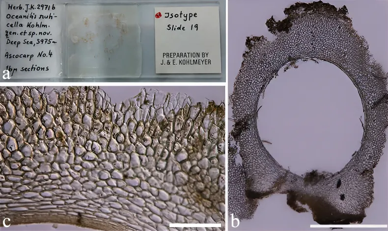
Oceanitis scuticella (NY 1348289, isotype). a. Herbarium material of Oceanitis scuticella; b. Section through ascomata; c. Peridium. Scale bars: 20 μm (b); 10 μm (c).

Jenis
Pelat ilmiah
Kredit
Monika C. Dayarathne, E. B. Gareth Jones, Ali H. Bahkali, Bandarupalli Devadatha, Kevin D. Hyde, Mohamed A. Abdel-Wahab, Mark S. Calabon, Pedro Correia, Faten A. Abdel-Aziz, Mahmoud S. Bakhit, Jariya Sakayaroj, Chasika Prematunga, Maria F. Caeiro, Egídia Azevedo, Raheleh Asghari, Carlo Chris S. Apurillo, Amuhenage Tharindu Bhagya, Herbert Dustin Aumentado, Samhita Mukhopadhyay, Chayanard Phukhamsakda, Yanpeng Chen, Sajeewa S. Maharachchikumbura, Sheng-Yu Guo, Ka-Lai Pang
Lisensi
CC BY 4.0
Sumber media
Buka entri sumber

Gambar menunjukkan objek menurut identifikasi sumber, bukan verifikasi independen spesimen oleh Fungi. Tidak ada skala baru yang ditambahkan.

06 / Rujukan

Sumber profil

  1. [1] Taxonomy and molecular evaluation of the Halosphaeriaceae (Microascales, Hypocreomycetidae) — Oceanitis scuticellaBagian sumber: PMC13329432_section_541
    Tanggal penghimpunan sumber: 2026-10-12
    Teks sumber: CC BY 4.0 · Dayarathne MC, Jones EBG, Bahkali AH, Devadatha B, Hyde KD, Abdel-Wahab MA, Calabon MS, Correia P, Abdel-Aziz FA, Bakhit MS, Sakayaroj J, Prematunga C, Caeiro MF, Azevedo E, Asghari R, Apurillo CCS, Bhagya AT, Aumentado HD, Mukhopadhyay S, Phukhamsakda C, Chen Y, Maharachchikumbura SS, Guo SY, Pang KL.
  2. [2] Catalogue of Life Base2026-09-11 — Oceanitis scuticellaBagian sumber: ColDP NameUsage.tsv ID 48C3L; accepted species; source 2073
    Tanggal penghimpunan sumber: 2026-10-12
  3. [3] Dokumentasi visual — Oceanitis scuticellaBagian sumber: Individual media record; identification follows the source, not independent determination
    Tanggal penghimpunan sumber: 2026-10-12
    Media sumber: CC BY 4.0 · Monika C. Dayarathne, E. B. Gareth Jones, Ali H. Bahkali, Bandarupalli Devadatha, Kevin D. Hyde, Mohamed A. Abdel-Wahab, Mark S. Calabon, Pedro Correia, Faten A. Abdel-Aziz, Mahmoud S. Bakhit, Jariya Sakayaroj, Chasika Prematunga, Maria F. Caeiro, Egídia Azevedo, Raheleh Asghari, Carlo Chris S. Apurillo, Amuhenage Tharindu Bhagya, Herbert Dustin Aumentado, Samhita Mukhopadhyay, Chayanard Phukhamsakda, Yanpeng Chen, Sajeewa S. Maharachchikumbura, Sheng-Yu Guo, Ka-Lai Pang

Adaptasi teks: CC BY 4.0. Dayarathne MC, Jones EBG, Bahkali AH, Devadatha B, Hyde KD, Abdel-Wahab MA, Calabon MS, Correia P, Abdel-Aziz FA, Bakhit MS, Sakayaroj J, Prematunga C, Caeiro MF, Azevedo E, Asghari R, Apurillo CCS, Bhagya AT, Aumentado HD, Mukhopadhyay S, Phukhamsakda C, Chen Y, Maharachchikumbura SS, Guo SY, Pang KL. — Taxonomy and molecular evaluation of the Halosphaeriaceae (Microascales, Hypocreomycetidae) — Oceanitis scuticella

Uraian asli dan riwayat sumber

Riwayat atau halaman sumber

Ringkasan sumber

Saprobic on wood. Sexual morph. Ascomata 1.4–2.03 µm high, 1.4–1.78 µm in diam., sub globose to ellipsoidal, round at the top, flat at the base, stalked or aggregated in a stroma, hypostroma 30–135 µm, ostiolate, papillate, fleshy, brown to dull orange-colored, gregarious. Hypostroma is 30–120 µm thick, light-colored, composed of small, polygonal to rounded cells with large lumina; masses of lilac-colored hyphae can be found in the wood vessels under the stromata. Peridium is 450–470 µm thick at the apex, 240–260 µm at the sides, composed of 17–35 layers of hyaline, thick-walled, polygonal, isodiametric cells with large lumina, forming a textura angularis; merging toward the center into flattened cells; outer 3–7-cells filled with small yellowish globules.

Revisi sumber

Morfologi sumber

Saprobic on wood. Sexual morph. Ascomata 1.4–2.03 µm high, 1.4–1.78 µm in diam., sub globose to ellipsoidal, round at the top, flat at the base, stalked or aggregated in a stroma, hypostroma 30–135 µm, ostiolate, papillate, fleshy, brown to dull orange-colored, gregarious. Hypostroma is 30–120 µm thick, light-colored, composed of small, polygonal to rounded cells with large lumina; masses of lilac-colored hyphae can be found in the wood vessels under the stromata. Peridium is 450–470 µm thick at the apex, 240–260 µm at the sides, composed of 17–35 layers of hyaline, thick-walled, polygonal, isodiametric cells with large lumina, forming a textura angularis; merging toward the center into flattened cells; outer 3–7-cells filled with small yellowish globules. Ostioles 25–30 µm in diam., periphysate. Paraphyses absent. Ascomata filled with hyaline, thin-walled, polygonal, isodiametric, pseudoparenchymatous cells, which eventually compressed by the asci. Asci are 70–90 × 12–17 µm, 8-spored, clavate, unitunicate, thin-walled, without apical apparatus, deliquescing. Ascospores 60–80 × 4–6 µm, filiform to elongate fusiform, 1-septate, hyaline, with one apical appendage: appendages 32–50 µm long, 2(-3) µm in diam. at the base, filamentous, tapering toward the apex, wavy, whip-like, hamate, closely adpressed to the spore wall, becoming detached from the wall, but adhering with its base to the apex of the ascospore (Based on Kohlmeyer and Kohlmeyer 1979; Dupont and Schwabe 2016). Asexual morph. Undetermined.

Revisi sumber

Ekologi sumber

Angola (Vanuatu archipelago, Pacific Ocean), South China Sea (Taiping Island, and Dongsha Island), Kuril-Kamchatka Trench (Pacific Ocean, Kamchatka Peninsula, the Kuril Islands, and Hokkaido island, Japan).

Revisi sumber

Struktur reproduksi sumber

Saprobic on wood. Sexual morph. Ascomata 1.4–2.03 µm high, 1.4–1.78 µm in diam., sub globose to ellipsoidal, round at the top, flat at the base, stalked or aggregated in a stroma, hypostroma 30–135 µm, ostiolate, papillate, fleshy, brown to dull orange-colored, gregarious. Hypostroma is 30–120 µm thick, light-colored, composed of small, polygonal to rounded cells with large lumina; masses of lilac-colored hyphae can be found in the wood vessels under the stromata. Peridium is 450–470 µm thick at the apex, 240–260 µm at the sides, composed of 17–35 layers of hyaline, thick-walled, polygonal, isodiametric cells with large lumina, forming a textura angularis; merging toward the center into flattened cells; outer 3–7-cells filled with small yellowish globules.

Revisi sumber

Rujukan halaman ini

Fungi. Oceanitis scuticella. Rilis data fungi-2026-10-12-expansion-2. https://fungi-atlas.mgadib.chatgpt.site/jamur/oceanitis-scuticella/.

Status: impor sumber; pemeriksaan editorial per profil belum selesai. Untuk klaim biologis, gunakan rujukan pada bagian terkait dan periksa konteks sumber aslinya.