01 / Morfologi

Ciri dan struktur

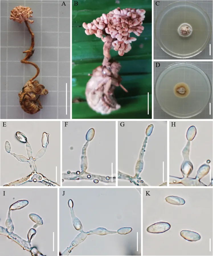
The colony grew slowly on PDA medium. Cultured at 25 °C for about 12 weeks, the diameter of the colony was 25–28 mm, pale pink, the edge white, hard texture. The back of the colony was white to brown. Surface hyphae rough, hyaline, septate. Conidiophores were cylindrical. Conidiogenous cells were solitary or whorled, ampuliform, smooth-walled, forming on conidiophores or colonies, hyaline, with swollen base, and slender top, 10.6–17.6 µm long, 2.9–4.3 µm wide at the swollen base, and 1.1–2.2 µm wide at the slender top. Conidia hyaline, ovoid or long oval, solitary, 6.8–10.1 × 3.3–4.5 µm.

Sebagian uraian ditampilkan dalam bahasa sumber; terjemahannya belum tersedia atau belum lolos pemeriksaan.

Rujukan: [1]

Pengukuran mengikuti objek, spesimen atau kultur, dan metode yang disebutkan sumber. Baca uraian asli untuk konteks lengkap.

02 / Kehidupan

Habitat dan peran ekologis

China, Yunnan Province, Jinghong City, Nabanhe National Nature Reserve, 22°8'21.32"N, 100°42'18.35"E, alt. 612 m, on cicada nymphs (Cicadidae, Hemiptera). The material was found in the soil of an evergreen broad-leaved forest, 18 August 2020, H. Yu (YHH 20081, holotype; YFCC 8834, ex-holotype culture). The colony grew slowly on PDA medium. Cultured at 25 °C for about 12 weeks, the diameter of the colony was 25–28 mm, pale pink, the edge white, hard texture. The back of the colony was white to brown. Surface hyphae rough, hyaline, septate. Conidiophores were cylindrical. Conidiogenous cells were solitary or whorled, ampuliform, smooth-walled, forming on conidiophores or colonies, hyaline, with swollen base, and slender top, 10.6–17.6 µm long, 2.9–4.3 µm wide at the swollen base, and 1.1–2.2 µm wide at the slender top. Conidia hyaline, ovoid or long oval, solitary, 6.8–10.1 × 3.3–4.5 µm. China, Yunnan Province, Jinghong City, Nabanhe National Nature Reserve, 22°8'21.32"N, 100°42'18.35"E, alt. 612 m, on cicada nymphs (Cicadidae, Hemiptera) was found in the soil an evergreen broad-leaved forest, 18 August 2020, H. Yu (YFCC 8832, YFCC 8833).

Sebagian uraian ditampilkan dalam bahasa sumber; terjemahannya belum tersedia atau belum lolos pemeriksaan.

Rujukan: [1]

Habitat atau konteks temuan
hutan; kultur yang didokumentasikan
Substrat
Informasi belum tersedia pada sumber rilis ini.
Peran atau konteks
Belum dikategorikan pada rilis ini.
Temukan hubungan serupa
03 / Siklus hidup

Reproduksi

The stroma was grown from the head of the host cicada nymph, solitary, the top of the stroma similar to hydrangea, pale pink, 1.6–6.4 cm long. Sexual morph was not observed. The colony grew slowly on PDA medium. Cultured at 25 °C for about 12 weeks, the diameter of the colony was 25–28 mm, pale pink, the edge white, hard texture. The back of the colony was white to brown. Surface hyphae rough, hyaline, septate. Conidiophores were cylindrical. Conidiogenous cells were solitary or whorled, ampuliform, smooth-walled, forming on conidiophores or colonies, hyaline, with swollen base, and slender top, 10.6–17.6 µm long, 2.9–4.3 µm wide at the swollen base, and 1.1–2.2 µm wide at the slender top. Conidia hyaline, ovoid or long oval, solitary, 6.8–10.1 × 3.3–4.5 µm.

Sebagian uraian ditampilkan dalam bahasa sumber; terjemahannya belum tersedia atau belum lolos pemeriksaan.

Rujukan: [1]

04 / Batas informasi

Catatan identitas dan cakupan

  • Nama diterima mengikuti Catalogue of Life Base2026-09-11; uraian mempertahankan konsep dan nama sumber aslinya.
  • Preserve original treatment localities, hosts and substrate scope. Materials examined describe documented specimens, not the full global range.
  • Impor dan terjemahan otomatis belum diperiksa editorial per profil. Struktur reproduktif yang disebutkan tidak membuktikan seluruh siklus hidup diketahui.
  • Pengukuran pada uraian mengikuti spesimen, kultur, dan metode sumber; angka tidak diperluas ke semua populasi.

Catatan distribusi dalam narasi mengikuti sumbernya. Dokumentasi spesimen atau foto mengikuti catatan sumber. Bukti wilayah Indonesia dan penilaian konservasi tidak disimpulkan dari gambar.

05 / Dokumentasi

Melihat dengan konteks

Ophiocordyceps hydrangea A, B fungus on a cicada nymph C, D colony on PDA medium E conidiophores, conidiogenous cells and conidia F–J conidiogenous cells and conidia K conidia. Scale bars: 1 cm (A, B); 2 cm (C, D); 10 µm (E, F, G, I, J); 5 µm (H, K).

Jenis
Pelat ilmiah
Kredit
Weiqiu Zou, Dexiang Tang, Zhihong Xu, Ou Huang, Yuanbing Wang, Ngoc-Lan Tran, Hong Yu
Lisensi
CC BY 4.0
Sumber media
Buka entri sumber

Gambar menunjukkan objek menurut identifikasi sumber, bukan verifikasi independen spesimen oleh Fungi. Tidak ada skala baru yang ditambahkan.

06 / Rujukan

Sumber profil

  1. [1] Multigene phylogeny and morphology reveal Ophiocordyceps hydrangea sp. nov. and Ophiocordyceps bidoupensis sp. nov. (Ophiocordycipitaceae) — Ophiocordyceps hydrangeaBagian sumber: PMC9849067_section_9
    Tanggal penghimpunan sumber: 2026-10-12
    Teks sumber: CC BY 4.0 · Zou W, Tang D, Xu Z, Huang O, Wang Y, Tran NL, Yu H.
  2. [2] Catalogue of Life Base2026-09-11 — Ophiocordyceps hydrangeaBagian sumber: ColDP NameUsage.tsv ID B2RKC; accepted species; source 2073
    Tanggal penghimpunan sumber: 2026-10-12
  3. [3] Dokumentasi visual — Ophiocordyceps hydrangeaBagian sumber: Individual media record; identification follows the source, not independent determination
    Tanggal penghimpunan sumber: 2026-10-12
    Media sumber: CC BY 4.0 · Weiqiu Zou, Dexiang Tang, Zhihong Xu, Ou Huang, Yuanbing Wang, Ngoc-Lan Tran, Hong Yu

Adaptasi teks: CC BY 4.0. Zou W, Tang D, Xu Z, Huang O, Wang Y, Tran NL, Yu H. — Multigene phylogeny and morphology reveal Ophiocordyceps hydrangea sp. nov. and Ophiocordyceps bidoupensis sp. nov. (Ophiocordycipitaceae) — Ophiocordyceps hydrangea

Uraian asli dan riwayat sumber

Riwayat atau halaman sumber

Ringkasan sumber

The colony grew slowly on PDA medium. Cultured at 25 °C for about 12 weeks, the diameter of the colony was 25–28 mm, pale pink, the edge white, hard texture. The back of the colony was white to brown. Surface hyphae rough, hyaline, septate. Conidiophores were cylindrical. Conidiogenous cells were solitary or whorled, ampuliform, smooth-walled, forming on conidiophores or colonies, hyaline, with swollen base, and slender top, 10.6–17.6 µm long, 2.9–4.3 µm wide at the swollen base, and 1.1–2.2 µm wide at the slender top.

Revisi sumber

Morfologi sumber

The colony grew slowly on PDA medium. Cultured at 25 °C for about 12 weeks, the diameter of the colony was 25–28 mm, pale pink, the edge white, hard texture. The back of the colony was white to brown. Surface hyphae rough, hyaline, septate. Conidiophores were cylindrical. Conidiogenous cells were solitary or whorled, ampuliform, smooth-walled, forming on conidiophores or colonies, hyaline, with swollen base, and slender top, 10.6–17.6 µm long, 2.9–4.3 µm wide at the swollen base, and 1.1–2.2 µm wide at the slender top. Conidia hyaline, ovoid or long oval, solitary, 6.8–10.1 × 3.3–4.5 µm.

Revisi sumber

Ekologi sumber

China, Yunnan Province, Jinghong City, Nabanhe National Nature Reserve, 22°8'21.32"N, 100°42'18.35"E, alt. 612 m, on cicada nymphs (Cicadidae, Hemiptera). The material was found in the soil of an evergreen broad-leaved forest, 18 August 2020, H. Yu (YHH 20081, holotype; YFCC 8834, ex-holotype culture). The colony grew slowly on PDA medium. Cultured at 25 °C for about 12 weeks, the diameter of the colony was 25–28 mm, pale pink, the edge white, hard texture. The back of the colony was white to brown. Surface hyphae rough, hyaline, septate. Conidiophores were cylindrical. Conidiogenous cells were solitary or whorled, ampuliform, smooth-walled, forming on conidiophores or colonies, hyaline, with swollen base, and slender top, 10.6–17.6 µm long, 2.9–4.3 µm wide at the swollen base, and 1.1–2.2 µm wide at the slender top. Conidia hyaline, ovoid or long oval, solitary, 6.8–10.1 × 3.3–4.5 µm. China, Yunnan Province, Jinghong City, Nabanhe National Nature Reserve, 22°8'21.32"N, 100°42'18.35"E, alt. 612 m, on cicada nymphs (Cicadidae, Hemiptera) was found in the soil an evergreen broad-leaved forest, 18 August 2020, H. Yu (YFCC 8832, YFCC 8833).

Revisi sumber

Struktur reproduksi sumber

The stroma was grown from the head of the host cicada nymph, solitary, the top of the stroma similar to hydrangea, pale pink, 1.6–6.4 cm long. Sexual morph was not observed. The colony grew slowly on PDA medium. Cultured at 25 °C for about 12 weeks, the diameter of the colony was 25–28 mm, pale pink, the edge white, hard texture. The back of the colony was white to brown. Surface hyphae rough, hyaline, septate. Conidiophores were cylindrical. Conidiogenous cells were solitary or whorled, ampuliform, smooth-walled, forming on conidiophores or colonies, hyaline, with swollen base, and slender top, 10.6–17.6 µm long, 2.9–4.3 µm wide at the swollen base, and 1.1–2.2 µm wide at the slender top. Conidia hyaline, ovoid or long oval, solitary, 6.8–10.1 × 3.3–4.5 µm.

Revisi sumber

Rujukan halaman ini

Fungi. Ophiocordyceps hydrangea. Rilis data fungi-2026-10-12-expansion-2. https://fungi-atlas.mgadib.chatgpt.site/jamur/ophiocordyceps-hydrangea/.

Status: impor sumber; pemeriksaan editorial per profil belum selesai. Untuk klaim biologis, gunakan rujukan pada bagian terkait dan periksa konteks sumber aslinya.