01 / Morfologi

Ciri dan struktur

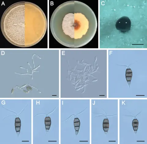
Sexual state not seen. Conidiomata in culture sporodochial, saucer-shaped, scattered or gregarious, superficial to immersed, shining, releasing black conidial masses on the surface. Conidiophores branched, subcylindrical, hyaline to light brown, indistinct, often reduced to conidiogenous cells. Conidiogenous cells cylindrical or ampulliform, hyaline, smooth-walled, solitary to aggregated, (5.5–)7–10(–11.5) × 2–3 μm (x ± SD = 8.4 ± 1.6 × 2.4 ± 0.4 μm). Conidia fusoid, ellipsoid, smooth, slightly constricted at the septa, 4-septate, (8–)17.5–25.5(–26) × (4–)6–7.5(–8) μm (x ± SD = 21.6 ± 4.1 × 6.7 ± 0.8 μm); apical cell with 2–3 tubular appendages (mostly three); apical appendages arising from an apical crest, unbranched, filiform, bent, (3.5–)10.5–16.5(–22) μm (x ± SD = 13.6 ± 3.1 μm) long; basal cell with one appendage; basal appendage tubular, centric appendage tubular, unbranched, occasionally swollen at the tip, (2–)2.5–4(–4.5) μm (x ± SD = 3.4 ± 0.7 μm) long.

Sebagian uraian ditampilkan dalam bahasa sumber; terjemahannya belum tersedia atau belum lolos pemeriksaan.

Rujukan: [1]

Pengukuran mengikuti objek, spesimen atau kultur, dan metode yang disebutkan sumber. Baca uraian asli untuk konteks lengkap.

02 / Kehidupan

Habitat dan peran ekologis

Pestalotiopsis disseminata was first reported from Eucalyptus botryoides in Portugal (Von Thümen 1881) and subsequently reported on a wide range of hosts and locations, including the genus Pinus (Hu et al. 2007; Liu et al. 2019; Silva et al. 2020). In this study, 17 strains were isolated from P. elliottii and P. thunbergii and clustered together with Pes. disseminata in the multi-locus phylogenetic tree (Fig. 3). Compared to the description of the isolate CBS 118552, ZXD579 exhibits smaller conidiogenous cells (5.5–11.5 × 2–3 μm vs. 7–24.5 × 2–5 μm), while other morphological characteristics are similar. This is the first report of Pes. disseminata on the host of P. elliottii and P. thunbergii.

Sebagian uraian ditampilkan dalam bahasa sumber; terjemahannya belum tersedia atau belum lolos pemeriksaan.

Rujukan: [1]

Habitat atau konteks temuan
Informasi belum tersedia pada sumber rilis ini.
Substrat
Informasi belum tersedia pada sumber rilis ini.
Peran atau konteks
Belum dikategorikan pada rilis ini.
Temukan hubungan serupa
03 / Siklus hidup

Reproduksi

Sexual state not seen. Conidiomata in culture sporodochial, saucer-shaped, scattered or gregarious, superficial to immersed, shining, releasing black conidial masses on the surface. Conidiophores branched, subcylindrical, hyaline to light brown, indistinct, often reduced to conidiogenous cells. Conidiogenous cells cylindrical or ampulliform, hyaline, smooth-walled, solitary to aggregated, (5.5–)7–10(–11.5) × 2–3 μm (x ± SD = 8.4 ± 1.6 × 2.4 ± 0.4 μm). Conidia fusoid, ellipsoid, smooth, slightly constricted at the septa, 4-septate, (8–)17.5–25.5(–26) × (4–)6–7.5(–8) μm (x ± SD = 21.6 ± 4.1 × 6.7 ± 0.8 μm); apical cell with 2–3 tubular appendages (mostly three); apical appendages arising from an apical crest, unbranched, filiform, bent, (3.5–)10.5–16.5(–22) μm (x ± SD = 13.6 ± 3.1 μm) long; basal cell with one appendage; basal appendage tubular, centric appendage tubular, unbranched, occasionally swollen at the tip, (2–)2.5–4(–4.5) μm (x ± SD = 3.4 ± 0.7 μm) long.

Sebagian uraian ditampilkan dalam bahasa sumber; terjemahannya belum tersedia atau belum lolos pemeriksaan.

Rujukan: [1]

Sinonim menurut acuan taksonomi rilis: Pestalotia disseminata
04 / Batas informasi

Catatan identitas dan cakupan

  • Nama diterima mengikuti Catalogue of Life Base2026-09-11; uraian mempertahankan konsep dan nama sumber aslinya.
  • Preserve original treatment localities, hosts and substrate scope. Materials examined describe documented specimens, not the full global range.
  • Impor dan terjemahan otomatis belum diperiksa editorial per profil. Struktur reproduktif yang disebutkan tidak membuktikan seluruh siklus hidup diketahui.
  • Pengukuran pada uraian mengikuti spesimen, kultur, dan metode sumber; angka tidak diperluas ke semua populasi.

Catatan distribusi dalam narasi mengikuti sumbernya. Dokumentasi spesimen atau foto mengikuti catatan sumber. Bukti wilayah Indonesia dan penilaian konservasi tidak disimpulkan dari gambar.

05 / Dokumentasi

Melihat dengan konteks

Morphology of Pestalotiopsis disseminata (ZXD579). A Colony on MEA after 10 days at 25°C; B colony on PDA after 10 days at 25°C; C conidiomata formed on MEA; D, E conidiogenous cells giving rise to conidia; F–K conidia. Scale bars: 500 μm (C); 20 μm (D–G).

Jenis
Pelat ilmiah
Kredit
Quan Chao Wang, Zhao Jie Zhan, Adil Sattar, Hao Nan Wang, Li Feng Zhou, Lori Eckhardt, Guo Qing Li, Fei Fei Liu, Hua Chao Xu, Xu Dong Zhou
Lisensi
CC BY 4.0
Sumber media
Buka entri sumber

Gambar menunjukkan objek menurut identifikasi sumber, bukan verifikasi independen spesimen oleh Fungi. Tidak ada skala baru yang ditambahkan.

06 / Rujukan

Sumber profil

  1. [1]  Pestalotiopsis (Amphisphaeriales, Sporocadaceae) species including six new taxa inhabiting pines from different climate zones in China — Pestalotiopsis disseminataBagian sumber: PMC12177513_section_13
    Tanggal penghimpunan sumber: 2026-10-12
    Teks sumber: CC BY 4.0 · Wang, Quan Chao & Zhan, Zhao Jie & Sattar, Adil & Wang, Hao Nan
  2. [2] Catalogue of Life Base2026-09-11 — Pestalotiopsis disseminataBagian sumber: ColDP NameUsage.tsv ID 4FC2K; accepted species; source 2073
    Tanggal penghimpunan sumber: 2026-10-12
  3. [3] Dokumentasi visual — Pestalotiopsis disseminataBagian sumber: Individual media record; identification follows the source, not independent determination
    Tanggal penghimpunan sumber: 2026-10-12
    Media sumber: CC BY 4.0 · Quan Chao Wang, Zhao Jie Zhan, Adil Sattar, Hao Nan Wang, Li Feng Zhou, Lori Eckhardt, Guo Qing Li, Fei Fei Liu, Hua Chao Xu, Xu Dong Zhou

Adaptasi teks: CC BY 4.0. Wang, Quan Chao & Zhan, Zhao Jie & Sattar, Adil & Wang, Hao Nan —  Pestalotiopsis (Amphisphaeriales, Sporocadaceae) species including six new taxa inhabiting pines from different climate zones in China — Pestalotiopsis disseminata

Uraian asli dan riwayat sumber

Riwayat atau halaman sumber

Ringkasan sumber

Sexual state not seen. Conidiomata in culture sporodochial, saucer-shaped, scattered or gregarious, superficial to immersed, shining, releasing black conidial masses on the surface. Conidiophores branched, subcylindrical, hyaline to light brown, indistinct, often reduced to conidiogenous cells. Conidiogenous cells cylindrical or ampulliform, hyaline, smooth-walled, solitary to aggregated, (5.5–)7–10(–11.5) × 2–3 μm (x ± SD = 8.4 ± 1.6 × 2.4 ± 0.4 μm). Conidia fusoid, ellipsoid, smooth, slightly constricted at the septa, 4-septate, (8–)17.5–25.5(–26) × (4–)6–7.5(–8) μm (x ± SD = 21.6 ± 4.1 × 6.7 ± 0.8 μm); apical cell with 2–3 tubular appendages (mostly three); apical appendages arising from an apical crest, unbranched, filiform, bent, (3.5–)10.5–16.5(–22) μm (x ± SD = 13.6 ± 3.1 μm) long; basal cell with one appendage; basal appendage tubular, centric appendage tubular, unbranched, occasionally swollen at the tip, (2–)2.5–4(–4.5) μm (x ± SD = 3.4 ± 0.7 μm) long.

Revisi sumber

Morfologi sumber

Sexual state not seen. Conidiomata in culture sporodochial, saucer-shaped, scattered or gregarious, superficial to immersed, shining, releasing black conidial masses on the surface. Conidiophores branched, subcylindrical, hyaline to light brown, indistinct, often reduced to conidiogenous cells. Conidiogenous cells cylindrical or ampulliform, hyaline, smooth-walled, solitary to aggregated, (5.5–)7–10(–11.5) × 2–3 μm (x ± SD = 8.4 ± 1.6 × 2.4 ± 0.4 μm). Conidia fusoid, ellipsoid, smooth, slightly constricted at the septa, 4-septate, (8–)17.5–25.5(–26) × (4–)6–7.5(–8) μm (x ± SD = 21.6 ± 4.1 × 6.7 ± 0.8 μm); apical cell with 2–3 tubular appendages (mostly three); apical appendages arising from an apical crest, unbranched, filiform, bent, (3.5–)10.5–16.5(–22) μm (x ± SD = 13.6 ± 3.1 μm) long; basal cell with one appendage; basal appendage tubular, centric appendage tubular, unbranched, occasionally swollen at the tip, (2–)2.5–4(–4.5) μm (x ± SD = 3.4 ± 0.7 μm) long.

Revisi sumber

Ekologi sumber

Pestalotiopsis disseminata was first reported from Eucalyptus botryoides in Portugal (Von Thümen 1881) and subsequently reported on a wide range of hosts and locations, including the genus Pinus (Hu et al. 2007; Liu et al. 2019; Silva et al. 2020). In this study, 17 strains were isolated from P. elliottii and P. thunbergii and clustered together with Pes. disseminata in the multi-locus phylogenetic tree (Fig. 3). Compared to the description of the isolate CBS 118552, ZXD579 exhibits smaller conidiogenous cells (5.5–11.5 × 2–3 μm vs. 7–24.5 × 2–5 μm), while other morphological characteristics are similar. This is the first report of Pes. disseminata on the host of P. elliottii and P. thunbergii.

Revisi sumber

Struktur reproduksi sumber

Sexual state not seen. Conidiomata in culture sporodochial, saucer-shaped, scattered or gregarious, superficial to immersed, shining, releasing black conidial masses on the surface. Conidiophores branched, subcylindrical, hyaline to light brown, indistinct, often reduced to conidiogenous cells. Conidiogenous cells cylindrical or ampulliform, hyaline, smooth-walled, solitary to aggregated, (5.5–)7–10(–11.5) × 2–3 μm (x ± SD = 8.4 ± 1.6 × 2.4 ± 0.4 μm). Conidia fusoid, ellipsoid, smooth, slightly constricted at the septa, 4-septate, (8–)17.5–25.5(–26) × (4–)6–7.5(–8) μm (x ± SD = 21.6 ± 4.1 × 6.7 ± 0.8 μm); apical cell with 2–3 tubular appendages (mostly three); apical appendages arising from an apical crest, unbranched, filiform, bent, (3.5–)10.5–16.5(–22) μm (x ± SD = 13.6 ± 3.1 μm) long; basal cell with one appendage; basal appendage tubular, centric appendage tubular, unbranched, occasionally swollen at the tip, (2–)2.5–4(–4.5) μm (x ± SD = 3.4 ± 0.7 μm) long.

Revisi sumber

Rujukan halaman ini

Fungi. Pestalotiopsis disseminata. Rilis data fungi-2026-10-12-expansion-2. https://fungi-atlas.mgadib.chatgpt.site/jamur/pestalotiopsis-disseminata/.

Status: impor sumber; pemeriksaan editorial per profil belum selesai. Untuk klaim biologis, gunakan rujukan pada bagian terkait dan periksa konteks sumber aslinya.