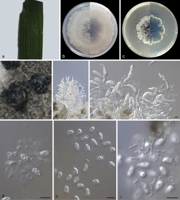
Ciri dan struktur
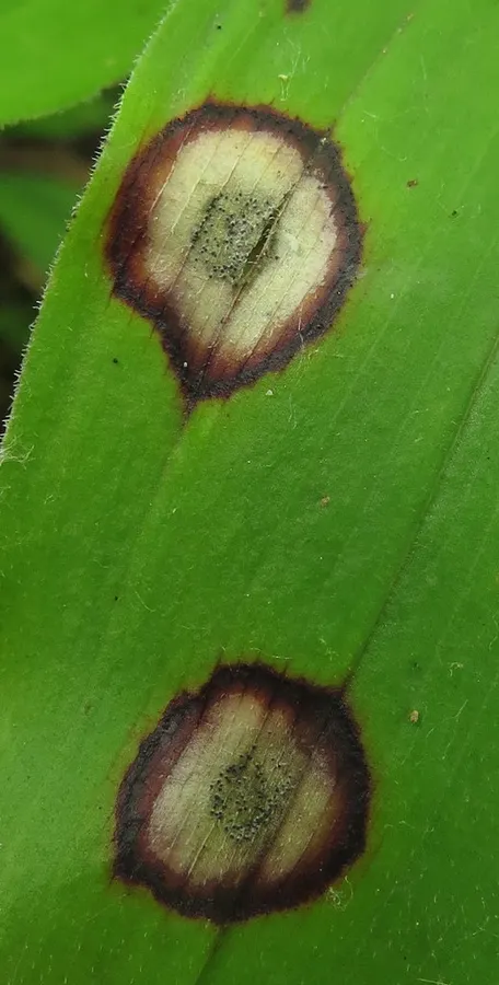
Leaf endogenic and associated with leaf spots. Asexual morph: Conidiomata pycnidial, mostly aggregated in clusters, black, erumpent. In MEA, cultures exuded colourless to opaque conidial masses, appeared on pycnidia after 10 days or longer. Pycnidial walls of multilayered, textura angularis, brown to dark brown, up to 35 μm thick; inner walls hyaline. Conidiophores subcylindrical to ampulliform, frequently reduced to conidiogenous cells or branching from a basal supporting cell, coated in mucoid layer, 8.0–14.0 × 3.0–5.0 μm. Conidiogenous cells terminal, subcylindrical to ampulliform, hyaline, smooth, 8.0–11.0 × 3.0–4.5 μm. Conidia 9.0–12.5 × 5.0–7.0 μm, mean ± SD = 10.6 ± 0.9 × 6.2 ± 0.5 μm, solitary, hyaline, aseptate, thin and smooth walled, coarsely guttulate or with a single large central guttule, ovoid, ampulliform, ellipsoidal to subglobose, enclosed in a thin mucoid sheath, 1.3–2.7 μm thick and bearing a hyaline, apical mucoid appendage, 3.0–8.5 × 1.0–1.5 μm, flexible, unbranched, tapering towards an acutely rounded tip. Spermatia hyaline, smooth, guttulate to granular, bacilliform, 6.0–8.2 × 1.3–2.0 μm, occurring in conidioma with conidia. Sexual morph: Ascomata shape and wall like those of the conidiomata. Asci bitunicate, hyaline, clavate to broadly fusoid-ellipsoid, with visible apical chamber, 2 μm diam., 45–85 × 9–13 μm. Ascospores bi- to multiseriate, hyaline, smooth, granular to guttulate, aseptate, straight, rarely curved, widest in the middle, limoniform with obtuse ends, 15–18 × 6–7 μm. Colonies on PDA occupying an entire 90 mm Petri dish in 14 days at 25 °C in darkness, with a growth rate of 6.0–6.5 mm/day, greenish-black in obverse and reverse.
Habitat dan peran ekologis
Colonies on PDA occupying an entire 90 mm Petri dish in 14 days at 25 °C in darkness, with a growth rate of 6.0–6.5 mm/day, greenish-black in obverse and reverse. Colonies on MEA 82–86 mm in diameter after 14 days at 25 °C in darkness, with a growth rate of 5.7–6.2 mm/day, undulate at edge, white to grey white in obverse and reverse, with moderate aerial mycelia on the surface, with black, gregarious conidiomata. Based on morphological features, Hennings (1908) described Phyllosticta capitalensis and Glienke et al. (2011) added molecular data. The holotype (CBS 128856) of P. capitalensis was collected from Stanhopea graveolens (Glienke et al. 2011). In our current study, two isolates (SAUCC 210144, SAUCC 210148), collected from diseased leaves of Rhapis excelsa, cluster in the P. capitalensis clade (Fig. 1). Although four other species are also in this clade, we consider these two isolates as P. capitalensis, based on their morphological characters, such as granular to guttulate ascospores (15–18 × 6–7 vs. 15–17 × 5–6 μm), subcylindrical to ampullate conidiogenous cells (8.0–11.0 × 3.0–4.5 vs. 7–10 × 3–5 μm), ellipsoidal to subglobose conidia (9–12.5 × 5–7 vs. 11–12 × 6–7 μm) and hyaline, apical mucoid appendages (3–8.5 × 1–1.5 vs. 6–8 × 1–1.5 μm).
- Habitat atau konteks temuan
- kultur yang didokumentasikan
- Substrat
- Informasi belum tersedia pada sumber rilis ini.
- Peran atau konteks
- Belum dikategorikan pada rilis ini.
Reproduksi
Leaf endogenic and associated with leaf spots. Asexual morph: Conidiomata pycnidial, mostly aggregated in clusters, black, erumpent. In MEA, cultures exuded colourless to opaque conidial masses, appeared on pycnidia after 10 days or longer. Pycnidial walls of multilayered, textura angularis, brown to dark brown, up to 35 μm thick; inner walls hyaline. Conidiophores subcylindrical to ampulliform, frequently reduced to conidiogenous cells or branching from a basal supporting cell, coated in mucoid layer, 8.0–14.0 × 3.0–5.0 μm. Conidiogenous cells terminal, subcylindrical to ampulliform, hyaline, smooth, 8.0–11.0 × 3.0–4.5 μm. Conidia 9.0–12.5 × 5.0–7.0 μm, mean ± SD = 10.6 ± 0.9 × 6.2 ± 0.5 μm, solitary, hyaline, aseptate, thin and smooth walled, coarsely guttulate or with a single large central guttule, ovoid, ampulliform, ellipsoidal to subglobose, enclosed in a thin mucoid sheath, 1.3–2.7 μm thick and bearing a hyaline, apical mucoid appendage, 3.0–8.5 × 1.0–1.5 μm, flexible, unbranched, tapering towards an acutely rounded tip.
Catatan identitas dan cakupan
- Nama diterima mengikuti Catalogue of Life Base2026-09-11; uraian mempertahankan konsep dan nama sumber aslinya.
- Preserve original treatment localities, hosts and substrate scope. Materials examined describe documented specimens, not the full global range.
- Impor dan terjemahan otomatis belum diperiksa editorial per profil. Struktur reproduktif yang disebutkan tidak membuktikan seluruh siklus hidup diketahui.
- Pengukuran pada uraian mengikuti spesimen, kultur, dan metode sumber; angka tidak diperluas ke semua populasi.
Catatan distribusi dalam narasi mengikuti sumbernya. Dokumentasi spesimen atau foto mengikuti catatan sumber. Bukti wilayah Indonesia dan penilaian konservasi tidak disimpulkan dari gambar.
Melihat dengan konteks
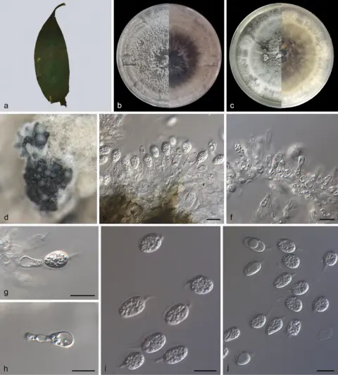
Phyllosticta capitalensis (holotype SAUCC 210144) a diseased leaf of Rhapis excelsa b, c colonies (left-above, right-reverse) after 15 days on PDA (b) and MEA (c) d conidiomata e asci and ascospores f asci, ascospores and conidia g conidiogenous cells with conidia h conidia i spermatia. Scale bars: 10 μm (e–i).
- Jenis
- Pelat ilmiah
- Kredit
- Zhaoxue Zhang, Xiaoyong Liu, Xiuguo Zhang, Zhe Meng
- Lisensi
- CC BY 4.0
- Sumber media
- Buka entri sumber
Dokumentasi tambahan
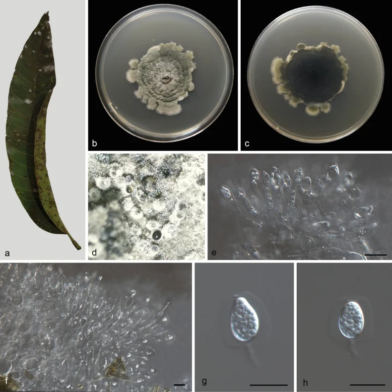
Phyllosticta capitalensis (CGMCC3.28668) a diseased leaf of Mangifera indica b, c colonies (left-above, right-reverse) after 14 d on PDA d conidiomata e, f conidiogenous cells with conidia g, h conidia. Scale bars: 10 μm (e–h).
Kredit dan penggunaan
Meng-Yuan Zhang, Zhao-Xue Zhang, Du-Hua Li, Xiu-Guo Zhang, Ji-Wen Xia, Zhuang Li
Ukuran dan format gambar disesuaikan tanpa pemotongan, retus, atau penambahan skala. Lisensi sumber dipertahankan.
Sumber profil
- [1] Morphological and phylogenetic analyses reveal two new species and a new record of Phyllosticta (Botryosphaeriales, Phyllostictaceae) from Hainan, China — Phyllosticta capitalensis
- [2] Catalogue of Life Base2026-09-11 — Phyllosticta capitalensis
- [3] Dokumentasi visual — Phyllosticta capitalensis
- [4] Dokumentasi visual — Phyllosticta capitalensis
Rujukan halaman ini
Fungi. Phyllosticta capitalensis. Rilis data fungi-2026-10-12-expansion-2. https://fungi-atlas.mgadib.chatgpt.site/jamur/phyllosticta-capitalensis/.
Status: impor sumber; pemeriksaan editorial per profil belum selesai. Untuk klaim biologis, gunakan rujukan pada bagian terkait dan periksa konteks sumber aslinya.