01 / Morfologi

Ciri dan struktur

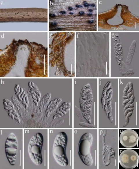
Saprobic on dead woody twigs. Sexual morph: Ascomata 300–380 μm high, 200–370 μm diam. (x̄ = 340 × 280 μm, n = 5), immersed to erumpent, solitary or scattered, uni-loculate, subglobose to obpyriform, fattened at the base, black, coriaceous, central ostiole. Peridium 13–20 μm wide, composed of brown, thick-walled cells of textura angularis that merge with host tissue. Hamathecium 3–6 μm wide, comprising abundant, branched, septate, hyphae-like paraphyses, anastomosed. Asci 60–87 × 16–24 μm (x̄ = 72 × 20 μm, n = 20), 8-spored, unitunicate, clavate to broadly clavate, with a short pedicel, rounded to truncate at the apex, with an inconspicuous apical ring. Ascospores 19–23 × 8–10 μm (x̄ = 21 × 9 μm, n = 30), overlapping biseriate, hyaline, mostly irregular ovoid, straight, tapered towards one end and rounded at other end, aseptate, thick-walled, guttulate, verrucose, without mucilaginous sheath or appendages. Asexual morph: Undetermined. Ascospores germinated on PDA within 24 h at room temperature (25 °C). Colonies on PDA, reaching 50 mm diameter after two weeks at 20–25 °C, mycelia superficial, circular, sparse mycelia, flat, fimbriate, white; reverse, dark brown.

Sebagian uraian ditampilkan dalam bahasa sumber; terjemahannya belum tersedia atau belum lolos pemeriksaan.

Rujukan: [1]

Pengukuran mengikuti objek, spesimen atau kultur, dan metode yang disebutkan sumber. Baca uraian asli untuk konteks lengkap.

02 / Kehidupan

Habitat dan peran ekologis

China, on dead branch of unidentified woody plant (Zhang et al. 2023b); on dead woody twigs of Trigonobalanus doichangensis (Fagaceae) (this study).

Sebagian uraian ditampilkan dalam bahasa sumber; terjemahannya belum tersedia atau belum lolos pemeriksaan.

Rujukan: [1]

Habitat atau konteks temuan
Informasi belum tersedia pada sumber rilis ini.
Substrat
Informasi belum tersedia pada sumber rilis ini.
Peran atau konteks
Belum dikategorikan pada rilis ini.
Temukan hubungan serupa
03 / Siklus hidup

Reproduksi

Saprobic on dead woody twigs. Sexual morph: Ascomata 300–380 μm high, 200–370 μm diam. (x̄ = 340 × 280 μm, n = 5), immersed to erumpent, solitary or scattered, uni-loculate, subglobose to obpyriform, fattened at the base, black, coriaceous, central ostiole. Peridium 13–20 μm wide, composed of brown, thick-walled cells of textura angularis that merge with host tissue. Hamathecium 3–6 μm wide, comprising abundant, branched, septate, hyphae-like paraphyses, anastomosed. Asci 60–87 × 16–24 μm (x̄ = 72 × 20 μm, n = 20), 8-spored, unitunicate, clavate to broadly clavate, with a short pedicel, rounded to truncate at the apex, with an inconspicuous apical ring. Ascospores 19–23 × 8–10 μm (x̄ = 21 × 9 μm, n = 30), overlapping biseriate, hyaline, mostly irregular ovoid, straight, tapered towards one end and rounded at other end, aseptate, thick-walled, guttulate, verrucose, without mucilaginous sheath or appendages.

Sebagian uraian ditampilkan dalam bahasa sumber; terjemahannya belum tersedia atau belum lolos pemeriksaan.

Rujukan: [1]

04 / Batas informasi

Catatan identitas dan cakupan

  • Nama diterima mengikuti Catalogue of Life Base2026-09-11; uraian mempertahankan konsep dan nama sumber aslinya.
  • Preserve original treatment localities, hosts and substrate scope. Materials examined describe documented specimens, not the full global range.
  • Impor dan terjemahan otomatis belum diperiksa editorial per profil. Struktur reproduktif yang disebutkan tidak membuktikan seluruh siklus hidup diketahui.
  • Pengukuran pada uraian mengikuti spesimen, kultur, dan metode sumber; angka tidak diperluas ke semua populasi.

Catatan distribusi dalam narasi mengikuti sumbernya. Dokumentasi spesimen atau foto mengikuti catatan sumber. Bukti wilayah Indonesia dan penilaian konservasi tidak disimpulkan dari gambar.

05 / Dokumentasi

Melihat dengan konteks

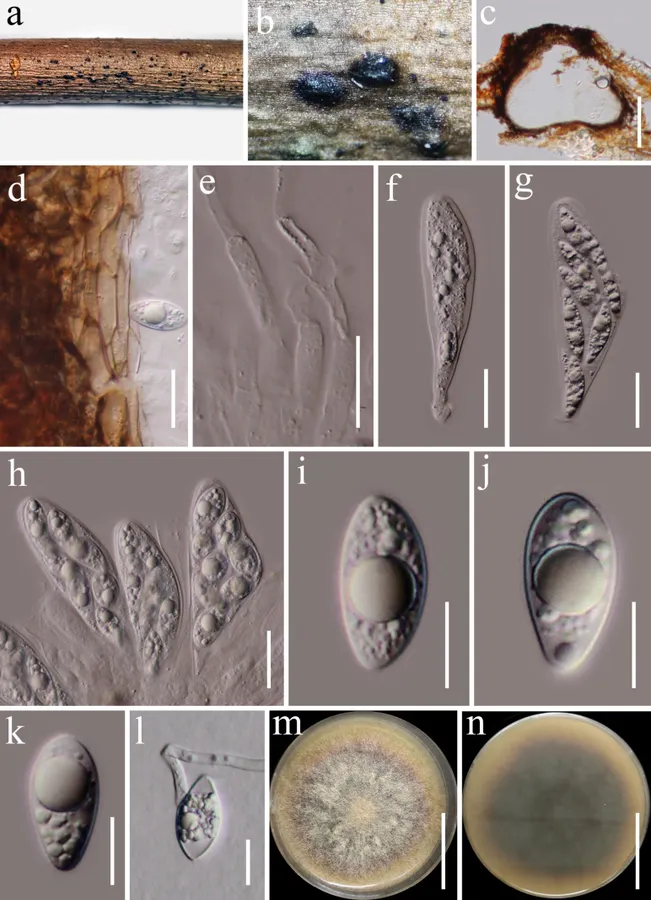
Planisphaeria reniformispora (HKAS 122722). a, b. Appearance of ascomata on host substrate; c. Section of ascoma; d. Peridium; e. Paraphyses; f–h. Asci; i–k. Ascospores; l. A germinating ascospore; m, n. Culture characters on PDA (m from above; n from below). Scale bars: 150 μm (c), 20 μm (d–h), 10 μm (i–l), 20 mm (m, n).

Jenis
Pelat ilmiah
Kredit
Guang-Cong Ren, Dhanushka Wanasinghe, Heng Gui, Zu-Yi Zhou, Xian Shi, Turki Kh. Faraj, Kai-Xuan Dong
Lisensi
CC BY 4.0
Sumber media
Buka entri sumber

Gambar menunjukkan objek menurut identifikasi sumber, bukan verifikasi independen spesimen oleh Fungi. Tidak ada skala baru yang ditambahkan.

06 / Rujukan

Sumber profil

  1. [1] Taxonomy and phylogeny of saprobic Sordariomycetes (Ascomycota) from woody litter in terrestrial ecosystems of Yunnan Province, China — Planisphaeria reniformisporaBagian sumber: PMC13019349_section_67
    Tanggal penghimpunan sumber: 2026-10-12
    Teks sumber: CC BY 4.0 · Ren GC, Wanasinghe D, Gui H, Zhou ZY, Shi X, Faraj TK, Dong KX.
  2. [2] Catalogue of Life Base2026-09-11 — Planisphaeria reniformisporaBagian sumber: ColDP NameUsage.tsv ID CBR2N; accepted species; source 2073
    Tanggal penghimpunan sumber: 2026-10-12
  3. [3] Dokumentasi visual — Planisphaeria reniformisporaBagian sumber: Individual media record; identification follows the source, not independent determination
    Tanggal penghimpunan sumber: 2026-10-12
    Media sumber: CC BY 4.0 · Guang-Cong Ren, Dhanushka Wanasinghe, Heng Gui, Zu-Yi Zhou, Xian Shi, Turki Kh. Faraj, Kai-Xuan Dong

Adaptasi teks: CC BY 4.0. Ren GC, Wanasinghe D, Gui H, Zhou ZY, Shi X, Faraj TK, Dong KX. — Taxonomy and phylogeny of saprobic Sordariomycetes (Ascomycota) from woody litter in terrestrial ecosystems of Yunnan Province, China — Planisphaeria reniformispora

Uraian asli dan riwayat sumber

Riwayat atau halaman sumber

Ringkasan sumber

Saprobic on dead woody twigs. Sexual morph: Ascomata 300–380 μm high, 200–370 μm diam. (x̄ = 340 × 280 μm, n = 5), immersed to erumpent, solitary or scattered, uni-loculate, subglobose to obpyriform, fattened at the base, black, coriaceous, central ostiole. Peridium 13–20 μm wide, composed of brown, thick-walled cells of textura angularis that merge with host tissue. Hamathecium 3–6 μm wide, comprising abundant, branched, septate, hyphae-like paraphyses, anastomosed. Asci 60–87 × 16–24 μm (x̄ = 72 × 20 μm, n = 20), 8-spored, unitunicate, clavate to broadly clavate, with a short pedicel, rounded to truncate at the apex, with an inconspicuous apical ring.

Revisi sumber

Morfologi sumber

Saprobic on dead woody twigs. Sexual morph: Ascomata 300–380 μm high, 200–370 μm diam. (x̄ = 340 × 280 μm, n = 5), immersed to erumpent, solitary or scattered, uni-loculate, subglobose to obpyriform, fattened at the base, black, coriaceous, central ostiole. Peridium 13–20 μm wide, composed of brown, thick-walled cells of textura angularis that merge with host tissue. Hamathecium 3–6 μm wide, comprising abundant, branched, septate, hyphae-like paraphyses, anastomosed. Asci 60–87 × 16–24 μm (x̄ = 72 × 20 μm, n = 20), 8-spored, unitunicate, clavate to broadly clavate, with a short pedicel, rounded to truncate at the apex, with an inconspicuous apical ring. Ascospores 19–23 × 8–10 μm (x̄ = 21 × 9 μm, n = 30), overlapping biseriate, hyaline, mostly irregular ovoid, straight, tapered towards one end and rounded at other end, aseptate, thick-walled, guttulate, verrucose, without mucilaginous sheath or appendages. Asexual morph: Undetermined. Ascospores germinated on PDA within 24 h at room temperature (25 °C). Colonies on PDA, reaching 50 mm diameter after two weeks at 20–25 °C, mycelia superficial, circular, sparse mycelia, flat, fimbriate, white; reverse, dark brown.

Revisi sumber

Ekologi sumber

China, on dead branch of unidentified woody plant (Zhang et al. 2023b); on dead woody twigs of Trigonobalanus doichangensis (Fagaceae) (this study).

Revisi sumber

Struktur reproduksi sumber

Saprobic on dead woody twigs. Sexual morph: Ascomata 300–380 μm high, 200–370 μm diam. (x̄ = 340 × 280 μm, n = 5), immersed to erumpent, solitary or scattered, uni-loculate, subglobose to obpyriform, fattened at the base, black, coriaceous, central ostiole. Peridium 13–20 μm wide, composed of brown, thick-walled cells of textura angularis that merge with host tissue. Hamathecium 3–6 μm wide, comprising abundant, branched, septate, hyphae-like paraphyses, anastomosed. Asci 60–87 × 16–24 μm (x̄ = 72 × 20 μm, n = 20), 8-spored, unitunicate, clavate to broadly clavate, with a short pedicel, rounded to truncate at the apex, with an inconspicuous apical ring. Ascospores 19–23 × 8–10 μm (x̄ = 21 × 9 μm, n = 30), overlapping biseriate, hyaline, mostly irregular ovoid, straight, tapered towards one end and rounded at other end, aseptate, thick-walled, guttulate, verrucose, without mucilaginous sheath or appendages.

Revisi sumber

Rujukan halaman ini

Fungi. Planisphaeria reniformispora. Rilis data fungi-2026-10-12-expansion-2. https://fungi-atlas.mgadib.chatgpt.site/jamur/planisphaeria-reniformispora/.

Status: impor sumber; pemeriksaan editorial per profil belum selesai. Untuk klaim biologis, gunakan rujukan pada bagian terkait dan periksa konteks sumber aslinya.