Ciri dan struktur
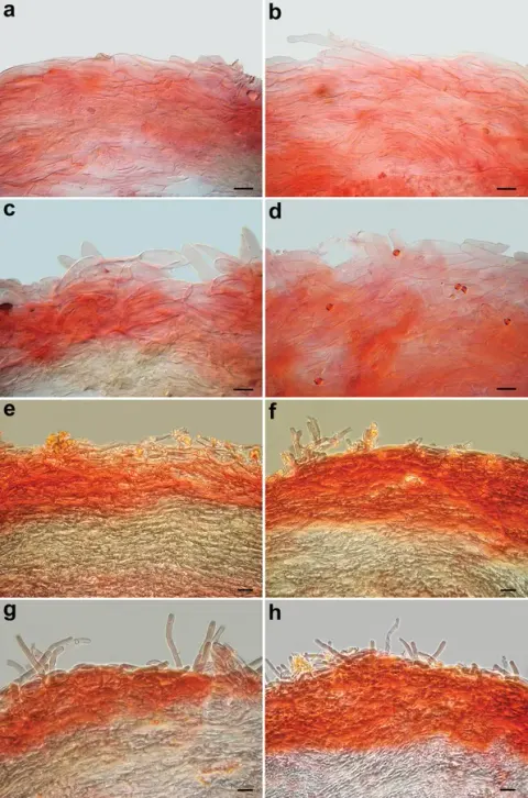
Arnolds et al. (2003: 66–68), Arauzo (2011a: 23, 25), Gisotti et al. (2021: 123–125). Spores (4.0–)4.5–4.8–5.2(–5.4) × (2.9–)3.5–3.7–4.0(–4.2) μm (64/1/1), Q = (1.10–)1.21–1.31–1.40(–1.51), V = (19.7–)29.2–35.2–41.3(–48.0) μm 3, broadly ellipsoid to ellipsoid, slightly amygdaliform in side view, walls up to 0.8 µm thick and dextrinoid at maturity, smooth, colorless in L4, hilar appendix prominent, 0.8–1 µm long. Basidia 21–25 × 6.5–8.0 µm, some with up to 1 μm thick wall (crassobasidia), tetrasporic, rarely bisporic to monosporic, clavate, sterigmata up to 4.5 µm long. Hymenophoral trama regular to subregular, composed of up to 12 µm wide cylindrical to inflated hyphae, colorless in L4. Cheilocystidia 15–30(–34) × 3–7(–11) µm, rare, scattered, basidiola-like to irregularly cylindrical, lageniform, sometimes lobed to furcate. Pleurocystidia present, very rare, similar to cheilocystidia but bigger, up to 56 × 12 µm. Pileipellis: suprapellis (at the pileus centre) a transition between trichoderm and epithelium consisting of loosely entangled ascending and erect pluriseptate hyphae, terminal elements broadly cylindrical, clavate to subglobose, up to 18 µm wide; towards the pileus margin it tends to become a cutis with repent hyphae; pigment abundant, brownish grey, bluish, intracellular and encrusting (extracellular), green in KOH; pileitrama consisting of up to 8 µm wide cylindrical hyphae. Stipitipellis consisting of 2–4 µm wide, multiseptate, parallel, cylindrical hyphae. Stipititrama similar to the stipitipellis structure but with 3–6 µm wide hyphae. Caulocystidia 10–70(–80) × 3.0–7 µm, filiform, sinuous, often multiseptate, sometimes irregularly lobed and furcate.
Habitat dan peran ekologis
SPAIN • Catalonia, Girona, La Selva, surroundings of Mas de Llagostera (UTM 31 T 0480 4622), 200 m, on acidic, humus-rich soil above granite, in forest with Pinus pinaster, Quercus suber, Arbutus unedo and Erica arborea, probably saprobic, 6 November 2002, leg. J. Carreras & M. Tabarés, (BCN SCM B-4742). The species was originally described from Spain (Arnolds et al. 2003; Arauzo 2011a) and then recently found in Liguria, Italy (Gisotti et al. 2021). Pseudobaeospora cyanea is clearly distinguished from the other species by a unique combination of features such as a bright bluish-purple pileus, pale lamellae and stipe, a trichodermic structure of the pileipellis (with inflated elements), green reaction of the pileipellis to KOH and well-developed cheilocystidia (Arnolds et al. 2003). Our microscopic analysis of the collection studied by Gisotti et al. (2021) (GDOR:M3986) revealed the presence of rare pleurocystidia which were not recorded by previous authors. Pleurocystidia were so far only reported for P. aciculifera Voto & Soop (Voto and Soop 2018), and P. taluna (Craig et al. 2023), two species of the southern hemisphere. Both Spanish and Italian collections were made in Mediterranean areas in winter, viz.
- Habitat atau konteks temuan
- Informasi belum tersedia pada sumber rilis ini.
- Substrat
- Informasi belum tersedia pada sumber rilis ini.
- Peran atau konteks
- Belum dikategorikan pada rilis ini.
Reproduksi
Spores (4.0–)4.5–4.8–5.2(–5.4) × (2.9–)3.5–3.7–4.0(–4.2) μm (64/1/1), Q = (1.10–)1.21–1.31–1.40(–1.51), V = (19.7–)29.2–35.2–41.3(–48.0) μm 3, broadly ellipsoid to ellipsoid, slightly amygdaliform in side view, walls up to 0.8 µm thick and dextrinoid at maturity, smooth, colorless in L4, hilar appendix prominent, 0.8–1 µm long. Basidia 21–25 × 6.5–8.0 µm, some with up to 1 μm thick wall (crassobasidia), tetrasporic, rarely bisporic to monosporic, clavate, sterigmata up to 4.5 µm long. Hymenophoral trama regular to subregular, composed of up to 12 µm wide cylindrical to inflated hyphae, colorless in L4. Cheilocystidia 15–30(–34) × 3–7(–11) µm, rare, scattered, basidiola-like to irregularly cylindrical, lageniform, sometimes lobed to furcate.
Catatan identitas dan cakupan
- Nama diterima mengikuti Catalogue of Life Base2026-09-11; uraian mempertahankan konsep dan nama sumber aslinya.
- Preserve original treatment localities, hosts and substrate scope. Materials examined describe documented specimens, not the full global range.
- Impor dan terjemahan otomatis belum diperiksa editorial per profil. Struktur reproduktif yang disebutkan tidak membuktikan seluruh siklus hidup diketahui.
- Pengukuran pada uraian mengikuti spesimen, kultur, dan metode sumber; angka tidak diperluas ke semua populasi.
Catatan distribusi dalam narasi mengikuti sumbernya. Dokumentasi spesimen atau foto mengikuti catatan sumber. Bukti wilayah Indonesia dan penilaian konservasi tidak disimpulkan dari gambar.
Melihat dengan konteks
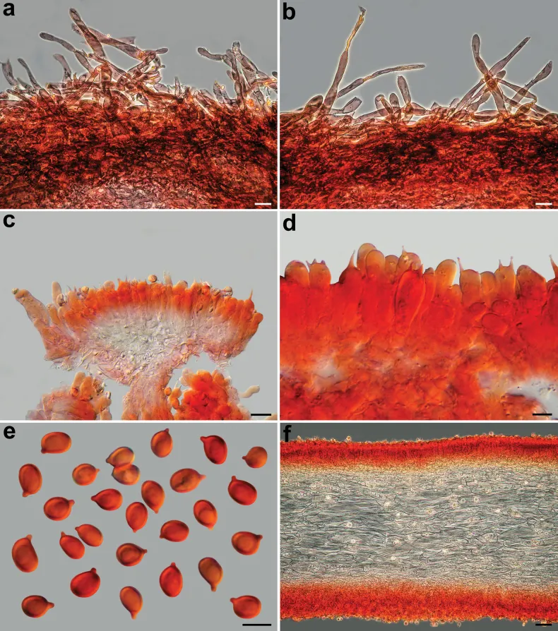
Pseudobaeospora cyanea, microscopic features (GDOR M3986). a, b Pileipellis; c hymenium (basidia and cheilocystidia); d basidia; e spores; f hymenophoral trama; a–f in ammoniacal Congo red. Scale bars: 20 µm (a, b, f); 10 µm (c); 5 µm (d, e). Photos by L. Setti.
- Jenis
- Pelat ilmiah
- Kredit
- Alfredo Vizzini, Giovanni Consiglio, Katarína Adamčíková, Ledo Setti, Slavomír Adamčík
- Lisensi
- CC BY 4.0
- Sumber media
- Buka entri sumber
Dokumentasi tambahan
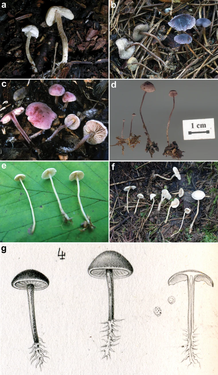
Basidiomes of some sequenced Pseudobaeospora collections. a P. calcarea (LIP PAM06090111-FR2013078); b P. cyanea (GDOR M3986); c P. laguncularis var. denudata (LIP PAM99101004-FR2013079); d P. pillodii (KRAM F-53314); e P. terrayi (SAV-F:3317, holotype); f P. terrayi (SAV-F:20813). Lectotype of. C. pillodii; g Quélet’s original plate, pl. XV, fig. 4. Photos: a, c by P.A. Moreau; b by D. Gisotti; d by A. Ronikier; e, f by S. Jančovičová.
Kredit dan penggunaan
Alfredo Vizzini, Giovanni Consiglio, Katarína Adamčíková, Ledo Setti, Slavomír Adamčík
Ukuran dan format gambar disesuaikan tanpa pemotongan, retus, atau penambahan skala. Lisensi sumber dipertahankan.
Sumber profil
- [1] Pseudobaeosporoideae, a new subfamily within the Tricholomataceae for the genus Pseudobaeospora (Agaricales, Tricholomatineae) based on morphological and molecular inference — Pseudobaeospora cyanea
- [2] Catalogue of Life Base2026-09-11 — Pseudobaeospora cyanea
- [3] Dokumentasi visual — Pseudobaeospora cyanea
- [4] Dokumentasi visual — Pseudobaeospora cyanea
Rujukan halaman ini
Fungi. Pseudobaeospora cyanea. Rilis data fungi-2026-10-12-expansion-2. https://fungi-atlas.mgadib.chatgpt.site/jamur/pseudobaeospora-cyanea/.
Status: impor sumber; pemeriksaan editorial per profil belum selesai. Untuk klaim biologis, gunakan rujukan pada bagian terkait dan periksa konteks sumber aslinya.