01 / Morfologi

Ciri dan struktur

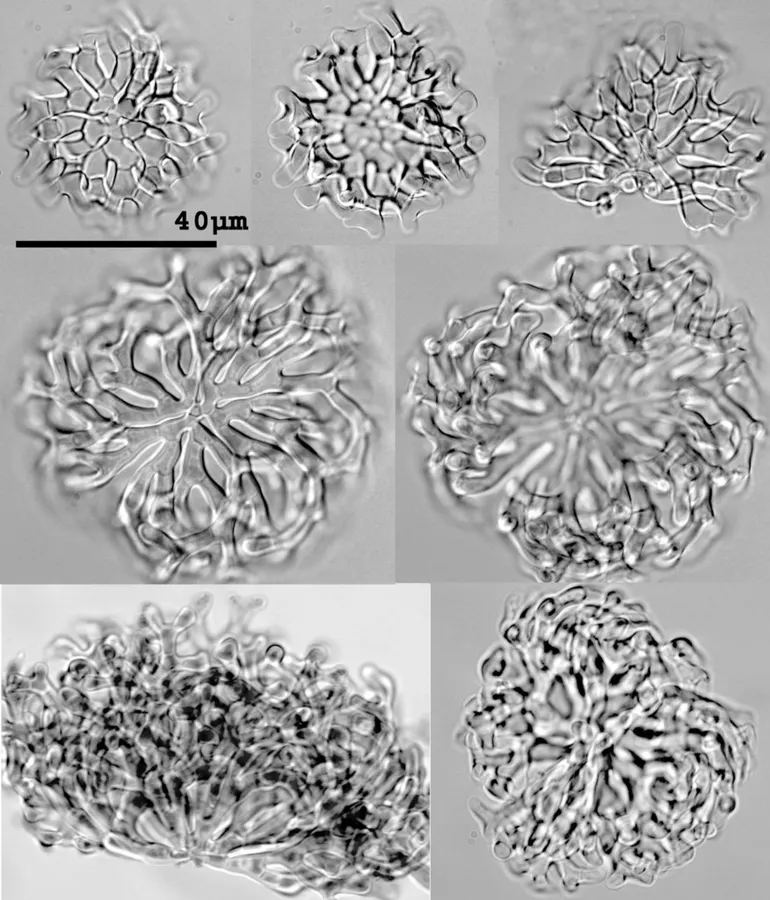
Conidiophores and conidiogenesis unknown. Conidia 60–85 µm diam., hyaline to pale yellow, filaments 4 µm wide, septate, consistently dichotomously branching from a basal plate, branches inter-leaved, finally tapering into a layer of curved filaments enclosing the conidium core. Air-trapping.

Sebagian uraian ditampilkan dalam bahasa sumber; terjemahannya belum tersedia atau belum lolos pemeriksaan.

Rujukan: [1]

Pengukuran mengikuti objek, spesimen atau kultur, dan metode yang disebutkan sumber. Baca uraian asli untuk konteks lengkap.

02 / Kehidupan

Habitat dan peran ekologis

Pseudoclathrosphaerina was erected as a monotypic genus by Voglmayr (1997) for P. evamariae, an aero-aquatic fungus isolated from leaves and known from the USA and Austria. The genus is distinguished from Clathrosphaerina because the terminal hyphae do not anastomose to form a clathrate structure and have a dichotomous, overlapping branching pattern arising from a cruciate basal plate. Voglmayr (2001) discussed the features of similar genera (excluding Nidulispora). The species described here agrees in morphology with Pseudoclathrosphaerina but differs from P. evamariae by the overall dimensions being considerably larger (P. evamariae 25–45 µm diam., and filament 2.5–3 µm), and the outer hyphae eventually becoming elongate and spirally curved to enclose the body. The monotypic genus Nidulispora was erected by Nawawi & Kuthubutheen (1990) for N. quadrifida. This similar fungus also develops from a small basal plate that develops dichotomously, terminating in elongated arms, but the developing branches do not overlap to form the pseudoclathrate structure characteristic of Pseudoclathrosphaerina. In addition the conidia in N. quadrifida are dematiaceous and smaller (32–52 × 40–60 µm). The outer spiral filaments of P. spiralis give it a superficial resemblance to the genus Spirosphaera.

Sebagian uraian ditampilkan dalam bahasa sumber; terjemahannya belum tersedia atau belum lolos pemeriksaan.

Rujukan: [1]

Habitat atau konteks temuan
Informasi belum tersedia pada sumber rilis ini.
Substrat
Informasi belum tersedia pada sumber rilis ini.
Peran atau konteks
Belum dikategorikan pada rilis ini.
Temukan hubungan serupa
03 / Siklus hidup

Reproduksi

Conidiophores and conidiogenesis unknown. Conidia 60–85 µm diam., hyaline to pale yellow, filaments 4 µm wide, septate, consistently dichotomously branching from a basal plate, branches inter-leaved, finally tapering into a layer of curved filaments enclosing the conidium core. Air-trapping.

Sebagian uraian ditampilkan dalam bahasa sumber; terjemahannya belum tersedia atau belum lolos pemeriksaan.

Rujukan: [1]

04 / Batas informasi

Catatan identitas dan cakupan

  • Nama diterima mengikuti Catalogue of Life Base2026-09-11; uraian mempertahankan konsep dan nama sumber aslinya.
  • Historical description as cited by BiotaNZ. Preserve explicit New Zealand/locality, host and specimen qualifiers. Distribution is the cited source scope, not a current global range.
  • Impor dan terjemahan otomatis belum diperiksa editorial per profil. Struktur reproduktif yang disebutkan tidak membuktikan seluruh siklus hidup diketahui.
  • Pengukuran pada uraian mengikuti spesimen, kultur, dan metode sumber; angka tidak diperluas ke semua populasi.

Catatan distribusi dalam narasi mengikuti sumbernya. Dokumentasi spesimen atau foto mengikuti catatan sumber. Bukti wilayah Indonesia dan penilaian konservasi tidak disimpulkan dari gambar.

05 / Dokumentasi

Melihat dengan konteks

Dokumentasi Pseudoclathrosphaerina spiralis menurut asosiasi foto pada record takson iNaturalist. Kredit: Jerry Cooper. Identitas mengikuti sumber; bukan penentuan spesimen independen.

Jenis
Fotografi
Kredit
(c) Jerry Cooper, some rights reserved (CC BY), uploaded by Jerry Cooper
Lisensi
CC BY 4.0
Sumber media
Buka entri sumber

Gambar menunjukkan objek menurut identifikasi sumber, bukan verifikasi independen spesimen oleh Fungi. Tidak ada skala baru yang ditambahkan.

06 / Rujukan

Sumber profil

  1. [1] Cooper, J.A. 2005: New Zealand hyphomycete fungi: additional records, new species, and notes on interesting collections. New Zealand Journal of Botany 43(1): 323-349. — Pseudoclathrosphaerina spiralisBagian sumber: biota:fcca4e11-ab62-44ac-8a3a-912c67176e86:8fb71af7-85b8-41bc-9dfc-b1272bb5b741
    Tanggal penghimpunan sumber: 2026-10-12
    Teks sumber: CC BY 4.0 · Cooper, J.A. 2005: New Zealand hyphomycete fungi: additional records, new species, and notes on interesting collections. New Zealand Journal of Botany 43(1): 323-349.
  2. [2] Catalogue of Life Base2026-09-11 — Pseudoclathrosphaerina spiralisBagian sumber: ColDP NameUsage.tsv ID 4NTSK; accepted species; source 2073
    Tanggal penghimpunan sumber: 2026-10-12
  3. [3] Dokumentasi visual — Pseudoclathrosphaerina spiralisBagian sumber: Individual media record; identification follows the source, not independent determination
    Tanggal penghimpunan sumber: 2026-10-12
    Media sumber: CC BY 4.0 · (c) Jerry Cooper, some rights reserved (CC BY), uploaded by Jerry Cooper

Adaptasi teks: CC BY 4.0. Cooper, J.A. 2005: New Zealand hyphomycete fungi: additional records, new species, and notes on interesting collections. New Zealand Journal of Botany 43(1): 323-349. — Cooper, J.A. 2005: New Zealand hyphomycete fungi: additional records, new species, and notes on interesting collections. New Zealand Journal of Botany 43(1): 323-349. — Pseudoclathrosphaerina spiralis

Uraian asli dan riwayat sumber

Riwayat atau halaman sumber

Ringkasan sumber

Conidiophores and conidiogenesis unknown. Conidia 60–85 µm diam., hyaline to pale yellow, filaments 4 µm wide, septate, consistently dichotomously branching from a basal plate, branches inter-leaved, finally tapering into a layer of curved filaments enclosing the conidium core. Air-trapping.

Revisi sumber

Morfologi sumber

Conidiophores and conidiogenesis unknown. Conidia 60–85 µm diam., hyaline to pale yellow, filaments 4 µm wide, septate, consistently dichotomously branching from a basal plate, branches inter-leaved, finally tapering into a layer of curved filaments enclosing the conidium core. Air-trapping.

Revisi sumber

Ekologi sumber

Pseudoclathrosphaerina was erected as a monotypic genus by Voglmayr (1997) for P. evamariae, an aero-aquatic fungus isolated from leaves and known from the USA and Austria. The genus is distinguished from Clathrosphaerina because the terminal hyphae do not anastomose to form a clathrate structure and have a dichotomous, overlapping branching pattern arising from a cruciate basal plate. Voglmayr (2001) discussed the features of similar genera (excluding Nidulispora). The species described here agrees in morphology with Pseudoclathrosphaerina but differs from P. evamariae by the overall dimensions being considerably larger (P. evamariae 25–45 µm diam., and filament 2.5–3 µm), and the outer hyphae eventually becoming elongate and spirally curved to enclose the body. The monotypic genus Nidulispora was erected by Nawawi & Kuthubutheen (1990) for N. quadrifida. This similar fungus also develops from a small basal plate that develops dichotomously, terminating in elongated arms, but the developing branches do not overlap to form the pseudoclathrate structure characteristic of Pseudoclathrosphaerina. In addition the conidia in N. quadrifida are dematiaceous and smaller (32–52 × 40–60 µm). The outer spiral filaments of P. spiralis give it a superficial resemblance to the genus Spirosphaera.

Revisi sumber

Struktur reproduksi sumber

Conidiophores and conidiogenesis unknown. Conidia 60–85 µm diam., hyaline to pale yellow, filaments 4 µm wide, septate, consistently dichotomously branching from a basal plate, branches inter-leaved, finally tapering into a layer of curved filaments enclosing the conidium core. Air-trapping.

Revisi sumber

Rujukan halaman ini

Fungi. Pseudoclathrosphaerina spiralis. Rilis data fungi-2026-10-12-expansion-2. https://fungi-atlas.mgadib.chatgpt.site/jamur/pseudoclathrosphaerina-spiralis/.

Status: impor sumber; pemeriksaan editorial per profil belum selesai. Untuk klaim biologis, gunakan rujukan pada bagian terkait dan periksa konteks sumber aslinya.