Ciri dan struktur
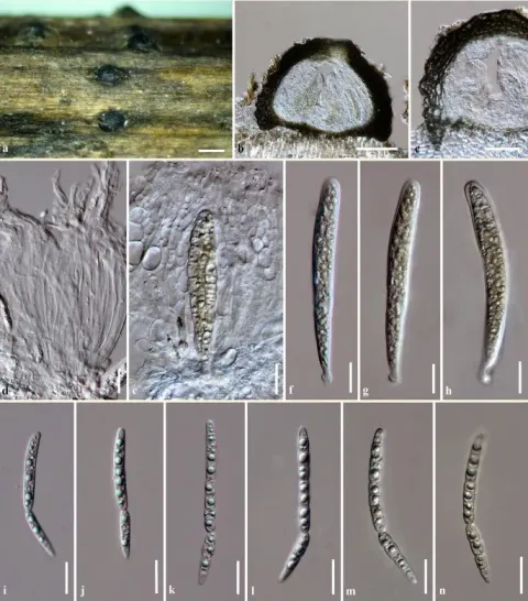
Saprobic on dead aerial stem of Artemisia sp. (Asterales, Asteraceae). Sexual morph: Ascomata (Fig. 4 a-b, c - with ostiole) 170–300 × 140–250 µm (x¯ = 200 × 177 µm, n = 10), solitary, scattered, dark brown to black, semi-immersed to erumpent, sessile, globose to subglobose, uni-loculate, coriaceous, ostiolate and papillate. Papilla (Fig. 4 d) 70–150 × 60–120 µm, mammiform to oblong, with a rounded to truncate apex, thick walled, composed of several layers, brown to dark brown cells of textura angularis, ostiole central, single and without periphyses. Peridium (Fig. 4 e) 15–35 µm (x¯ = 20 µm, n = 15), brown to black, thick-walled, pseudoparenchymatous cells, composed of 4–6 cell layers, outer layers composed of dark brown loosely packed cells of textura angularis, inner layers composed of light brown to hyaline flattened cells of textura prismatica. Hamathecium comprising numerous, 1.5–2.5 µm wide (x¯ = 2 µm, n = 15), filamentous, distinctly septate, cellular pseudoparaphyses (Fig. 4 f) with guttules, slightly constricted at the septa, anastomosing at the apex, embedded in a hyaline gelatinous matrix. Asci (Fig. 4 g-j) 100–150 × 6–9 µm (x¯ = 132 × 8 µm, n = 15), 8-spored, bitunicate, fissitunicate, cylindrical to cylindrical-clavate, short furcate pedicel, apically rounded, well-developed ocular chamber. Ascospores (Fig. 4 k-m) 120–150 × 2–3 µm (x¯ = 131 × 2.8 µm, n = 25), fasciculate, lying parallel or spiral at the centre, scolecosporous, filiform or filamentous, narrowly rounded towards the ends, slightly swollen at the middle of 4 th or 5 th cell from the apex (Fig. 4 n), yellowish to yellowish brown, 15–18 septate and not constricted at the septa, smooth-walled.
Habitat dan peran ekologis
Saprobic on dead aerial stem of Artemisia sp. (Asterales, Asteraceae). Sexual morph: Ascomata (Fig. 4 a-b, c - with ostiole) 170–300 × 140–250 µm (x¯ = 200 × 177 µm, n = 10), solitary, scattered, dark brown to black, semi-immersed to erumpent, sessile, globose to subglobose, uni-loculate, coriaceous, ostiolate and papillate. Papilla (Fig. 4 d) 70–150 × 60–120 µm, mammiform to oblong, with a rounded to truncate apex, thick walled, composed of several layers, brown to dark brown cells of textura angularis, ostiole central, single and without periphyses. Peridium (Fig. 4 e) 15–35 µm (x¯ = 20 µm, n = 15), brown to black, thick-walled, pseudoparenchymatous cells, composed of 4–6 cell layers, outer layers composed of dark brown loosely packed cells of textura angularis, inner layers composed of light brown to hyaline flattened cells of textura prismatica. Hamathecium comprising numerous, 1.5–2.5 µm wide (x¯ = 2 µm, n = 15), filamentous, distinctly septate, cellular pseudoparaphyses (Fig. 4 f) with guttules, slightly constricted at the septa, anastomosing at the apex, embedded in a hyaline gelatinous matrix. Asci (Fig. 4 g-j) 100–150 × 6–9 µm (x¯ = 132 × 8 µm, n = 15), 8-spored, bitunicate, fissitunicate, cylindrical to cylindrical-clavate, short furcate pedicel, apically rounded, well-developed ocular chamber.
- Habitat atau konteks temuan
- Informasi belum tersedia pada sumber rilis ini.
- Substrat
- Informasi belum tersedia pada sumber rilis ini.
- Peran atau konteks
- Pengurai
Reproduksi
Saprobic on dead aerial stem of Artemisia sp. (Asterales, Asteraceae). Sexual morph: Ascomata (Fig. 4 a-b, c - with ostiole) 170–300 × 140–250 µm (x¯ = 200 × 177 µm, n = 10), solitary, scattered, dark brown to black, semi-immersed to erumpent, sessile, globose to subglobose, uni-loculate, coriaceous, ostiolate and papillate. Papilla (Fig. 4 d) 70–150 × 60–120 µm, mammiform to oblong, with a rounded to truncate apex, thick walled, composed of several layers, brown to dark brown cells of textura angularis, ostiole central, single and without periphyses. Peridium (Fig. 4 e) 15–35 µm (x¯ = 20 µm, n = 15), brown to black, thick-walled, pseudoparenchymatous cells, composed of 4–6 cell layers, outer layers composed of dark brown loosely packed cells of textura angularis, inner layers composed of light brown to hyaline flattened cells of textura prismatica.
Catatan identitas dan cakupan
- Nama diterima mengikuti Catalogue of Life Base2026-09-11; uraian mempertahankan konsep dan nama sumber aslinya.
- Preserve original treatment localities, hosts and substrate scope. Materials examined describe documented specimens, not the full global range.
- Impor dan terjemahan otomatis belum diperiksa editorial per profil. Struktur reproduktif yang disebutkan tidak membuktikan seluruh siklus hidup diketahui.
- Pengukuran pada uraian mengikuti spesimen, kultur, dan metode sumber; angka tidak diperluas ke semua populasi.
Catatan distribusi dalam narasi mengikuti sumbernya. Dokumentasi spesimen atau foto mengikuti catatan sumber. Bukti wilayah Indonesia dan penilaian konservasi tidak disimpulkan dari gambar.
Melihat dengan konteks
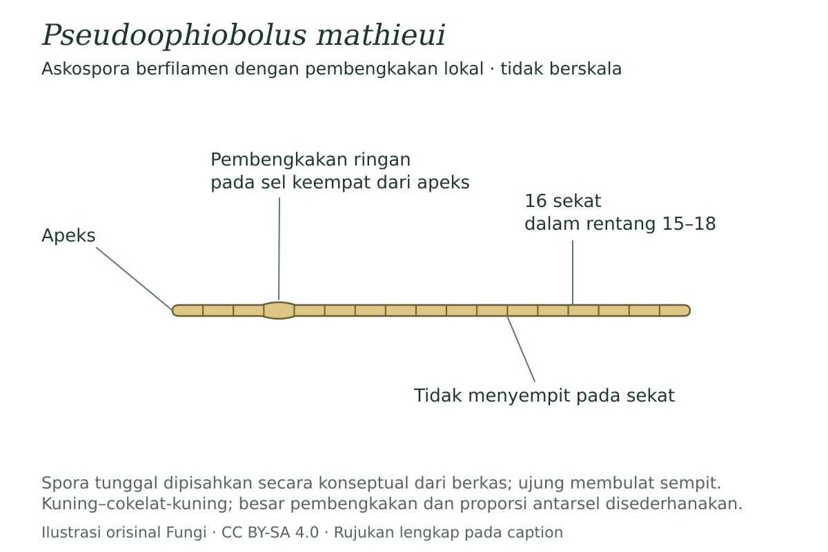
Skema satu askospora Pseudoophiobolus mathieui yang dipisahkan secara konseptual dari berkas: berfilamen, berujung membulat sempit, berwarna kuning–cokelat-kuning, dan memiliki 15–18 sekat tanpa penyempitan pada sekat. Enam belas sekat serta pembengkakan ringan pada sel keempat dari apeks ditampilkan sebagai varian yang dinyatakan sumber. Tidak berskala; proporsi sel dan besar pembengkakan disederhanakan.
- Jenis
- Ilustrasi skematis
- Kredit
- Ilustrasi orisinal Fungi berdasarkan uraian: Wijesinghe SN, Wang Y, Zucconi L, Dayarathne MC, Boonmee S, Camporesi E, Wanasinghe DN, Hyde KD. — Additions to Italian Pleosporinae, including Italica heraclei sp. nov. — Pseudoophiobolus mathieui. Sumber: CC BY 4.0. Perubahan: skema anatomi konseptual, label dan caption Indonesia; bukan penyalinan gambar sumber.
- Lisensi
- CC BY-SA 4.0
- Sumber media
- Buka entri sumber
Sumber profil
- [1] Additions to Italian Pleosporinae, including Italica heraclei sp. nov. — Pseudoophiobolus mathieui
- [2] Catalogue of Life Base2026-09-11 — Pseudoophiobolus mathieui
Rujukan halaman ini
Fungi. Pseudoophiobolus mathieui. Rilis data fungi-2026-10-12-expansion-2. https://fungi-atlas.mgadib.chatgpt.site/jamur/pseudoophiobolus-mathieui/.
Status: impor sumber; pemeriksaan editorial per profil belum selesai. Untuk klaim biologis, gunakan rujukan pada bagian terkait dan periksa konteks sumber aslinya.