01 / Morfologi

Ciri dan struktur

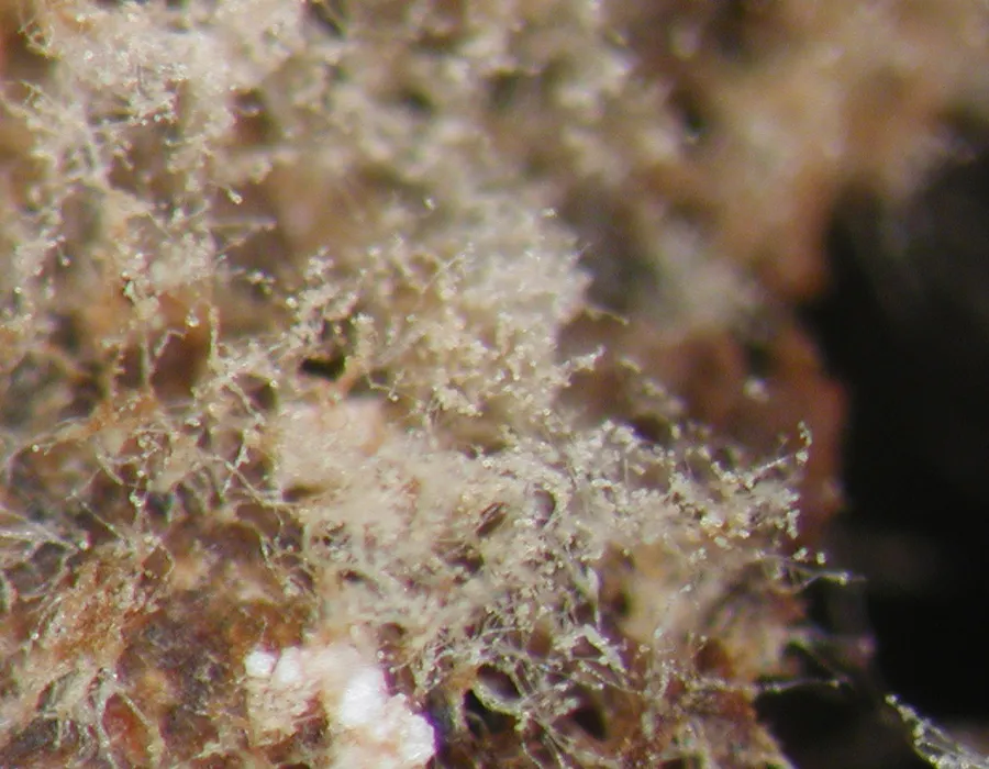
Sporulating colonies are loose and tufted. Young growth is white, becoming pale ochre or yellowish brown with age. The vegetative hyphae are branched, septate, smooth and thin-walled, generally 2–5 μm wide and occasionally reaching about 7.5 μm. The conidiophores are erect, straight and septate. Ukrainian specimens examined by Akulov measured about 350–650 μm long. They are usually unbranched, although one or two branches may develop near the base. Their width decreases from approximately 6–8 μm at the base to 3–5 μm toward the apex. The fertile region is cylindrical and somewhat curved, commonly 150–180 μm long. Continued sympodial growth during conidium production may extend it to around 400 μm. Its surface bears numerous pointed conidiogenous denticles about 1–1.5 μm long. The conidia develop both apically and laterally. They are pale ochre, thick-walled and globose to nearly globose, with a smooth to finely ornamented surface. Akulov recorded diameters of 8–10 μm, occasionally reaching 12 μm. Material compared in 2026 had conidia approximately 9–12 μm in diameter and a smooth to finely verruculose surface.

Sebagian uraian ditampilkan dalam bahasa sumber; terjemahannya belum tersedia atau belum lolos pemeriksaan.

Rujukan: [1]

Pengukuran mengikuti objek, spesimen atau kultur, dan metode yang disebutkan sumber. Baca uraian asli untuk konteks lengkap.

02 / Kehidupan

Habitat dan peran ekologis

Most published collections of R. globulifera have been associated with fruiting bodies of wood-decaying fungi. Reported substrates include Fomes fomentarius, Inonotus dryadeus and Ganoderma tsugae. Akulov reported the species from Ukraine for the first time in 2011. The collections occurred on several fungal substrates, including a fruit body of Ganoderma applanatum, decaying fruit bodies of Oxyporus latemarginatus, an aethalium of a Fuligo species, and fruit bodies of Nemania serpens. Earlier culture observations found that the fungus could be isolated from polypore fruit bodies that appeared externally undamaged. This led to the suggestion that it may begin growing internally within its fungal substrate before producing visible surface growth. The proposal concerns its possible mode of development and does not represent a demonstrated complete life cycle. Records reviewed in 2011 included Georgia, the Netherlands, European Russia, Ukraine, France and the Czech Republic, as well as Japan, the United States and Canada.

Sebagian uraian ditampilkan dalam bahasa sumber; terjemahannya belum tersedia atau belum lolos pemeriksaan.

Rujukan: [1]

Habitat atau konteks temuan
kultur yang didokumentasikan
Substrat
Informasi belum tersedia pada sumber rilis ini.
Peran atau konteks
Pengurai
Temukan hubungan serupa
03 / Siklus hidup

Reproduksi

Sporulating colonies are loose and tufted. Young growth is white, becoming pale ochre or yellowish brown with age. The vegetative hyphae are branched, septate, smooth and thin-walled, generally 2–5 μm wide and occasionally reaching about 7.5 μm. The conidiophores are erect, straight and septate. Ukrainian specimens examined by Akulov measured about 350–650 μm long. They are usually unbranched, although one or two branches may develop near the base. Their width decreases from approximately 6–8 μm at the base to 3–5 μm toward the apex. The fertile region is cylindrical and somewhat curved, commonly 150–180 μm long. Continued sympodial growth during conidium production may extend it to around 400 μm.

Sebagian uraian ditampilkan dalam bahasa sumber; terjemahannya belum tersedia atau belum lolos pemeriksaan.

Rujukan: [1]

04 / Batas informasi

Catatan identitas dan cakupan

  • Nama diterima mengikuti Catalogue of Life Base2026-09-11; uraian mempertahankan konsep dan nama sumber aslinya.
  • Original geographic, specimen and life-cycle scope retained; source import awaits individual editorial review.
  • Impor dan terjemahan otomatis belum diperiksa editorial per profil. Struktur reproduktif yang disebutkan tidak membuktikan seluruh siklus hidup diketahui.
  • Pengukuran pada uraian mengikuti spesimen, kultur, dan metode sumber; angka tidak diperluas ke semua populasi.

Catatan distribusi dalam narasi mengikuti sumbernya. Dokumentasi spesimen atau foto mengikuti catatan sumber. Bukti wilayah Indonesia dan penilaian konservasi tidak disimpulkan dari gambar.

05 / Dokumentasi

Melihat dengan konteks

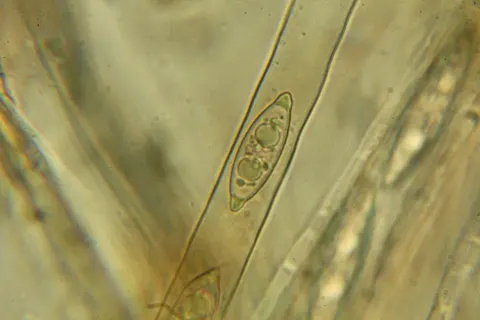
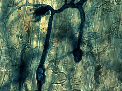
Dokumentasi Rhinotrichella globulifera menurut asosiasi foto pada record takson iNaturalist. Kredit: Jerry Cooper. Identitas mengikuti sumber; bukan penentuan spesimen independen.

Jenis
Fotografi
Kredit
(c) Jerry Cooper, some rights reserved (CC BY), uploaded by Jerry Cooper
Lisensi
CC BY 4.0
Sumber media
Buka entri sumber

Gambar menunjukkan objek menurut identifikasi sumber, bukan verifikasi independen spesimen oleh Fungi. Tidak ada skala baru yang ditambahkan.

06 / Rujukan

Sumber profil

  1. [1] Rhinotrichella globulifera — Wikipedia (en)Bagian sumber: Frozen revision 1375578420
    Tanggal penghimpunan sumber: 2026-10-12
    Teks sumber: CC BY-SA4.0 · Wikipedia contributors
  2. [2] Catalogue of Life Base2026-09-11 — Rhinotrichella globuliferaBagian sumber: ColDP NameUsage.tsv ID 78TCF; accepted species; source 2073
    Tanggal penghimpunan sumber: 2026-10-12
  3. [3] Dokumentasi visual — Rhinotrichella globuliferaBagian sumber: Individual media record; identification follows the source, not independent determination
    Tanggal penghimpunan sumber: 2026-10-12
    Media sumber: CC BY 4.0 · (c) Jerry Cooper, some rights reserved (CC BY), uploaded by Jerry Cooper

Adaptasi teks: CC BY-SA 4.0. Wikipedia contributors — Rhinotrichella globulifera — Wikipedia (en)

Uraian asli dan riwayat sumber

Riwayat atau halaman sumber

Ringkasan sumber

Rhinotrichella globulifera is a fungicolous ascomycete fungus known primarily from its asexual state. It has most often been recorded growing on the fruiting bodies of other fungi, particularly polypores, although it has also been found on plant remains. The species produces long erect conidiophores bearing approximately spherical, pale ochre conidia. Records compiled in 2011 indicated its presence in Europe, Asia and North America.

Revisi sumber

Morfologi sumber

Sporulating colonies are loose and tufted. Young growth is white, becoming pale ochre or yellowish brown with age. The vegetative hyphae are branched, septate, smooth and thin-walled, generally 2–5 μm wide and occasionally reaching about 7.5 μm. The conidiophores are erect, straight and septate. Ukrainian specimens examined by Akulov measured about 350–650 μm long. They are usually unbranched, although one or two branches may develop near the base. Their width decreases from approximately 6–8 μm at the base to 3–5 μm toward the apex. The fertile region is cylindrical and somewhat curved, commonly 150–180 μm long. Continued sympodial growth during conidium production may extend it to around 400 μm. Its surface bears numerous pointed conidiogenous denticles about 1–1.5 μm long. The conidia develop both apically and laterally. They are pale ochre, thick-walled and globose to nearly globose, with a smooth to finely ornamented surface. Akulov recorded diameters of 8–10 μm, occasionally reaching 12 μm. Material compared in 2026 had conidia approximately 9–12 μm in diameter and a smooth to finely verruculose surface.

Revisi sumber

Ekologi sumber

Most published collections of R. globulifera have been associated with fruiting bodies of wood-decaying fungi. Reported substrates include Fomes fomentarius, Inonotus dryadeus and Ganoderma tsugae. Akulov reported the species from Ukraine for the first time in 2011. The collections occurred on several fungal substrates, including a fruit body of Ganoderma applanatum, decaying fruit bodies of Oxyporus latemarginatus, an aethalium of a Fuligo species, and fruit bodies of Nemania serpens. Earlier culture observations found that the fungus could be isolated from polypore fruit bodies that appeared externally undamaged. This led to the suggestion that it may begin growing internally within its fungal substrate before producing visible surface growth. The proposal concerns its possible mode of development and does not represent a demonstrated complete life cycle. Records reviewed in 2011 included Georgia, the Netherlands, European Russia, Ukraine, France and the Czech Republic, as well as Japan, the United States and Canada.

Revisi sumber

Struktur reproduksi sumber

Sporulating colonies are loose and tufted. Young growth is white, becoming pale ochre or yellowish brown with age. The vegetative hyphae are branched, septate, smooth and thin-walled, generally 2–5 μm wide and occasionally reaching about 7.5 μm. The conidiophores are erect, straight and septate. Ukrainian specimens examined by Akulov measured about 350–650 μm long. They are usually unbranched, although one or two branches may develop near the base. Their width decreases from approximately 6–8 μm at the base to 3–5 μm toward the apex. The fertile region is cylindrical and somewhat curved, commonly 150–180 μm long. Continued sympodial growth during conidium production may extend it to around 400 μm.

Revisi sumber

Rujukan halaman ini

Fungi. Rhinotrichella globulifera. Rilis data fungi-2026-10-12-expansion-2. https://fungi-atlas.mgadib.chatgpt.site/jamur/rhinotrichella-globulifera/.

Status: impor sumber; pemeriksaan editorial per profil belum selesai. Untuk klaim biologis, gunakan rujukan pada bagian terkait dan periksa konteks sumber aslinya.