01 / Morfologi

Ciri dan struktur

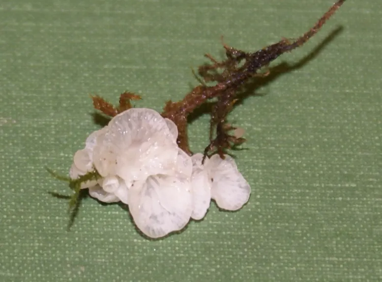
Basidiome very small (1-6 mm diam.), dorsally attached to host moss plant or to rhizomorphs, pendent, attached by a short pseudostipe, usually more or less centrally; young specimens resembling a minute tobacco pipe. Pileus narrowly to broadly parabolic, becoming convex, pure white, dry, floccose to villose near the pseudostipe, grooved following the outline of the lamellae, margin strongly inrolled when young, becoming almost straight at maturity and sometimes slightly undulating. Hymenophore white to pale salmon pink when dry, smooth in very small basidiomes, but more usually lamellate, lamellae fold-like, thick, shallow, becoming more distant in dried material, 3-4 principal veins meeting at a central to sub-central point, forking or anastomosing with 1-2 series of lamellae. Taste and odour not noticeable. Spore print not known. Spores 5-6.6 x 4.5-5.5 (5.2 x 4.8) µm, Q = 1.09, spherical to subspherical with a conspicuous apiculus often with a noticeable scar, hyaline, inamyloid, not dextrinoid, acyanophilic, very thinwalled. Basidia 20-23 x 6-8 µm, 4-spored, with relatively long sterigmata (-6 µm), which are so well separated on the rather broad basidia that the basidia may very often appear 2-spored. Cheilocystidia and pleurocystidia absent. Trama subparallel to somewhat interwoven, hyphae 3-8 µm diam., many inflated. Subhymenium also of loosely packed, inflated cells equal to or greater in diameter than those of the trama. Context of interwoven, narrow (5 µm diam.) to inflated (-11 µm diam.) hyphae, those near the surface repent and faintly encrusted. Pileipellis a tangle of erect hyphae, simple to sparsely diverticulate-nodulose, 2-5 µm diam.

Sebagian uraian ditampilkan dalam bahasa sumber; terjemahannya belum tersedia atau belum lolos pemeriksaan.

Rujukan: [1]

Pengukuran mengikuti objek, spesimen atau kultur, dan metode yang disebutkan sumber. Baca uraian asli untuk konteks lengkap.

02 / Kehidupan

Habitat dan peran ekologis

Basidiomes found in quantity on cushions of the umbrella moss Hypnodendron colensoi on the ground in mixed podocarp dicotyledonous forest and associated with fine, white rhizomorphs along the stem of the moss. Fresh basidiomes were found mainly on apparently living moss plants, but adjacent patches of dead moss plants in the cushions were mantled with dark-coloured (dead?) fungal rhizomorphs, suggesting that the fungus could be parasitic. Basidiomes were also found on pieces of dead frond of the fern Diplazium sp. in the moss cushion, attached to more luxuriant rhizomorphs and mycelium.

Sebagian uraian ditampilkan dalam bahasa sumber; terjemahannya belum tersedia atau belum lolos pemeriksaan.

Rujukan: [1]

Habitat atau konteks temuan
hutan
Substrat
tanah
Peran atau konteks
Belum dikategorikan pada rilis ini.
Temukan hubungan serupa
03 / Siklus hidup

Reproduksi

Basidiome very small (1-6 mm diam.), dorsally attached to host moss plant or to rhizomorphs, pendent, attached by a short pseudostipe, usually more or less centrally; young specimens resembling a minute tobacco pipe. Pileus narrowly to broadly parabolic, becoming convex, pure white, dry, floccose to villose near the pseudostipe, grooved following the outline of the lamellae, margin strongly inrolled when young, becoming almost straight at maturity and sometimes slightly undulating. Hymenophore white to pale salmon pink when dry, smooth in very small basidiomes, but more usually lamellate, lamellae fold-like, thick, shallow, becoming more distant in dried material, 3-4 principal veins meeting at a central to sub-central point, forking or anastomosing with 1-2 series of lamellae.

Sebagian uraian ditampilkan dalam bahasa sumber; terjemahannya belum tersedia atau belum lolos pemeriksaan.

Rujukan: [1]

Sinonim menurut acuan taksonomi rilis: Agaricus bryophilus; Cantharellus bryophilus; Dictyolus bryophilus; Leptoglossum bryophilum; Leptotus bryophilus; Merulius bryophilus; Mniopetalum bryophilum
04 / Batas informasi

Catatan identitas dan cakupan

  • Nama diterima mengikuti Catalogue of Life Base2026-09-11; uraian mempertahankan konsep dan nama sumber aslinya.
  • Historical description as cited by BiotaNZ. Preserve explicit New Zealand/locality, host and specimen qualifiers. Distribution is the cited source scope, not a current global range.
  • Impor dan terjemahan otomatis belum diperiksa editorial per profil. Struktur reproduktif yang disebutkan tidak membuktikan seluruh siklus hidup diketahui.
  • Pengukuran pada uraian mengikuti spesimen, kultur, dan metode sumber; angka tidak diperluas ke semua populasi.

Catatan distribusi dalam narasi mengikuti sumbernya. Dokumentasi spesimen atau foto mengikuti catatan sumber. Bukti wilayah Indonesia dan penilaian konservasi tidak disimpulkan dari gambar.

05 / Dokumentasi

Melihat dengan konteks

Dokumentasi Rimbachia bryophila menurut asosiasi foto pada record takson iNaturalist. Kredit: user: smilacina. Identitas mengikuti sumber; bukan penentuan spesimen independen.

Jenis
Fotografi
Kredit
(c) user: smilacina, some rights reserved (CC BY-SA)
Sumber media
Buka entri sumber

Gambar menunjukkan objek menurut identifikasi sumber, bukan verifikasi independen spesimen oleh Fungi. Tidak ada skala baru yang ditambahkan.

06 / Rujukan

Sumber profil

  1. [1] Segedin, B.P. 1994: Studies in the Agaricales of New Zealand: new records and new species of the genera Cheimonophyllum, Mniopetalum, and Anthracophyllum (Tricholomataceae, Collybieae). New Zealand Journal of Botany 32: 61-72. — Mniopetalum bryophilumBagian sumber: biota:1cb0faea-36b9-11d5-9548-00d0592d548c:1cb193e2-36b9-11d5-9548-00d0592d548c
    Tanggal penghimpunan sumber: 2026-10-12
    Teks sumber: CC BY 4.0 · Segedin, B.P. 1994: Studies in the Agaricales of New Zealand: new records and new species of the genera Cheimonophyllum, Mniopetalum, and Anthracophyllum (Tricholomataceae, Collybieae). New Zealand Journal of Botany 32: 61-72.
  2. [2] Catalogue of Life Base2026-09-11 — Rimbachia bryophilaBagian sumber: ColDP NameUsage.tsv ID 4T3GP; accepted species; source 2073
    Tanggal penghimpunan sumber: 2026-10-12
  3. [3] Dokumentasi visual — Rimbachia bryophilaBagian sumber: Individual media record; identification follows the source, not independent determination
    Tanggal penghimpunan sumber: 2026-10-12
    Media sumber: CC BY-SA 4.0 · (c) user: smilacina, some rights reserved (CC BY-SA)

Adaptasi teks: CC BY 4.0. Segedin, B.P. 1994: Studies in the Agaricales of New Zealand: new records and new species of the genera Cheimonophyllum, Mniopetalum, and Anthracophyllum (Tricholomataceae, Collybieae). New Zealand Journal of Botany 32: 61-72. — Segedin, B.P. 1994: Studies in the Agaricales of New Zealand: new records and new species of the genera Cheimonophyllum, Mniopetalum, and Anthracophyllum (Tricholomataceae, Collybieae). New Zealand Journal of Botany 32: 61-72. — Mniopetalum bryophilum

Uraian asli dan riwayat sumber

Riwayat atau halaman sumber

Ringkasan sumber

Basidiome very small (1-6 mm diam.), dorsally attached to host moss plant or to rhizomorphs, pendent, attached by a short pseudostipe, usually more or less centrally; young specimens resembling a minute tobacco pipe. Pileus narrowly to broadly parabolic, becoming convex, pure white, dry, floccose to villose near the pseudostipe, grooved following the outline of the lamellae, margin strongly inrolled when young, becoming almost straight at maturity and sometimes slightly undulating. Hymenophore white to pale salmon pink when dry, smooth in very small basidiomes, but more usually lamellate, lamellae fold-like, thick, shallow, becoming more distant in dried material, 3-4 principal veins meeting at a central to sub-central point, forking or anastomosing with 1-2 series of lamellae.

Revisi sumber

Morfologi sumber

Basidiome very small (1-6 mm diam.), dorsally attached to host moss plant or to rhizomorphs, pendent, attached by a short pseudostipe, usually more or less centrally; young specimens resembling a minute tobacco pipe. Pileus narrowly to broadly parabolic, becoming convex, pure white, dry, floccose to villose near the pseudostipe, grooved following the outline of the lamellae, margin strongly inrolled when young, becoming almost straight at maturity and sometimes slightly undulating. Hymenophore white to pale salmon pink when dry, smooth in very small basidiomes, but more usually lamellate, lamellae fold-like, thick, shallow, becoming more distant in dried material, 3-4 principal veins meeting at a central to sub-central point, forking or anastomosing with 1-2 series of lamellae. Taste and odour not noticeable. Spore print not known. Spores 5-6.6 x 4.5-5.5 (5.2 x 4.8) µm, Q = 1.09, spherical to subspherical with a conspicuous apiculus often with a noticeable scar, hyaline, inamyloid, not dextrinoid, acyanophilic, very thinwalled. Basidia 20-23 x 6-8 µm, 4-spored, with relatively long sterigmata (-6 µm), which are so well separated on the rather broad basidia that the basidia may very often appear 2-spored. Cheilocystidia and pleurocystidia absent. Trama subparallel to somewhat interwoven, hyphae 3-8 µm diam., many inflated. Subhymenium also of loosely packed, inflated cells equal to or greater in diameter than those of the trama. Context of interwoven, narrow (5 µm diam.) to inflated (-11 µm diam.) hyphae, those near the surface repent and faintly encrusted. Pileipellis a tangle of erect hyphae, simple to sparsely diverticulate-nodulose, 2-5 µm diam.

Revisi sumber

Ekologi sumber

Basidiomes found in quantity on cushions of the umbrella moss Hypnodendron colensoi on the ground in mixed podocarp dicotyledonous forest and associated with fine, white rhizomorphs along the stem of the moss. Fresh basidiomes were found mainly on apparently living moss plants, but adjacent patches of dead moss plants in the cushions were mantled with dark-coloured (dead?) fungal rhizomorphs, suggesting that the fungus could be parasitic. Basidiomes were also found on pieces of dead frond of the fern Diplazium sp. in the moss cushion, attached to more luxuriant rhizomorphs and mycelium.

Revisi sumber

Struktur reproduksi sumber

Basidiome very small (1-6 mm diam.), dorsally attached to host moss plant or to rhizomorphs, pendent, attached by a short pseudostipe, usually more or less centrally; young specimens resembling a minute tobacco pipe. Pileus narrowly to broadly parabolic, becoming convex, pure white, dry, floccose to villose near the pseudostipe, grooved following the outline of the lamellae, margin strongly inrolled when young, becoming almost straight at maturity and sometimes slightly undulating. Hymenophore white to pale salmon pink when dry, smooth in very small basidiomes, but more usually lamellate, lamellae fold-like, thick, shallow, becoming more distant in dried material, 3-4 principal veins meeting at a central to sub-central point, forking or anastomosing with 1-2 series of lamellae.

Revisi sumber

Rujukan halaman ini

Fungi. Rimbachia bryophila. Rilis data fungi-2026-10-12-expansion-2. https://fungi-atlas.mgadib.chatgpt.site/jamur/rimbachia-bryophila/.

Status: impor sumber; pemeriksaan editorial per profil belum selesai. Untuk klaim biologis, gunakan rujukan pada bagian terkait dan periksa konteks sumber aslinya.