Ciri dan struktur
Asexual morph: Mycosed hosts covered by dense white to lavender mycelia, produces numerous white, powdery conidia. Colonies on PDA fast-growing, 47–50 mm in diameter after 14 days at 25 °C, villiform, light yellow in the middle with a white edge, middle hyphae thickening, reverse light yellow. Hypha smooth-walled, hyaline, septate, 0.7–1.8 µm wide. Conidiophores smooth-walled, cylindrical, solitary or verticillate, 2.4–14.0 × 0.9–1.8 µm. Phialides verticillate, usually in whorls of two to four or solitary on hyphae, 3.0–13.5 µm long, basal portion cylindrical to narrowly lageniform, tapering gradually or abruptly toward the apex, from 0.6–1.6 µm wide (base). Conidia smooth and hyaline, oblong to cylindrical, one-celled, 1.6–2.8 × 0.6–1.2 µm, often in chains. Size and shape of phialides and conidia similar in culture and on natural substratum. Notes: Morphologically, S. farinospora resembled the phylogenetically sister species S. hepiali. They had the same host, the Hepialid larva, and isaria-like asexual conidiogenous structures, producing synnemata with powdery conidia at the apex. However, S. farinospora was also found to occur on a spider. Parasitic Samsoniella species on spiders had rarely been reported. In addition, our morphological observation revealed a significant difference in conidia sizes between S. farinospora (1.6–2.8 × 0.6–1.2 µm) and S. hepiali (1.8–3.3 × 1.4–2.2 µm). Both the morphological study and phylogenetic analyses of combined nr SSU, nr LSU, tef-1α, rpb1 and rpb2 sequence data supported the idea that this fungus was a distinctive species in the genus of Samsoniella.
Habitat dan peran ekologis
Distribution: Currently only known in Chu Yang Sin National Park, Dole Province, Vietnam.
- Habitat atau konteks temuan
- Informasi belum tersedia pada sumber rilis ini.
- Substrat
- Informasi belum tersedia pada sumber rilis ini.
- Peran atau konteks
- Belum dikategorikan pada rilis ini.
Reproduksi
Asexual morph: Mycosed hosts covered by dense white to lavender mycelia, produces numerous white, powdery conidia. Colonies on PDA fast-growing, 47–50 mm in diameter after 14 days at 25 °C, villiform, light yellow in the middle with a white edge, middle hyphae thickening, reverse light yellow. Hypha smooth-walled, hyaline, septate, 0.7–1.8 µm wide. Conidiophores smooth-walled, cylindrical, solitary or verticillate, 2.4–14.0 × 0.9–1.8 µm. Phialides verticillate, usually in whorls of two to four or solitary on hyphae, 3.0–13.5 µm long, basal portion cylindrical to narrowly lageniform, tapering gradually or abruptly toward the apex, from 0.6–1.6 µm wide (base). Conidia smooth and hyaline, oblong to cylindrical, one-celled, 1.6–2.8 × 0.6–1.2 µm, often in chains.
Catatan identitas dan cakupan
- Nama diterima mengikuti Catalogue of Life Base2026-09-11; uraian mempertahankan konsep dan nama sumber aslinya.
- Preserve original treatment localities, hosts and substrate scope. Materials examined describe documented specimens, not the full global range.
- Impor dan terjemahan otomatis belum diperiksa editorial per profil. Struktur reproduktif yang disebutkan tidak membuktikan seluruh siklus hidup diketahui.
- Pengukuran pada uraian mengikuti spesimen, kultur, dan metode sumber; angka tidak diperluas ke semua populasi.
Catatan distribusi dalam narasi mengikuti sumbernya. Dokumentasi spesimen atau foto mengikuti catatan sumber. Bukti wilayah Indonesia dan penilaian konservasi tidak disimpulkan dari gambar.
Melihat dengan konteks
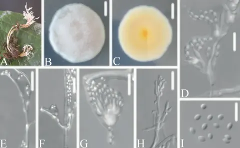
Morphology of Samsoniella farinospora. (A) Spider infected by S. farinospora. (B, C) Culture character on PDA medium. (D – F) Conidiogenous cells (conidiophores, phialides) and conidia on PDA. (G) Conidia on PDA. Scale A: 2 mm; B, C: 20 mm; D, E: 5 µm; F, G: 3 µm.
- Jenis
- Pelat ilmiah
- Kredit
- © 2022 by the authors.
- Lisensi
- CC BY 4.0
- Sumber media
- Buka entri sumber
Sumber profil
- [1] Morphological and Phylogenetic Characterization Reveals Five New Species of Samsoniella (Cordycipitaceae, Hypocreales) — Samsoniella farinospora
- [2] Catalogue of Life Base2026-09-11 — Samsoniella farinospora
- [3] Dokumentasi visual — Samsoniella farinospora
Rujukan halaman ini
Fungi. Samsoniella farinospora. Rilis data fungi-2026-10-12-expansion-2. https://fungi-atlas.mgadib.chatgpt.site/jamur/samsoniella-farinospora/.
Status: impor sumber; pemeriksaan editorial per profil belum selesai. Untuk klaim biologis, gunakan rujukan pada bagian terkait dan periksa konteks sumber aslinya.