01 / Morfologi

Ciri dan struktur

Parasitic on living branches of bamboo. Sexual morph: Ascostromata 1–6 cm long × 1–4 cm wide, solitary, superficial, subglobose, long ellipsoid to irregular, tuberculate, fleshy, white to pinkish, with locules lining the periphery, with dark ostiolate points appearing on surface. Ascostromatic tissue thick, pinkish, composed of wide, woven hyphae of textura intricata. Locules in vertical section 370–700 µm high × 370–700 µm diam. (x̄ = 541 × 513 µm, n = 20), globose to subglobose, immersed in the peripheral layer of ascostromata, with 100–200 µm wide ostioles. Peridium 20–45 µm thick, composed of several layers of hyaline to light brown, small cells of textura angularis to textura intricata. Hamathecium composed of interthecial, hyaline septate, branched pseudoparaphyses, 1–2.5 µm wide. Asci 200–370 × 20–35 µm (x̄ = 291.6 × 26.6 µm, n = 20), 4–6-spored, thick-walled, bitunicate, fissitunicate, cylindrical, short-pedicellate, with an ocular chamber. Ascospores 50–77 × 15–24 µm (x̄ = 62.3 × 18.1 µm, n = 20), 1-seriate, overlapped, fusiform, muriform, hyaline, with 7 transverse septa, constricted at the septum, smooth-walled. Asexual morph: Conidiomata 200–500 µm high, 300–400 µm wide, loculate, forming within ascostromata, globose to subglobose or irregular. Wall of locules 20–40 µm thick, composed of several layers of hyaline to light brown, small cells of textura intricata. Conidiophores reduced to conidiogenous cells. Conidiogenous cells 3–6 × 2–3 µm (x̄ = 4.7 × 2.1 µm, n = 10), blastic, cylindrical, hyaline, smooth-walled. Conidia 60–80 × 19–25 µm (x̄ = 75.4 × 23.1 µm, n = 20), fusiform, muriform, hyaline, with irregularly transverse and longitudinal septa, straight to curved, smooth-walled.

Sebagian uraian ditampilkan dalam bahasa sumber; terjemahannya belum tersedia atau belum lolos pemeriksaan.

Rujukan: [1]

Pengukuran mengikuti objek, spesimen atau kultur, dan metode yang disebutkan sumber. Baca uraian asli untuk konteks lengkap.

02 / Kehidupan

Habitat dan peran ekologis

Parasitic on living branches of bamboo. Sexual morph: Ascostromata 1–6 cm long × 1–4 cm wide, solitary, superficial, subglobose, long ellipsoid to irregular, tuberculate, fleshy, white to pinkish, with locules lining the periphery, with dark ostiolate points appearing on surface. Ascostromatic tissue thick, pinkish, composed of wide, woven hyphae of textura intricata. Locules in vertical section 370–700 µm high × 370–700 µm diam. (x̄ = 541 × 513 µm, n = 20), globose to subglobose, immersed in the peripheral layer of ascostromata, with 100–200 µm wide ostioles. Peridium 20–45 µm thick, composed of several layers of hyaline to light brown, small cells of textura angularis to textura intricata. Hamathecium composed of interthecial, hyaline septate, branched pseudoparaphyses, 1–2.5 µm wide. Asci 200–370 × 20–35 µm (x̄ = 291.6 × 26.6 µm, n = 20), 4–6-spored, thick-walled, bitunicate, fissitunicate, cylindrical, short-pedicellate, with an ocular chamber. Ascospores 50–77 × 15–24 µm (x̄ = 62.3 × 18.1 µm, n = 20), 1-seriate, overlapped, fusiform, muriform, hyaline, with 7 transverse septa, constricted at the septum, smooth-walled. Asexual morph: Conidiomata 200–500 µm high, 300–400 µm wide, loculate, forming within ascostromata, globose to subglobose or irregular.

Sebagian uraian ditampilkan dalam bahasa sumber; terjemahannya belum tersedia atau belum lolos pemeriksaan.

Rujukan: [1]

Habitat atau konteks temuan
Informasi belum tersedia pada sumber rilis ini.
Substrat
Informasi belum tersedia pada sumber rilis ini.
Peran atau konteks
Belum dikategorikan pada rilis ini.
Temukan hubungan serupa
03 / Siklus hidup

Reproduksi

Parasitic on living branches of bamboo. Sexual morph: Ascostromata 1–6 cm long × 1–4 cm wide, solitary, superficial, subglobose, long ellipsoid to irregular, tuberculate, fleshy, white to pinkish, with locules lining the periphery, with dark ostiolate points appearing on surface. Ascostromatic tissue thick, pinkish, composed of wide, woven hyphae of textura intricata. Locules in vertical section 370–700 µm high × 370–700 µm diam. (x̄ = 541 × 513 µm, n = 20), globose to subglobose, immersed in the peripheral layer of ascostromata, with 100–200 µm wide ostioles. Peridium 20–45 µm thick, composed of several layers of hyaline to light brown, small cells of textura angularis to textura intricata.

Sebagian uraian ditampilkan dalam bahasa sumber; terjemahannya belum tersedia atau belum lolos pemeriksaan.

Rujukan: [1]

04 / Batas informasi

Catatan identitas dan cakupan

  • Nama diterima mengikuti Catalogue of Life Base2026-09-11; uraian mempertahankan konsep dan nama sumber aslinya.
  • Preserve original treatment localities, hosts and substrate scope. Materials examined describe documented specimens, not the full global range.
  • Impor dan terjemahan otomatis belum diperiksa editorial per profil. Struktur reproduktif yang disebutkan tidak membuktikan seluruh siklus hidup diketahui.
  • Pengukuran pada uraian mengikuti spesimen, kultur, dan metode sumber; angka tidak diperluas ke semua populasi.

Catatan distribusi dalam narasi mengikuti sumbernya. Dokumentasi spesimen atau foto mengikuti catatan sumber. Bukti wilayah Indonesia dan penilaian konservasi tidak disimpulkan dari gambar.

05 / Dokumentasi

Melihat dengan konteks

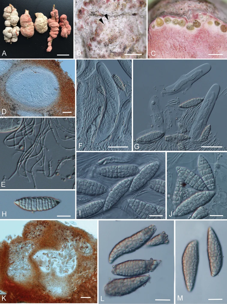
Shiraia bambusicola A–J sexual morph A fruiting bodies (HKAS102253, HKAS102254, HKAS102257, HKAS102261, HKAS102262) B–J photographs from material HKAS102253 B Surface of ascostromata showing the dark openings of ostiole C vertical section of ascostromata D vertical section of locule E pseudoparaphyses F, G asci (G Showing the fissitunicate asci) H–J ascospores K–M asexual morph K vertical section of asexual locules L–M conidia. Scale bars: 2 cm (A), 5 mm (B), 1 mm (C), 100 μm (D, K), 50 μm (F, G), 20 μm (H–J, L, M).

Jenis
Pelat ilmiah
Kredit
Dong-Qin Dai, Nalin N. Wijayawardene, Li-Zhou Tang, Chao Liu, Li-Hong Han, Hong-Long Chu, Hai-Bo Wang, Chun-Fang Liao, Er-Fu Yang, Rui-Fang Xu, Yun-Min Li, Kevin D. Hyde, D. Jayarama Bhat, Paul F. Cannon
Lisensi
CC BY 4.0
Sumber media
Buka entri sumber

Gambar menunjukkan objek menurut identifikasi sumber, bukan verifikasi independen spesimen oleh Fungi. Tidak ada skala baru yang ditambahkan.

06 / Rujukan

Sumber profil

  1. [1] Rubroshiraia gen. nov., a second hypocrellin-producing genus in Shiraiaceae (Pleosporales) — Shiraia bambusicolaBagian sumber: PMC6726671_section_14
    Tanggal penghimpunan sumber: 2026-10-12
    Teks sumber: CC BY 4.0 · Dai DQ, Wijayawardene NN, Tang LZ, Liu C, Han LH, Chu HL, Wang HB, Liao CF, Yang EF, Xu RF, Li YM, Hyde KD, Bhat DJ, Cannon PF.
  2. [2] Catalogue of Life Base2026-09-11 — Shiraia bambusicolaBagian sumber: ColDP NameUsage.tsv ID 4X4QK; accepted species; source 2073
    Tanggal penghimpunan sumber: 2026-10-12
  3. [3] Dokumentasi visual — Shiraia bambusicolaBagian sumber: Identitas mengikuti sumber dokumentasi; bukan penentuan spesimen independen.
    Tanggal penghimpunan sumber: 2026-10-17
    Media sumber: CC BY 4.0 · Dong-Qin Dai, Nalin N. Wijayawardene, Li-Zhou Tang, Chao Liu, Li-Hong Han, Hong-Long Chu, Hai-Bo Wang, Chun-Fang Liao, Er-Fu Yang, Rui-Fang Xu, Yun-Min Li, Kevin D. Hyde, D. Jayarama Bhat, Paul F. Cannon

Adaptasi teks: CC BY 4.0. Dai DQ, Wijayawardene NN, Tang LZ, Liu C, Han LH, Chu HL, Wang HB, Liao CF, Yang EF, Xu RF, Li YM, Hyde KD, Bhat DJ, Cannon PF. — Rubroshiraia gen. nov., a second hypocrellin-producing genus in Shiraiaceae (Pleosporales) — Shiraia bambusicola

Uraian asli dan riwayat sumber

Riwayat atau halaman sumber

Ringkasan sumber

Parasitic on living branches of bamboo. Sexual morph: Ascostromata 1–6 cm long × 1–4 cm wide, solitary, superficial, subglobose, long ellipsoid to irregular, tuberculate, fleshy, white to pinkish, with locules lining the periphery, with dark ostiolate points appearing on surface. Ascostromatic tissue thick, pinkish, composed of wide, woven hyphae of textura intricata. Locules in vertical section 370–700 µm high × 370–700 µm diam. (x̄ = 541 × 513 µm, n = 20), globose to subglobose, immersed in the peripheral layer of ascostromata, with 100–200 µm wide ostioles.

Revisi sumber

Morfologi sumber

Parasitic on living branches of bamboo. Sexual morph: Ascostromata 1–6 cm long × 1–4 cm wide, solitary, superficial, subglobose, long ellipsoid to irregular, tuberculate, fleshy, white to pinkish, with locules lining the periphery, with dark ostiolate points appearing on surface. Ascostromatic tissue thick, pinkish, composed of wide, woven hyphae of textura intricata. Locules in vertical section 370–700 µm high × 370–700 µm diam. (x̄ = 541 × 513 µm, n = 20), globose to subglobose, immersed in the peripheral layer of ascostromata, with 100–200 µm wide ostioles. Peridium 20–45 µm thick, composed of several layers of hyaline to light brown, small cells of textura angularis to textura intricata. Hamathecium composed of interthecial, hyaline septate, branched pseudoparaphyses, 1–2.5 µm wide. Asci 200–370 × 20–35 µm (x̄ = 291.6 × 26.6 µm, n = 20), 4–6-spored, thick-walled, bitunicate, fissitunicate, cylindrical, short-pedicellate, with an ocular chamber. Ascospores 50–77 × 15–24 µm (x̄ = 62.3 × 18.1 µm, n = 20), 1-seriate, overlapped, fusiform, muriform, hyaline, with 7 transverse septa, constricted at the septum, smooth-walled. Asexual morph: Conidiomata 200–500 µm high, 300–400 µm wide, loculate, forming within ascostromata, globose to subglobose or irregular. Wall of locules 20–40 µm thick, composed of several layers of hyaline to light brown, small cells of textura intricata. Conidiophores reduced to conidiogenous cells. Conidiogenous cells 3–6 × 2–3 µm (x̄ = 4.7 × 2.1 µm, n = 10), blastic, cylindrical, hyaline, smooth-walled. Conidia 60–80 × 19–25 µm (x̄ = 75.4 × 23.1 µm, n = 20), fusiform, muriform, hyaline, with irregularly transverse and longitudinal septa, straight to curved, smooth-walled.

Revisi sumber

Ekologi sumber

Parasitic on living branches of bamboo. Sexual morph: Ascostromata 1–6 cm long × 1–4 cm wide, solitary, superficial, subglobose, long ellipsoid to irregular, tuberculate, fleshy, white to pinkish, with locules lining the periphery, with dark ostiolate points appearing on surface. Ascostromatic tissue thick, pinkish, composed of wide, woven hyphae of textura intricata. Locules in vertical section 370–700 µm high × 370–700 µm diam. (x̄ = 541 × 513 µm, n = 20), globose to subglobose, immersed in the peripheral layer of ascostromata, with 100–200 µm wide ostioles. Peridium 20–45 µm thick, composed of several layers of hyaline to light brown, small cells of textura angularis to textura intricata. Hamathecium composed of interthecial, hyaline septate, branched pseudoparaphyses, 1–2.5 µm wide. Asci 200–370 × 20–35 µm (x̄ = 291.6 × 26.6 µm, n = 20), 4–6-spored, thick-walled, bitunicate, fissitunicate, cylindrical, short-pedicellate, with an ocular chamber. Ascospores 50–77 × 15–24 µm (x̄ = 62.3 × 18.1 µm, n = 20), 1-seriate, overlapped, fusiform, muriform, hyaline, with 7 transverse septa, constricted at the septum, smooth-walled. Asexual morph: Conidiomata 200–500 µm high, 300–400 µm wide, loculate, forming within ascostromata, globose to subglobose or irregular.

Revisi sumber

Struktur reproduksi sumber

Parasitic on living branches of bamboo. Sexual morph: Ascostromata 1–6 cm long × 1–4 cm wide, solitary, superficial, subglobose, long ellipsoid to irregular, tuberculate, fleshy, white to pinkish, with locules lining the periphery, with dark ostiolate points appearing on surface. Ascostromatic tissue thick, pinkish, composed of wide, woven hyphae of textura intricata. Locules in vertical section 370–700 µm high × 370–700 µm diam. (x̄ = 541 × 513 µm, n = 20), globose to subglobose, immersed in the peripheral layer of ascostromata, with 100–200 µm wide ostioles. Peridium 20–45 µm thick, composed of several layers of hyaline to light brown, small cells of textura angularis to textura intricata.

Revisi sumber

Rujukan halaman ini

Fungi. Shiraia bambusicola. Rilis data fungi-2026-10-12-expansion-2. https://fungi-atlas.mgadib.chatgpt.site/jamur/shiraia-bambusicola/.

Status: impor sumber; pemeriksaan editorial per profil belum selesai. Untuk klaim biologis, gunakan rujukan pada bagian terkait dan periksa konteks sumber aslinya.