01 / Morfologi

Ciri dan struktur

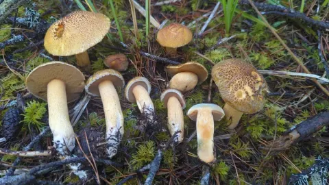
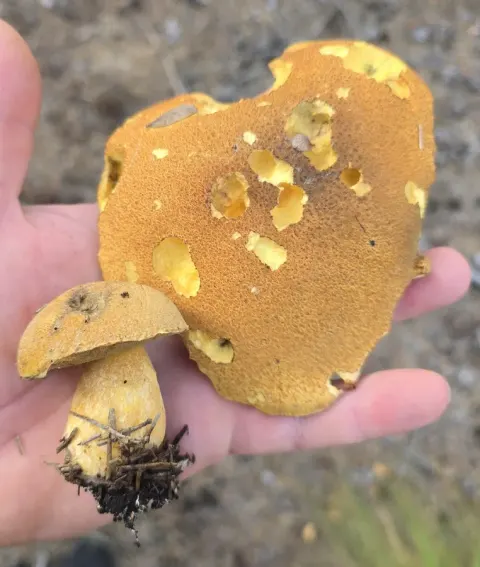
The cap of the fruit body is 3 to 12 cm in diameter, and depending on its age, is either conic to convex, to somewhat flattened at maturity. The cap margin is initially rolled downward before straightening out, often with hanging remnants of partial veil (appendiculate). The cap surface is covered with densely matted filaments that are rough and scale-like. The scales are pink to brownish red, fading to a pale brown-gray or dull yellow in maturity. Under the scales, the cap surface is yellow to pale yellow-orange. While many other Suillus species have a sticky or slimy cap, S. spraguei is dry. The flesh is yellow. The pores on the underside of the cap are yellowish and angular, measuring 0.5 to 5 mm wide, and formed by tubes that extend 4 to 8 mm deep. These pores have a slightly decurrent attachment to the stem (extending down its length). Young specimens have a whitish fibrous partial veil that protects the developing pores; as the cap expands it rips the veil, which remains as a grayish ring on the stem. The stem is 4 to 12 cm long, and 1 to 2.5 cm thick, roughly cylindrical in shape, or sometimes with a bulbous bottom so as to be somewhat club-shaped. The stem surface is tomentose, with scales at the top, and a ring on the upper half of the stem. Below the ring the stem is fibrillose, covered with a mat of soft hairs. Its color at the top is yellow, but with wine-red to reddish-brown scales below, underlaid with a pale yellow to grayish color. The stem is usually solid, rarely hollow. The tissue of all parts of the fruit body—cap, pores, and stem—will turn brownish shortly after being bruised or injured.

Sebagian uraian ditampilkan dalam bahasa sumber; terjemahannya belum tersedia atau belum lolos pemeriksaan.

Rujukan: [1]

Pengukuran mengikuti objek, spesimen atau kultur, dan metode yang disebutkan sumber. Baca uraian asli untuk konteks lengkap.

02 / Kehidupan

Habitat dan peran ekologis

Dalam alam, S. spraguei membentuk hubungan ektomikoriza dengan spesies pinus lima jarum. tetapi mereka dapat ditemukan selambat pada bulan Oktober. Jamur dapat menjadi parasit oleh jamur Hypomyces completus. Pada tahap asexual H. completus, awalnya muncul sebagai bintik-bintik jamur putih pada permukaan topi atau batang yang dengan cepat menyebar untuk menutupi seluruh permukaan jamur dan menghasilkan konidia (spora asexual). Pada tahap seksual, jamur berubah warna, berkembang dari kuning-kecoklatan ke coklat, hijau-kecoklatan dan akhirnya hitam saat membuat perithecia, askus yang mengandung struktur seksual yang menghasilkan askospora. Peritesi adalah jerawat dan memberikan permukaan tekstur kasar. Sebuah studi Japanese field menemukan bahwa S. spraguei adalah jamur dominan dalam kondisi 21 tahun Korean pine, baik dalam hal ektomikoriza (diukur sebagai persentase biomassa yang ada dalam sampel tanah) dan produksi tubuh buah (yang terdiri dari 90% dari berat kering total tubuh buah yang dikumpulkan dari semua spesies). Produksi S. spraguei buah tubuh rata-rata sekitar satu per meter persegi, tanpa banyak variasi selama periode penelitian empat tahun. Jamur muncul sebagian besar dari August to November, cenderung tumbuh dalam kelompok, dan distribusi spasial kelompok secara acak ⁇ lokasi kelompok tidak berkorelasi dengan penampilan di tahun sebelumnya.

Rujukan: [1]

Habitat atau konteks temuan
Informasi belum tersedia pada sumber rilis ini.
Substrat
tanah
Peran atau konteks
Mitra tumbuhan
Temukan hubungan serupa
03 / Siklus hidup

Reproduksi

In deposit, such as with a spore print, the spores of S. spraguei appear olive-brown in color; this changes to clay or tawny-olive after drying. Microscopically, the spores have smooth surfaces, measuring 9–11 by 3–4.5 μm; in side profile they have asymmetrical sides and a suprahilar depression (a surface indentation formed where the spore attaches to the basidia), while in face view they appear oblong. The spores are not amyloid, meaning that they do not absorb iodine when stained with Melzer's reagent. The basidia (the spore-bearing cells in the hymenium) are thin-walled, four-spored, and have dimensions of 17–19 by 5–7.8 μm. In the presence of potassium hydroxide, they appear hyaline (translucent), and they become pale yellow to nearly hyaline in Melzer's reagent.

Sebagian uraian ditampilkan dalam bahasa sumber; terjemahannya belum tersedia atau belum lolos pemeriksaan.

Rujukan: [1]

Sinonim menurut acuan taksonomi rilis: Boletinus pictus; Boletus murrayi; Boletus pictus; Boletus spraguei; Suillus murrayi; Suillus pictus
04 / Batas informasi

Catatan identitas dan cakupan

  • Nama diterima mengikuti Catalogue of Life Base2026-09-11; uraian mempertahankan konsep dan nama sumber aslinya.
  • Original geographic, specimen and life-cycle scope retained; source import awaits individual editorial review.
  • Impor dan terjemahan otomatis belum diperiksa editorial per profil. Struktur reproduktif yang disebutkan tidak membuktikan seluruh siklus hidup diketahui.
  • Pengukuran pada uraian mengikuti spesimen, kultur, dan metode sumber; angka tidak diperluas ke semua populasi.

Catatan distribusi dalam narasi mengikuti sumbernya. Dokumentasi spesimen atau foto mengikuti catatan sumber. Bukti wilayah Indonesia dan penilaian konservasi tidak disimpulkan dari gambar.

05 / Dokumentasi

Melihat dengan konteks

Dokumentasi Suillus spraguei menurut asosiasi foto pada record takson iNaturalist. Kredit: Kurt Miller. Identitas mengikuti sumber; bukan penentuan spesimen independen.

Jenis
Fotografi
Kredit
(c) Kurt Miller, some rights reserved (CC BY-SA), uploaded by Kurt Miller
Sumber media
Buka entri sumber

Gambar menunjukkan objek menurut identifikasi sumber, bukan verifikasi independen spesimen oleh Fungi. Tidak ada skala baru yang ditambahkan.

06 / Rujukan

Sumber profil

  1. [1] Suillus spraguei — Wikipedia (en)Bagian sumber: Frozen revision 1339028131
    Tanggal penghimpunan sumber: 2026-10-12
    Teks sumber: CC BY-SA4.0 · Wikipedia contributors
  2. [2] Catalogue of Life Base2026-09-11 — Suillus spragueiBagian sumber: ColDP NameUsage.tsv ID 53F8L; accepted species; source 2073
    Tanggal penghimpunan sumber: 2026-10-12
  3. [3] Dokumentasi visual — Suillus spragueiBagian sumber: Individual media record; identification follows the source, not independent determination
    Tanggal penghimpunan sumber: 2026-10-12
    Media sumber: CC BY-SA 4.0 · (c) Kurt Miller, some rights reserved (CC BY-SA), uploaded by Kurt Miller

Adaptasi teks: CC BY-SA 4.0. Wikipedia contributors — Suillus spraguei — Wikipedia (en)

Uraian asli dan riwayat sumber

Riwayat atau halaman sumber

Ringkasan sumber

Suillus spraguei is a species of fungus in the family Suillaceae. It is known by a variety of common names, including the painted slipperycap, the painted suillus or the red and yellow suillus. Suillus spraguei has had a complex taxonomical history and is also frequently referred to as Suillus pictus. The readily identifiable fruit bodies have caps that are dark red when fresh, dry to the touch, and covered with mats of hairs and scales that are separated by yellow cracks. On the underside of the cap are small, yellow, angular pores that become brownish as the mushroom ages.

Revisi sumber

Morfologi sumber

The cap of the fruit body is 3 to 12 cm in diameter, and depending on its age, is either conic to convex, to somewhat flattened at maturity. The cap margin is initially rolled downward before straightening out, often with hanging remnants of partial veil (appendiculate). The cap surface is covered with densely matted filaments that are rough and scale-like. The scales are pink to brownish red, fading to a pale brown-gray or dull yellow in maturity. Under the scales, the cap surface is yellow to pale yellow-orange. While many other Suillus species have a sticky or slimy cap, S. spraguei is dry. The flesh is yellow. The pores on the underside of the cap are yellowish and angular, measuring 0.5 to 5 mm wide, and formed by tubes that extend 4 to 8 mm deep. These pores have a slightly decurrent attachment to the stem (extending down its length). Young specimens have a whitish fibrous partial veil that protects the developing pores; as the cap expands it rips the veil, which remains as a grayish ring on the stem. The stem is 4 to 12 cm long, and 1 to 2.5 cm thick, roughly cylindrical in shape, or sometimes with a bulbous bottom so as to be somewhat club-shaped. The stem surface is tomentose, with scales at the top, and a ring on the upper half of the stem. Below the ring the stem is fibrillose, covered with a mat of soft hairs. Its color at the top is yellow, but with wine-red to reddish-brown scales below, underlaid with a pale yellow to grayish color. The stem is usually solid, rarely hollow. The tissue of all parts of the fruit body—cap, pores, and stem—will turn brownish shortly after being bruised or injured.

Revisi sumber

Ekologi sumber

In nature, S. spraguei forms ectomycorrhizal relationships with five-needled pine species. but they may be found as late as October. The mushrooms can be parasitized by the fungus Hypomyces completus. In the asexual stage of H. completus, it appears initially as patches of whitish mold on the surface of the cap or stem that rapidly spread to cover the entire mushroom surface and produce conidia (asexual spores). In the sexual stage, the mold changes color, progressing from yellow-brown to brown, greenish-brown and eventually black as it makes perithecia, asci-containing sexual structures that produce ascospores. The perithecia are pimply and give the surface a roughened texture. A Japanese field study found that S. spraguei was the dominant fungus in a 21-year-old stand of Korean pine, both in terms of ectomycorrhizae (measured as percentage of biomass present in soil samples) and by fruit body production (comprising over 90% of dry weight of total fruit bodies collected of all species). The production of S. spraguei fruit bodies averaged about one per square meter, without much variance during the four-year study period. The mushrooms appeared mostly from August to November, tended to grow in clumps, and the spatial distribution of clumps was random—the location of the clumps was not correlatable with appearances in previous years.

Revisi sumber

Struktur reproduksi sumber

In deposit, such as with a spore print, the spores of S. spraguei appear olive-brown in color; this changes to clay or tawny-olive after drying. Microscopically, the spores have smooth surfaces, measuring 9–11 by 3–4.5 μm; in side profile they have asymmetrical sides and a suprahilar depression (a surface indentation formed where the spore attaches to the basidia), while in face view they appear oblong. The spores are not amyloid, meaning that they do not absorb iodine when stained with Melzer's reagent. The basidia (the spore-bearing cells in the hymenium) are thin-walled, four-spored, and have dimensions of 17–19 by 5–7.8 μm. In the presence of potassium hydroxide, they appear hyaline (translucent), and they become pale yellow to nearly hyaline in Melzer's reagent.

Revisi sumber

Rujukan halaman ini

Fungi. Suillus spraguei. Rilis data fungi-2026-10-12-expansion-2. https://fungi-atlas.mgadib.chatgpt.site/jamur/suillus-spraguei/.

Status: impor sumber; pemeriksaan editorial per profil belum selesai. Untuk klaim biologis, gunakan rujukan pada bagian terkait dan periksa konteks sumber aslinya.