01 / Morfologi

Ciri dan struktur

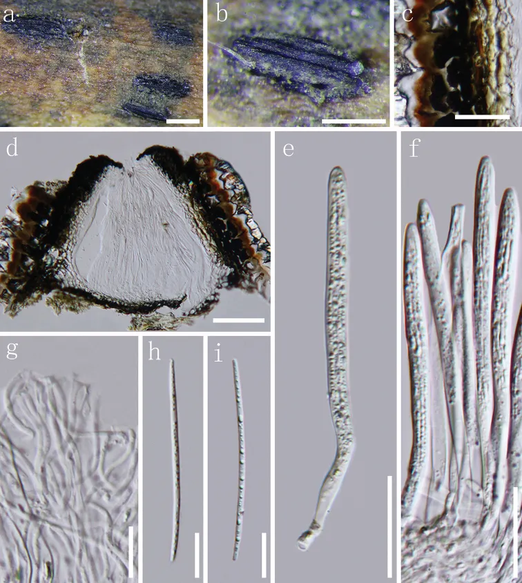
Apothecia developing on dead branch, elliptical or oblong-elliptical in outline, ends slightly acute to obtuse. Apothecia surface black, matt or slightly glossy, moderately raising the substratum surface, opening by a single longitudinal split that extends to the ends of the apothecium (Fig. 3a, b). Lips absent. In median vertical section (Fig. 3d), apothecia deeply embedded in host tissue, with host cells becoming filled with fungal tissue as the apothecium develops. Covering stroma (Fig. 3c) 30–45 µm thick, composed of blackish-brown to black, thick-walled cells of textura angularis towards the exterior and several layers of pale to nearly hyaline, thin-walled cells towards the interior. Along the edge of the apothecial opening, there is a flattened, 12–20 µm thick extension adjacent to the covering stroma that is composed of strongly melanised tissue with no obvious cellular structure. Basal stroma 8–18 µm thick, dark brown or blackish-brown, composed of angular to globose, thick-walled cells, 2.5–4 µm diam. A triangular space between the covering stroma and basal stroma consists of thin-walled, nearly hyaline to grey-brown cells arranged in textura prismatica. Paraphyses 1–2 µm, filiform, hyaline, septate, gradually swollen or branching once at the apex, embedded in gelatinous sheaths. Asci (103–)110–122.5 × 5.5–7 µm (x ¯ = 113 × 6 µm, n = 20), 8-spored, unitunicate, cylindrical, long stalk, thin-walled, apex truncate to somewhat round, J-, without circumapical thickening. Ascospores 55–66 × 1.5–2.0 µm (x ¯ = 61 × 1.8 µm, n = 25), fascicle, but not coiled, filiform, gradually tapering toward the ends, hyaline, aseptate, smooth-walled, straight or slightly curved, lacking gelatinous sheath.

Sebagian uraian ditampilkan dalam bahasa sumber; terjemahannya belum tersedia atau belum lolos pemeriksaan.

Rujukan: [1]

Pengukuran mengikuti objek, spesimen atau kultur, dan metode yang disebutkan sumber. Baca uraian asli untuk konteks lengkap.

02 / Kehidupan

Habitat dan peran ekologis

Apothecia developing on dead branch, elliptical or oblong-elliptical in outline, ends slightly acute to obtuse. Apothecia surface black, matt or slightly glossy, moderately raising the substratum surface, opening by a single longitudinal split that extends to the ends of the apothecium (Fig. 3a, b). Lips absent. In median vertical section (Fig. 3d), apothecia deeply embedded in host tissue, with host cells becoming filled with fungal tissue as the apothecium develops. Covering stroma (Fig. 3c) 30–45 µm thick, composed of blackish-brown to black, thick-walled cells of textura angularis towards the exterior and several layers of pale to nearly hyaline, thin-walled cells towards the interior. Along the edge of the apothecial opening, there is a flattened, 12–20 µm thick extension adjacent to the covering stroma that is composed of strongly melanised tissue with no obvious cellular structure. Basal stroma 8–18 µm thick, dark brown or blackish-brown, composed of angular to globose, thick-walled cells, 2.5–4 µm diam. A triangular space between the covering stroma and basal stroma consists of thin-walled, nearly hyaline to grey-brown cells arranged in textura prismatica. Paraphyses 1–2 µm, filiform, hyaline, septate, gradually swollen or branching once at the apex, embedded in gelatinous sheaths.

Sebagian uraian ditampilkan dalam bahasa sumber; terjemahannya belum tersedia atau belum lolos pemeriksaan.

Rujukan: [1]

Habitat atau konteks temuan
Informasi belum tersedia pada sumber rilis ini.
Substrat
Informasi belum tersedia pada sumber rilis ini.
Peran atau konteks
Belum dikategorikan pada rilis ini.
Temukan hubungan serupa
03 / Siklus hidup

Reproduksi

Apothecia developing on dead branch, elliptical or oblong-elliptical in outline, ends slightly acute to obtuse. Apothecia surface black, matt or slightly glossy, moderately raising the substratum surface, opening by a single longitudinal split that extends to the ends of the apothecium (Fig. 3a, b). Lips absent. In median vertical section (Fig. 3d), apothecia deeply embedded in host tissue, with host cells becoming filled with fungal tissue as the apothecium develops. Covering stroma (Fig. 3c) 30–45 µm thick, composed of blackish-brown to black, thick-walled cells of textura angularis towards the exterior and several layers of pale to nearly hyaline, thin-walled cells towards the interior. Along the edge of the apothecial opening, there is a flattened, 12–20 µm thick extension adjacent to the covering stroma that is composed of strongly melanised tissue with no obvious cellular structure.

Sebagian uraian ditampilkan dalam bahasa sumber; terjemahannya belum tersedia atau belum lolos pemeriksaan.

Rujukan: [1]

04 / Batas informasi

Catatan identitas dan cakupan

  • Nama diterima mengikuti Catalogue of Life Base2026-09-11; uraian mempertahankan konsep dan nama sumber aslinya.
  • Preserve original treatment localities, hosts and substrate scope. Materials examined describe documented specimens, not the full global range.
  • Impor dan terjemahan otomatis belum diperiksa editorial per profil. Struktur reproduktif yang disebutkan tidak membuktikan seluruh siklus hidup diketahui.
  • Pengukuran pada uraian mengikuti spesimen, kultur, dan metode sumber; angka tidak diperluas ke semua populasi.

Catatan distribusi dalam narasi mengikuti sumbernya. Dokumentasi spesimen atau foto mengikuti catatan sumber. Bukti wilayah Indonesia dan penilaian konservasi tidak disimpulkan dari gambar.

05 / Dokumentasi

Melihat dengan konteks

Terriera karsti a, b apothecia observed under the dissecting microscope c detail of covering stroma in vertical section d vertical section through an apothecium e, f asci in various states of maturity g apices of paraphyses h, i ascospores. Note: c–i mounted in water. Scale bar: 1 mm (a), 500 µm (b), 20 µm (c, e, f), 100 µm (d), 10 µm (g, i).

Jenis
Pelat ilmiah
Kredit
Jin-Feng Zhang, Jian-Kui Liu, Kevin D. Hyde, Anusha H. Ekanayaka, Zuo-Yi Liu
Lisensi
CC BY 4.0
Sumber media
Buka entri sumber

Gambar menunjukkan objek menurut identifikasi sumber, bukan verifikasi independen spesimen oleh Fungi. Tidak ada skala baru yang ditambahkan.

06 / Rujukan

Sumber profil

  1. [1] Morpho-phylogenetic evidence reveals new species in Rhytismataceae (Rhytismatales, Leotiomycetes, Ascomycota) from Guizhou Province, China — Terriera karstiBagian sumber: PMC7790812_section_15
    Tanggal penghimpunan sumber: 2026-10-12
    Teks sumber: CC BY 4.0 · Zhang JF, Liu JK, Hyde KD, Ekanayaka AH, Liu ZY.
  2. [2] Catalogue of Life Base2026-09-11 — Terriera karstiBagian sumber: ColDP NameUsage.tsv ID 9ZGH3; accepted species; source 2073
    Tanggal penghimpunan sumber: 2026-10-12
  3. [3] Dokumentasi visual — Terriera karstiBagian sumber: Individual media record; identification follows the source, not independent determination
    Tanggal penghimpunan sumber: 2026-10-12
    Media sumber: CC BY 4.0 · Jin-Feng Zhang, Jian-Kui Liu, Kevin D. Hyde, Anusha H. Ekanayaka, Zuo-Yi Liu

Adaptasi teks: CC BY 4.0. Zhang JF, Liu JK, Hyde KD, Ekanayaka AH, Liu ZY. — Morpho-phylogenetic evidence reveals new species in Rhytismataceae (Rhytismatales, Leotiomycetes, Ascomycota) from Guizhou Province, China — Terriera karsti

Uraian asli dan riwayat sumber

Riwayat atau halaman sumber

Ringkasan sumber

Apothecia developing on dead branch, elliptical or oblong-elliptical in outline, ends slightly acute to obtuse. Apothecia surface black, matt or slightly glossy, moderately raising the substratum surface, opening by a single longitudinal split that extends to the ends of the apothecium (Fig. 3a, b). Lips absent. In median vertical section (Fig. 3d), apothecia deeply embedded in host tissue, with host cells becoming filled with fungal tissue as the apothecium develops. Covering stroma (Fig. 3c) 30–45 µm thick, composed of blackish-brown to black, thick-walled cells of textura angularis towards the exterior and several layers of pale to nearly hyaline, thin-walled cells towards the interior.

Revisi sumber

Morfologi sumber

Apothecia developing on dead branch, elliptical or oblong-elliptical in outline, ends slightly acute to obtuse. Apothecia surface black, matt or slightly glossy, moderately raising the substratum surface, opening by a single longitudinal split that extends to the ends of the apothecium (Fig. 3a, b). Lips absent. In median vertical section (Fig. 3d), apothecia deeply embedded in host tissue, with host cells becoming filled with fungal tissue as the apothecium develops. Covering stroma (Fig. 3c) 30–45 µm thick, composed of blackish-brown to black, thick-walled cells of textura angularis towards the exterior and several layers of pale to nearly hyaline, thin-walled cells towards the interior. Along the edge of the apothecial opening, there is a flattened, 12–20 µm thick extension adjacent to the covering stroma that is composed of strongly melanised tissue with no obvious cellular structure. Basal stroma 8–18 µm thick, dark brown or blackish-brown, composed of angular to globose, thick-walled cells, 2.5–4 µm diam. A triangular space between the covering stroma and basal stroma consists of thin-walled, nearly hyaline to grey-brown cells arranged in textura prismatica. Paraphyses 1–2 µm, filiform, hyaline, septate, gradually swollen or branching once at the apex, embedded in gelatinous sheaths. Asci (103–)110–122.5 × 5.5–7 µm (x ¯ = 113 × 6 µm, n = 20), 8-spored, unitunicate, cylindrical, long stalk, thin-walled, apex truncate to somewhat round, J-, without circumapical thickening. Ascospores 55–66 × 1.5–2.0 µm (x ¯ = 61 × 1.8 µm, n = 25), fascicle, but not coiled, filiform, gradually tapering toward the ends, hyaline, aseptate, smooth-walled, straight or slightly curved, lacking gelatinous sheath.

Revisi sumber

Ekologi sumber

Apothecia developing on dead branch, elliptical or oblong-elliptical in outline, ends slightly acute to obtuse. Apothecia surface black, matt or slightly glossy, moderately raising the substratum surface, opening by a single longitudinal split that extends to the ends of the apothecium (Fig. 3a, b). Lips absent. In median vertical section (Fig. 3d), apothecia deeply embedded in host tissue, with host cells becoming filled with fungal tissue as the apothecium develops. Covering stroma (Fig. 3c) 30–45 µm thick, composed of blackish-brown to black, thick-walled cells of textura angularis towards the exterior and several layers of pale to nearly hyaline, thin-walled cells towards the interior. Along the edge of the apothecial opening, there is a flattened, 12–20 µm thick extension adjacent to the covering stroma that is composed of strongly melanised tissue with no obvious cellular structure. Basal stroma 8–18 µm thick, dark brown or blackish-brown, composed of angular to globose, thick-walled cells, 2.5–4 µm diam. A triangular space between the covering stroma and basal stroma consists of thin-walled, nearly hyaline to grey-brown cells arranged in textura prismatica. Paraphyses 1–2 µm, filiform, hyaline, septate, gradually swollen or branching once at the apex, embedded in gelatinous sheaths.

Revisi sumber

Struktur reproduksi sumber

Apothecia developing on dead branch, elliptical or oblong-elliptical in outline, ends slightly acute to obtuse. Apothecia surface black, matt or slightly glossy, moderately raising the substratum surface, opening by a single longitudinal split that extends to the ends of the apothecium (Fig. 3a, b). Lips absent. In median vertical section (Fig. 3d), apothecia deeply embedded in host tissue, with host cells becoming filled with fungal tissue as the apothecium develops. Covering stroma (Fig. 3c) 30–45 µm thick, composed of blackish-brown to black, thick-walled cells of textura angularis towards the exterior and several layers of pale to nearly hyaline, thin-walled cells towards the interior. Along the edge of the apothecial opening, there is a flattened, 12–20 µm thick extension adjacent to the covering stroma that is composed of strongly melanised tissue with no obvious cellular structure.

Revisi sumber

Rujukan halaman ini

Fungi. Terriera karsti. Rilis data fungi-2026-10-12-expansion-2. https://fungi-atlas.mgadib.chatgpt.site/jamur/terriera-karsti/.

Status: impor sumber; pemeriksaan editorial per profil belum selesai. Untuk klaim biologis, gunakan rujukan pada bagian terkait dan periksa konteks sumber aslinya.