Ciri dan struktur
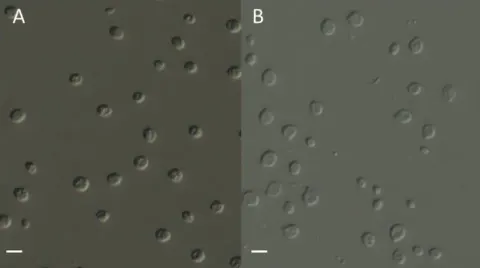
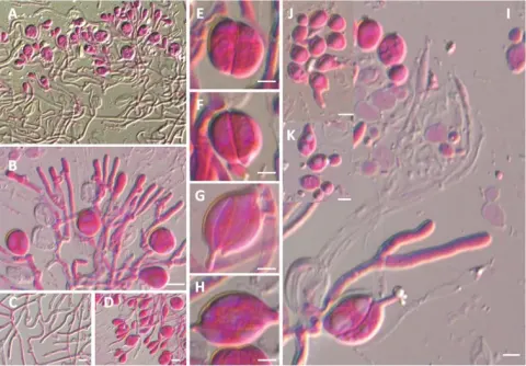
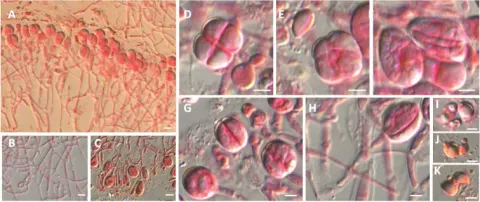
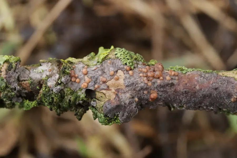
Fruit bodies are gelatinous, orange-red to brownish red, initially up to 2.5 mm across, and discoid to pustular. Eventually they coalesce and become effused cerebriform (brain-like), up to 50 mm across. Microscopically, the hyphae have clamp connections and the basidia are tremelloid (ellipsoid, with oblique to vertical septa), 2 to 4-celled, 14 to 30 by 8 to 11 μm. Sterigmata and basidiospores are not formed in the initial, discoid to pustular stage; instead, clusters of small, ellipsoid conidiospores are released, typically with a thin wisp of ribbon-like hypha still attached. In the effused, cerebriform stage, smooth, globose to subglobose basidiospores are produced measuring 5.5 to 8 by 7.5 to 10 μm.
Habitat dan peran ekologis
Tremella versicolor is a parasite on basidiocarps of the lignicolous, corticioid genus Peniophora, including Peniophora lycii, P. cinerea, P. quercina, P. nuda, P. violaceolivida, P. reidii, and P. incarnata. The hosts typically grow on dead, attached or recently fallen branches of deciduous trees. The species was originally described from England and has been recorded in Europe from Belgium, Denmark, Germany, Italy, the Netherlands, and Northern Ireland. Old reports from Sweden refer to Tremella subencephala. Tremella versicolor has also been reported from the USA.
- Habitat atau konteks temuan
- Informasi belum tersedia pada sumber rilis ini.
- Substrat
- Informasi belum tersedia pada sumber rilis ini.
- Peran atau konteks
- Belum dikategorikan pada rilis ini.
Reproduksi
Fruit bodies are gelatinous, orange-red to brownish red, initially up to 2.5 mm across, and discoid to pustular. Eventually they coalesce and become effused cerebriform (brain-like), up to 50 mm across. Microscopically, the hyphae have clamp connections and the basidia are tremelloid (ellipsoid, with oblique to vertical septa), 2 to 4-celled, 14 to 30 by 8 to 11 μm. Sterigmata and basidiospores are not formed in the initial, discoid to pustular stage; instead, clusters of small, ellipsoid conidiospores are released, typically with a thin wisp of ribbon-like hypha still attached. In the effused, cerebriform stage, smooth, globose to subglobose basidiospores are produced measuring 5.5 to 8 by 7.5 to 10 μm.
Catatan identitas dan cakupan
- Nama diterima mengikuti Catalogue of Life Base2026-09-11; uraian mempertahankan konsep dan nama sumber aslinya.
- Original geographic, specimen and life-cycle scope retained; source import awaits individual editorial review.
- Impor dan terjemahan otomatis belum diperiksa editorial per profil. Struktur reproduktif yang disebutkan tidak membuktikan seluruh siklus hidup diketahui.
- Pengukuran pada uraian mengikuti spesimen, kultur, dan metode sumber; angka tidak diperluas ke semua populasi.
Catatan distribusi dalam narasi mengikuti sumbernya. Dokumentasi spesimen atau foto mengikuti catatan sumber. Bukti wilayah Indonesia dan penilaian konservasi tidak disimpulkan dari gambar.
Melihat dengan konteks
Dokumentasi Tremella versicolor menurut asosiasi foto pada record takson iNaturalist. Kredit: Björn Sothmann. Identitas mengikuti sumber; bukan penentuan spesimen independen.
- Jenis
- Fotografi
- Kredit
- (c) Björn Sothmann, niektoré práva vyhradené (CC BY-SA), uploaded by Björn Sothmann
- Lisensi
- CC BY-SA 4.0
- Sumber media
- Buka entri sumber
Dokumentasi tambahan
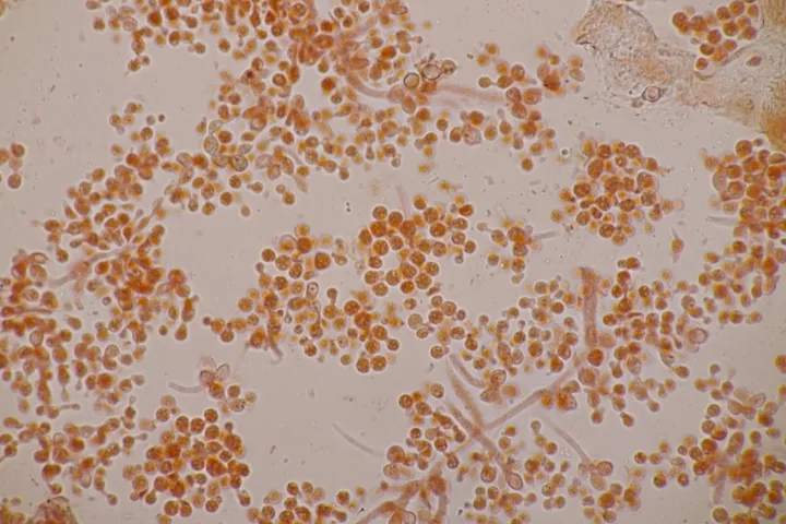
Foto yang diasosiasikan dengan Tremella versicolor pada iNaturalist. Identifikasi mengikuti sumber; bukan penentuan spesimen independen.
Kredit dan penggunaan
(c) Björn Sothmann, niektoré práva vyhradené (CC BY-SA), uploaded by Björn Sothmann
Ukuran dan format gambar disesuaikan tanpa pemotongan, retus, atau penambahan skala. Lisensi sumber dipertahankan.
Sumber profil
- [1] Tremella versicolor — Wikipedia (en)
- [2] Catalogue of Life Base2026-09-11 — Tremella versicolor
- [3] Dokumentasi visual — Tremella versicolor
- [4] Dokumentasi visual — Tremella versicolor
Rujukan halaman ini
Fungi. Tremella versicolor. Rilis data fungi-2026-10-12-expansion-2. https://fungi-atlas.mgadib.chatgpt.site/jamur/tremella-versicolor/.
Status: impor sumber; pemeriksaan editorial per profil belum selesai. Untuk klaim biologis, gunakan rujukan pada bagian terkait dan periksa konteks sumber aslinya.