Saprobic on batang mati dari Galium sp. Morph seksual tidak ditentukan. Aseksual morph konidiomata 195–220 µm diam., 210–250 µm tinggi, pycnidial, semi-merendam dalam jaringan inang, bercahaya, kesepian, non-papilate, cokelat. Pycnidial walls 22–28 µm tebal, terdiri dari beberapa lapisan sel coklat terang hingga coklat gelap, diatur dalam tekstura angularis hingga tekstura prismatica. Conidiophores absent. Conidiogenous cells terbentuk pada lapisan dalam konidiomata, ampulliform, phialidic, halus, cokelat pucat. konidia 30–42 × 5–7 µm (\(\bar{x} \) = 35.8 × 6.2 µm, n = 20), silinder, lurus hingga sedikit melengkung, secara bertahap menyusut menuju puncak bulat, dengan puncak bulat, awalnya hialin menjadi cokelat pucat, setelah matang cokelat emas, 7–10-euseptate, dinding tipis, dengan lampiran pad-seperti di puncak.
Sebagian uraian ditampilkan dalam bahasa sumber; terjemahannya belum tersedia atau belum lolos pemeriksaan.
Profil ini diimpor dari uraian spesies pada sumber berlisensi dan sebagian teks diterjemahkan otomatis. Pemeriksaan editorial per profil belum selesai. Bagian yang belum diterjemahkan atau belum lolos pemeriksaan terjemahan tetap ditampilkan dalam bahasa sumber. Angka dan nama ilmiah dipertahankan.
Saprobic on dead stem of Galium sp. Sexual morph Undetermined. Asexual morph Conidiomata 195–220 µm diam., 210–250 µm high, pycnidial, semi-immersed in the host tissue, scattered, solitary, non-papillate, brown. Pycnidial walls 22–28 µm thick, composed of several layers of light brown to dark brown cells, arranged in textura angularis to textura prismatica. Conidiophores absent. Conidiogenous cells formed on the inner layer of conidiomata, ampulliform, phialidic, smooth, pale brown. Conidia 30–42 × 5–7 µm (\(\bar{x} \) = 35.8 × 6.2 µm, n = 20), cylindrical, straight to slightly curved, gradually tapering towards the rounded apex, with rounded apex, initially hyaline to pale brown, after maturity golden brown, 7–10-euseptate, thin-walled, with pad-like appendages at the apex. Culture characteristics: Colonies on PDA reaching 8 mm diam. after 2 weeks at 25–30 °C, colonies medium dense, circular, convex, surface slightly rough with edge entire, effuse, velvety to hairy, margin well-defined, colony from above: white to grayish at the margin, white at the centre reverse, white to grayish at the margin, white to greyish at the centre, mycelium white to greyish with tufting, not producing pigments in PDA.
Sebagian uraian ditampilkan dalam bahasa sumber; terjemahannya belum tersedia atau belum lolos pemeriksaan.
Saprobic on dead stem of Galium sp. Sexual morph Undetermined. Asexual morph Conidiomata 195–220 µm diam., 210–250 µm high, pycnidial, semi-immersed in the host tissue, scattered, solitary, non-papillate, brown. Pycnidial walls 22–28 µm thick, composed of several layers of light brown to dark brown cells, arranged in textura angularis to textura prismatica. Conidiophores absent. Conidiogenous cells formed on the inner layer of conidiomata, ampulliform, phialidic, smooth, pale brown. Conidia 30–42 × 5–7 µm (\(\bar{x} \) = 35.8 × 6.2 µm, n = 20), cylindrical, straight to slightly curved, gradually tapering towards the rounded apex, with rounded apex, initially hyaline to pale brown, after maturity golden brown, 7–10-euseptate, thin-walled, with pad-like appendages at the apex.
Sebagian uraian ditampilkan dalam bahasa sumber; terjemahannya belum tersedia atau belum lolos pemeriksaan.
Sinonim menurut acuan taksonomi rilis: Wojnowicia dactylidis
04 / Batas informasi
Catatan identitas dan cakupan
Nama diterima mengikuti Catalogue of Life Base2026-09-11; uraian mempertahankan konsep dan nama sumber aslinya.
Preserve original monograph treatment localities, hosts and specimen/substrate scope. Materials examined document specimens, not the full global range.
Impor dan terjemahan otomatis belum diperiksa editorial per profil. Struktur reproduktif yang disebutkan tidak membuktikan seluruh siklus hidup diketahui.
Pengukuran pada uraian mengikuti spesimen, kultur, dan metode sumber; angka tidak diperluas ke semua populasi.
Catatan distribusi dalam narasi mengikuti sumbernya. Dokumentasi spesimen atau foto mengikuti catatan sumber. Bukti wilayah Indonesia dan penilaian konservasi tidak disimpulkan dari gambar.
05 / Dokumentasi
Melihat dengan konteks
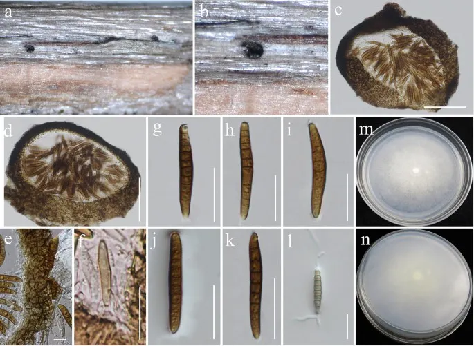
Wojnowiciella dactylidis (MFLU 16-1366). a Appearance of conidiomata on host. b Close-up of conidioma. c, d Section of conidiomata. e Pycnidial wall. f Developing conidia attached to conidiogenous cell. g – k Conidia. l Germinating conidium. m Colony from above. n Colony from below. Scale bars: c, d = 75 µm, e = 10 µm, f = 20 µm, g – l = 20 µm
Jenis
Pelat ilmiah
Kredit
Penulis dan sumber artikel: Hyde, Kevin D.; Dong, Yang; Phookamsak, Rungtiwa; Jeewon, Rajesh; Bhat, D. Jayarama; Jones, E. B. Gareth; Liu, Ning-Guo; Abeywickrama, Pranami D.; Mapook, Ausana; Wei, Deping; Perera, Rekhani H.; Manawasinghe, Ishara S.; Pem, Dhandevi; Bundhun, Digvijayini; Karunarathna, Anuruddha; Ekanayaka, Anusha H.; Bao, Dan-Feng; Li, Junfu; Samarakoon, Milan C.; Chaiwan, Napalai; Lin, Chuan-Gen; Phutthacharoen, Kunthida; Zhang, Sheng-Nan; Senanayake, Indunil C.; Goonasekara, Ishani D.; Thambugala, Kasun M.; Phukhamsakda, Chayanard; Tennakoon, Danushka S.; Jiang, Hong-Bo; Yang, Jing; Zeng, Ming; Huanraluek, Naruemon; Liu, Jian-Kui (Jack); Wijesinghe, Subodini N.; Tian, Qing; Tibpromma, Saowaluck; Brahmanage, Rashika S.; Boonmee, Saranyaphat; Huang, Shi-Ke; Thiyagaraja, Vinodhini; Lu, Yong-Zhong; Jayawardena, Ruvishika S.; Dong, Wei; Yang, Er-Fu; Singh, Sanjay K.; Singh, Shiv Mohan; Rana, Shiwali; Lad, Sneha S.; Anand, Garima; Devadatha, Bandarupalli; Niranjan, M.; Sarma, V. Venkateswara; Liimatainen, Kare; Aguirre-Hudson, Begoña; Niskanen, Tuula; Overall, Andy; Alvarenga, Renato Lúcio Mendes; Gibertoni, Tatiana Baptista; Pfliegler, Walter P.; Horváth, Enikő; Imre, Alexandra; Alves, Amanda Lucia; da Silva Santos, Ana Carla; Tiago, Patricia Vieira; Bulgakov, Timur S.; Wanasinghe, Dhanushaka N.; Bahkali, Ali H.; Doilom, Mingkwan; Elgorban, Abdallah M.; Maharachchikumbura, Sajeewa S. N.; Rajeshkumar, Kunhiraman C.; Haelewaters, Danny; Mortimer, Peter E.; Zhao, Qi; Lumyong, Saisamorn; Xu, Jianchu; Sheng, Jun
Saprobic on dead stem of Galium sp.
Sexual morph Undetermined.
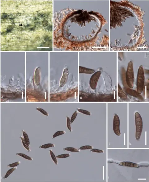
Asexual morph Conidiomata 195–220 µm diam., 210–250 µm high, pycnidial, semi-immersed in the host tissue, scattered, solitary, non-papillate, brown.
Pycnidial walls 22–28 µm thick, composed of several layers of light brown to dark brown cells, arranged in textura angularis to textura prismatica.
Conidiophores absent.
Conidiogenous cells formed on the inner layer of conidiomata, ampulliform, phialidic, smooth, pale brown.
Conidia 30–42 × 5–7 µm (\(\bar{x} \) = 35.8 × 6.2 µm, n = 20), cylindrical, straight to slightly curved, gradually tapering towards the rounded apex, with rounded apex, initially hyaline to pale brown, after maturity golden brown, 7–10-euseptate, thin-walled, with pad-like appendages at the apex.
Saprobic on dead stem of Galium sp. Sexual morph Undetermined. Asexual morph Conidiomata 195–220 µm diam., 210–250 µm high, pycnidial, semi-immersed in the host tissue, scattered, solitary, non-papillate, brown. Pycnidial walls 22–28 µm thick, composed of several layers of light brown to dark brown cells, arranged in textura angularis to textura prismatica. Conidiophores absent. Conidiogenous cells formed on the inner layer of conidiomata, ampulliform, phialidic, smooth, pale brown. Conidia 30–42 × 5–7 µm (\(\bar{x} \) = 35.8 × 6.2 µm, n = 20), cylindrical, straight to slightly curved, gradually tapering towards the rounded apex, with rounded apex, initially hyaline to pale brown, after maturity golden brown, 7–10-euseptate, thin-walled, with pad-like appendages at the apex.
Culture characteristics: Colonies on PDA reaching 8 mm diam. after 2 weeks at 25–30 °C, colonies medium dense, circular, convex, surface slightly rough with edge entire, effuse, velvety to hairy, margin well-defined, colony from above: white to grayish at the margin, white at the centre reverse, white to grayish at the margin, white to greyish at the centre, mycelium white to greyish with tufting, not producing pigments in PDA.
Saprobic on dead stem of Galium sp.
Sexual morph Undetermined.
Asexual morph Conidiomata 195–220 µm diam., 210–250 µm high, pycnidial, semi-immersed in the host tissue, scattered, solitary, non-papillate, brown.
Pycnidial walls 22–28 µm thick, composed of several layers of light brown to dark brown cells, arranged in textura angularis to textura prismatica.
Conidiophores absent.
Conidiogenous cells formed on the inner layer of conidiomata, ampulliform, phialidic, smooth, pale brown.
Conidia 30–42 × 5–7 µm (\(\bar{x} \) = 35.8 × 6.2 µm, n = 20), cylindrical, straight to slightly curved, gradually tapering towards the rounded apex, with rounded apex, initially hyaline to pale brown, after maturity golden brown, 7–10-euseptate, thin-walled, with pad-like appendages at the apex.
Status: impor sumber; pemeriksaan editorial per profil belum selesai. Untuk klaim biologis, gunakan rujukan pada bagian terkait dan periksa konteks sumber aslinya.