Saprobic on Lonicera maackii. Morph seksual tidak ditentukan. Transformasi aseksual Coelomycetous. konidiomata 110–190 μm tinggi, 110–190 μm berlian, pikinidia, bercacah, sepi hingga gregaris, tenggelam, sedikit diangkat, terlihat sebagai titik hitam kecil di permukaan inang, uni-lokulat, globosa hingga subglobosa, glabrous, cokelat gelap hingga hitam, ostioles pusat, apapillate, dengan bukaan seperti pori. Conidiomata walls 5–12 μm lebar, dinding tipis, tebal yang sama, terdiri dari lapisan sel 1–3, sel-sel berwarna coklat gelap, berwarna pseudoparenkimat, yang disusun dalam tekstura angularis.
Profil ini diimpor dari uraian spesies pada sumber berlisensi dan sebagian teks diterjemahkan otomatis. Pemeriksaan editorial per profil belum selesai. Bagian yang belum diterjemahkan atau belum lolos pemeriksaan terjemahan tetap ditampilkan dalam bahasa sumber. Angka dan nama ilmiah dipertahankan.
Saprobic on Lonicera maackii. Sexual morph Undetermined. Asexual morph Coelomycetous. Conidiomata 110–190 μm high, 110–190 μm diam., pycnidial, scattered, solitary to gregarious, immersed, slightly raised, visible as small black dot on host surface, uni-loculate, globose to subglobose, glabrous, dark brown to black, ostioles central, apapillate, with pore-like opening. Conidiomata walls 5–12 μm wide, thin-walled, of equal thickness, composed of 1–3 cell layers, of flattened, dark brown, pseudoparenchymatous cells, arranged in a textura angularis. Conidiophores reduced to conidiogenous cells. Conidiogenous cells (3–)4–6.5(–9) × (2.5–)3.5–6(–8) μm l/w (\(\bar{x} \) = 5 × 5.3 µm, n = 40), holoblastic, phialidic, discrete, determinate, ampulliform to doliiform, unbranched, aseptate, hyaline, smooth, arising from the inner cavity of the conidioma wall. Conidia (16–)18–24(–27.5) × 4–6(–7) μm l/w (\(\bar{x} \) = 22 × 5.6 µm, n = 50), dark brown, subcylindrical to fusiform, or reniform, slightly curved, with acute to rounded apex, and rounded to truncate base, 3–7-septate, not constricted at the septa, thick-walled, smooth-walled, with guttules, having flattened, mucous caps at both ends. Culture characteristics: Colonies on PDA reaching 35–38 mm diam. after 2 weeks at room temperature (20–30 °C). Colony medium dense, slightly irregular in shape, flattened to slightly raised, surface slightly rough with greyish-green hyphal tufts, edge undulate, with entire margin, floccose; from above, slightly radiating, white to cream at the margin, pale brown at the middle, separated from the margin with brown to dark green concentric ring near the edge, greyish green to dark green at the centre; from below, white to cream margin, brown to dark brown at the centre; not producing pigmentation on agar medium.
Sebagian uraian ditampilkan dalam bahasa sumber; terjemahannya belum tersedia atau belum lolos pemeriksaan.
Pengukuran mengikuti objek, spesimen atau kultur, dan metode yang disebutkan sumber. Baca uraian asli untuk konteks lengkap.
02 / Kehidupan
Habitat dan peran ekologis
Saprobic on Lonicera maackii. Sexual morph Undetermined. Asexual morph Coelomycetous. Conidiomata 110–190 μm high, 110–190 μm diam., pycnidial, scattered, solitary to gregarious, immersed, slightly raised, visible as small black dot on host surface, uni-loculate, globose to subglobose, glabrous, dark brown to black, ostioles central, apapillate, with pore-like opening. Conidiomata walls 5–12 μm wide, thin-walled, of equal thickness, composed of 1–3 cell layers, of flattened, dark brown, pseudoparenchymatous cells, arranged in a textura angularis. Conidiophores reduced to conidiogenous cells. Conidiogenous cells (3–)4–6.5(–9) × (2.5–)3.5–6(–8) μm l/w (\(\bar{x} \) = 5 × 5.3 µm, n = 40), holoblastic, phialidic, discrete, determinate, ampulliform to doliiform, unbranched, aseptate, hyaline, smooth, arising from the inner cavity of the conidioma wall. Conidia (16–)18–24(–27.5) × 4–6(–7) μm l/w (\(\bar{x} \) = 22 × 5.6 µm, n = 50), dark brown, subcylindrical to fusiform, or reniform, slightly curved, with acute to rounded apex, and rounded to truncate base, 3–7-septate, not constricted at the septa, thick-walled, smooth-walled, with guttules, having flattened, mucous caps at both ends. Culture characteristics: Colonies on PDA reaching 35–38 mm diam. after 2 weeks at room temperature (20–30 °C).
Sebagian uraian ditampilkan dalam bahasa sumber; terjemahannya belum tersedia atau belum lolos pemeriksaan.
Saprobic on Lonicera maackii. Sexual morph Undetermined. Asexual morph Coelomycetous. Conidiomata 110–190 μm high, 110–190 μm diam., pycnidial, scattered, solitary to gregarious, immersed, slightly raised, visible as small black dot on host surface, uni-loculate, globose to subglobose, glabrous, dark brown to black, ostioles central, apapillate, with pore-like opening. Conidiomata walls 5–12 μm wide, thin-walled, of equal thickness, composed of 1–3 cell layers, of flattened, dark brown, pseudoparenchymatous cells, arranged in a textura angularis. Conidiophores reduced to conidiogenous cells. Conidiogenous cells (3–)4–6.5(–9) × (2.5–)3.5–6(–8) μm l/w (\(\bar{x} \) = 5 × 5.3 µm, n = 40), holoblastic, phialidic, discrete, determinate, ampulliform to doliiform, unbranched, aseptate, hyaline, smooth, arising from the inner cavity of the conidioma wall.
Sebagian uraian ditampilkan dalam bahasa sumber; terjemahannya belum tersedia atau belum lolos pemeriksaan.
Nama diterima mengikuti Catalogue of Life Base2026-09-11; uraian mempertahankan konsep dan nama sumber aslinya.
Preserve original monograph treatment localities, hosts and specimen/substrate scope. Materials examined document specimens, not the full global range.
Impor dan terjemahan otomatis belum diperiksa editorial per profil. Struktur reproduktif yang disebutkan tidak membuktikan seluruh siklus hidup diketahui.
Pengukuran pada uraian mengikuti spesimen, kultur, dan metode sumber; angka tidak diperluas ke semua populasi.
Catatan distribusi dalam narasi mengikuti sumbernya. Dokumentasi spesimen atau foto mengikuti catatan sumber. Bukti wilayah Indonesia dan penilaian konservasi tidak disimpulkan dari gambar.
05 / Dokumentasi
Melihat dengan konteks
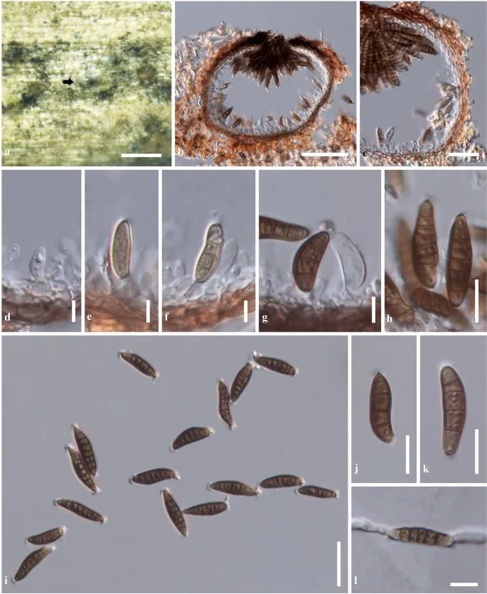
Wojnowiciella kunmingensis (KUN-HKAS 102233, holotype). a Appearance of conidiomata on host. b Section through conidioma. c Section through conidioma wall. d–g Conidiogenous cells. h–k Conidia. l Germinated conidium. Scale bars a = 200 μm, b = 50 μm, c, i = 20 μm, h, j–l = 10 μm, d–g = 5 μm
Jenis
Pelat ilmiah
Kredit
Penulis dan sumber artikel: Phookamsak, Rungtiwa; Hyde, Kevin D.; Jeewon, Rajesh; Bhat, D. Jayarama; Jones, E. B. Gareth; Maharachchikumbura, Sajeewa S. N.; Raspé, Olivier; Karunarathna, Samantha C.; Wanasinghe, Dhanushka N.; Hongsanan, Sinang; Doilom, Mingkwan; Tennakoon, Danushka S.; Machado, Alexandre R.; Firmino, André L.; Ghosh, Aniket; Karunarathna, Anuruddha; Mešić, Armin; Dutta, Arun Kumar; Thongbai, Benjarong; Devadatha, Bandarupalli; Norphanphoun, Chada; Senwanna, Chanokned; Wei, Deping; Pem, Dhandevi; Ackah, Frank Kwekucher; Wang, Gen-Nuo; Jiang, Hong-Bo; Madrid, Hugo; Lee, Hyang Burm; Goonasekara, Ishani D.; Manawasinghe, Ishara S.; Kušan, Ivana; Cano, Josep; Gené, Josepa; Li, Junfu; Das, Kanad; Acharya, Krishnendu; Raj, K. N. Anil; Latha, K. P. Deepna; Chethana, K. W. Thilini; He, Mao-Qiang; Dueñas, Margarita; Jadan, Margita; Martín, María P.; Samarakoon, Milan C.; Dayarathne, Monika C.; Raza, Mubashar; Park, Myung Soo; Telleria, M. Teresa; Chaiwan, Napalai; Matočec, Neven; de Silva, Nimali I.; Pereira, Olinto L.; Singh, Paras Nath; Manimohan, Patinjareveettil; Uniyal, Priyanka; Shang, Qiu-Ju; Bhatt, Rajendra P.; Perera, Rekhani H.; Alvarenga, Renato Lúcio Mendes; Nogal-Prata, Sandra; Singh, Sanjay K.; Vadthanarat, Santhiti; Oh, Seung-Yoon; Huang, Shi-Ke; Rana, Shiwali; Konta, Sirinapa; Paloi, Soumitra; Jayasiri, Subashini C.; Jeon, Sun Jeong; Mehmood, Tahir; Gibertoni, Tatiana Baptista; Nguyen, Thuong T. T.; Singh, Upendra; Thiyagaraja, Vinodhini; Sarma, V. Venkateswara; Dong, Wei; Yu, Xian-Dong; Lu, Yong-Zhong; Lim, Young Woon; Chen, Yun; Tkalčec, Zdenko; Zhang, Zhi-Feng; Luo, Zong-Long; Daranagama, Dinushani A.; Thambugala, Kasun M.; Tibpromma, Saowaluck; Camporesi, Erio; Bulgakov, Timur S.; Dissanayake, Asha J.; Senanayake, Indunil C.; Dai, Dong Qin; Tang, Li-Zhou; Khan, Sehroon; Zhang, Huang; Promputtha, Itthayakorn; Cai, Lei; Chomnunti, Putarak; Zhao, Rui-Lin; Lumyong, Saisamorn; Boonmee, Saranyaphat; Wen, Ting-Chi; Mortimer, Peter E.; Xu, Jianchu
Saprobic on Lonicera maackii.
Sexual morph Undetermined.
Asexual morph Coelomycetous.
Conidiomata 110–190 μm high, 110–190 μm diam., pycnidial, scattered, solitary to gregarious, immersed, slightly raised, visible as small black dot on host surface, uni-loculate, globose to subglobose, glabrous, dark brown to black, ostioles central, apapillate, with pore-like opening.
Conidiomata walls 5–12 μm wide, thin-walled, of equal thickness, composed of 1–3 cell layers, of flattened, dark brown, pseudoparenchymatous cells, arranged in a textura angularis.
Saprobic on Lonicera maackii.
Sexual morph Undetermined.
Asexual morph Coelomycetous.
Conidiomata 110–190 μm high, 110–190 μm diam., pycnidial, scattered, solitary to gregarious, immersed, slightly raised, visible as small black dot on host surface, uni-loculate, globose to subglobose, glabrous, dark brown to black, ostioles central, apapillate, with pore-like opening.
Conidiomata walls 5–12 μm wide, thin-walled, of equal thickness, composed of 1–3 cell layers, of flattened, dark brown, pseudoparenchymatous cells, arranged in a textura angularis.
Conidiophores reduced to conidiogenous cells.
Conidiogenous cells (3–)4–6.5(–9) × (2.5–)3.5–6(–8) μm l/w (\(\bar{x} \) = 5 × 5.3 µm, n = 40), holoblastic, phialidic, discrete, determinate, ampulliform to doliiform, unbranched, aseptate, hyaline, smooth, arising from the inner cavity of the conidioma wall.
Conidia (16–)18–24(–27.5) × 4–6(–7) μm l/w (\(\bar{x} \) = 22 × 5.6 µm, n = 50), dark brown, subcylindrical to fusiform, or reniform, slightly curved, with acute to rounded apex, and rounded to truncate base, 3–7-septate, not constricted at the septa, thick-walled, smooth-walled, with guttules, having flattened, mucous caps at both ends.
Culture characteristics: Colonies on PDA reaching 35–38 mm diam. after 2 weeks at room temperature (20–30 °C).
Colony medium dense, slightly irregular in shape, flattened to slightly raised, surface slightly rough with greyish-green hyphal tufts, edge undulate, with entire margin, floccose; from above, slightly radiating, white to cream at the margin, pale brown at the middle, separated from the margin with brown to dark green concentric ring near the edge, greyish green to dark green at the centre; from below, white to cream margin, brown to dark brown at the centre; not producing pigmentation on agar medium.
Saprobic on Lonicera maackii.
Sexual morph Undetermined.
Asexual morph Coelomycetous.
Conidiomata 110–190 μm high, 110–190 μm diam., pycnidial, scattered, solitary to gregarious, immersed, slightly raised, visible as small black dot on host surface, uni-loculate, globose to subglobose, glabrous, dark brown to black, ostioles central, apapillate, with pore-like opening.
Conidiomata walls 5–12 μm wide, thin-walled, of equal thickness, composed of 1–3 cell layers, of flattened, dark brown, pseudoparenchymatous cells, arranged in a textura angularis.
Conidiophores reduced to conidiogenous cells.
Conidiogenous cells (3–)4–6.5(–9) × (2.5–)3.5–6(–8) μm l/w (\(\bar{x} \) = 5 × 5.3 µm, n = 40), holoblastic, phialidic, discrete, determinate, ampulliform to doliiform, unbranched, aseptate, hyaline, smooth, arising from the inner cavity of the conidioma wall.
Conidia (16–)18–24(–27.5) × 4–6(–7) μm l/w (\(\bar{x} \) = 22 × 5.6 µm, n = 50), dark brown, subcylindrical to fusiform, or reniform, slightly curved, with acute to rounded apex, and rounded to truncate base, 3–7-septate, not constricted at the septa, thick-walled, smooth-walled, with guttules, having flattened, mucous caps at both ends.
Culture characteristics: Colonies on PDA reaching 35–38 mm diam. after 2 weeks at room temperature (20–30 °C).
Saprobic on Lonicera maackii.
Sexual morph Undetermined.
Asexual morph Coelomycetous.
Conidiomata 110–190 μm high, 110–190 μm diam., pycnidial, scattered, solitary to gregarious, immersed, slightly raised, visible as small black dot on host surface, uni-loculate, globose to subglobose, glabrous, dark brown to black, ostioles central, apapillate, with pore-like opening.
Conidiomata walls 5–12 μm wide, thin-walled, of equal thickness, composed of 1–3 cell layers, of flattened, dark brown, pseudoparenchymatous cells, arranged in a textura angularis.
Conidiophores reduced to conidiogenous cells.
Conidiogenous cells (3–)4–6.5(–9) × (2.5–)3.5–6(–8) μm l/w (\(\bar{x} \) = 5 × 5.3 µm, n = 40), holoblastic, phialidic, discrete, determinate, ampulliform to doliiform, unbranched, aseptate, hyaline, smooth, arising from the inner cavity of the conidioma wall.
Status: impor sumber; pemeriksaan editorial per profil belum selesai. Untuk klaim biologis, gunakan rujukan pada bagian terkait dan periksa konteks sumber aslinya.
Saprobic on batang mati dari Galium sp. Morph seksual tidak ditentukan. Aseksual morph konidiomata 195–220 µm diam., 210–250 µm tinggi, pycnidial, semi-merendam dalam jaringan inang, bercahaya, kesepian, non-papilate, cokelat. Pycnidial…