01 / Morfologi

Ciri dan struktur

Saprobic on dead wood culms. Sexual morph: see Mapook et al. (2020). Asexual morph: Pycnidia on natural host, stromatic, developing above the epidermis and becoming raised when mature. Stromata 60–786 × 43–306 µm (x̄ = 320.8 × 172.0 µm, n = 10), with a color ranging from dark brown to black, rough surface with tiny protrusions, some exhibit a certain degree of gloss. and exhibit circular, oval, or irregular shapes, adhering tightly to the surface of decaying wood. On PDA medium, Conidiophores reduced to conidiogenous cells. Conidiogenous cell 8–17 × 2–5 µm (x̄ = 11.5 × 3 µm, n = 10), enteroblastic, indeterminate, discrete, unbranched, clavate or ampulliform, aseptate, hyaline, smooth-walled. Conidia (3.3–) 3.7–3.8 (–4.7) × (2.8–) 3.1–3.2 (3.6) µm (x̄ = 3.8 × 3.2 µm, n = 50), oblong to ellipsoid or rounded to obtuse ends, aseptate, hyaline to pale yellow, smooth-walled. Under dark conditions at 25 °C, the colony reaches a diameter of 15 mm within seven days on PDA medium. The colony is dense, smooth, and irregularly shaped, with a lace-like margin and compact structure. The center is grayish-white, bordered by a white lace-like margin, while the outer edge is grayish-black. The reverse side of the colony is black, producing a dark pigment.

Sebagian uraian ditampilkan dalam bahasa sumber; terjemahannya belum tersedia atau belum lolos pemeriksaan.

Rujukan: [1]

Pengukuran mengikuti objek, spesimen atau kultur, dan metode yang disebutkan sumber. Baca uraian asli untuk konteks lengkap.

02 / Kehidupan

Habitat dan peran ekologis

Saprobic on dead wood culms. Sexual morph: see Mapook et al. (2020). Asexual morph: Pycnidia on natural host, stromatic, developing above the epidermis and becoming raised when mature. Stromata 60–786 × 43–306 µm (x̄ = 320.8 × 172.0 µm, n = 10), with a color ranging from dark brown to black, rough surface with tiny protrusions, some exhibit a certain degree of gloss. and exhibit circular, oval, or irregular shapes, adhering tightly to the surface of decaying wood. On PDA medium, Conidiophores reduced to conidiogenous cells. Conidiogenous cell 8–17 × 2–5 µm (x̄ = 11.5 × 3 µm, n = 10), enteroblastic, indeterminate, discrete, unbranched, clavate or ampulliform, aseptate, hyaline, smooth-walled. Conidia (3.3–) 3.7–3.8 (–4.7) × (2.8–) 3.1–3.2 (3.6) µm (x̄ = 3.8 × 3.2 µm, n = 50), oblong to ellipsoid or rounded to obtuse ends, aseptate, hyaline to pale yellow, smooth-walled. Under dark conditions at 25 °C, the colony reaches a diameter of 15 mm within seven days on PDA medium. The colony is dense, smooth, and irregularly shaped, with a lace-like margin and compact structure. The center is grayish-white, bordered by a white lace-like margin, while the outer edge is grayish-black. The reverse side of the colony is black, producing a dark pigment.

Sebagian uraian ditampilkan dalam bahasa sumber; terjemahannya belum tersedia atau belum lolos pemeriksaan.

Rujukan: [1]

Habitat atau konteks temuan
kultur yang didokumentasikan
Substrat
kayu
Peran atau konteks
Pengurai
Temukan hubungan serupa
03 / Siklus hidup

Reproduksi

Saprobic on dead wood culms. Sexual morph: see Mapook et al. (2020). Asexual morph: Pycnidia on natural host, stromatic, developing above the epidermis and becoming raised when mature. Stromata 60–786 × 43–306 µm (x̄ = 320.8 × 172.0 µm, n = 10), with a color ranging from dark brown to black, rough surface with tiny protrusions, some exhibit a certain degree of gloss. and exhibit circular, oval, or irregular shapes, adhering tightly to the surface of decaying wood. On PDA medium, Conidiophores reduced to conidiogenous cells. Conidiogenous cell 8–17 × 2–5 µm (x̄ = 11.5 × 3 µm, n = 10), enteroblastic, indeterminate, discrete, unbranched, clavate or ampulliform, aseptate, hyaline, smooth-walled.

Sebagian uraian ditampilkan dalam bahasa sumber; terjemahannya belum tersedia atau belum lolos pemeriksaan.

Rujukan: [1]

04 / Batas informasi

Catatan identitas dan cakupan

  • Nama diterima mengikuti Catalogue of Life Base2026-09-11; uraian mempertahankan konsep dan nama sumber aslinya.
  • Preserve original treatment localities, hosts and substrate scope. Materials examined describe documented specimens, not the full global range.
  • Impor dan terjemahan otomatis belum diperiksa editorial per profil. Struktur reproduktif yang disebutkan tidak membuktikan seluruh siklus hidup diketahui.
  • Pengukuran pada uraian mengikuti spesimen, kultur, dan metode sumber; angka tidak diperluas ke semua populasi.

Catatan distribusi dalam narasi mengikuti sumbernya. Dokumentasi spesimen atau foto mengikuti catatan sumber. Bukti wilayah Indonesia dan penilaian konservasi tidak disimpulkan dari gambar.

05 / Dokumentasi

Melihat dengan konteks

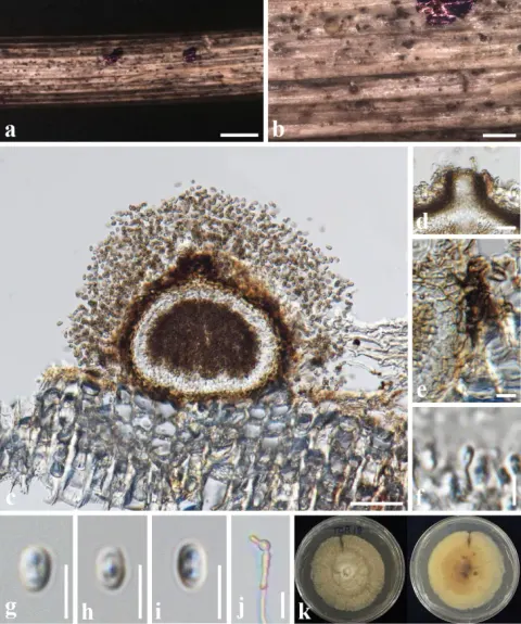
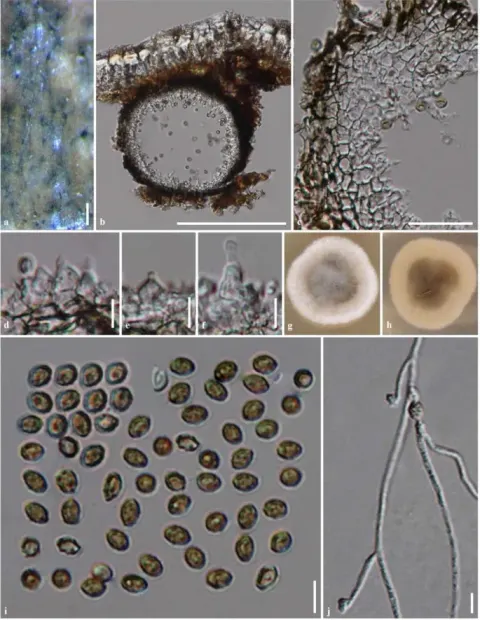
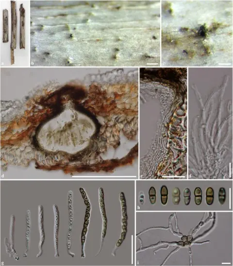
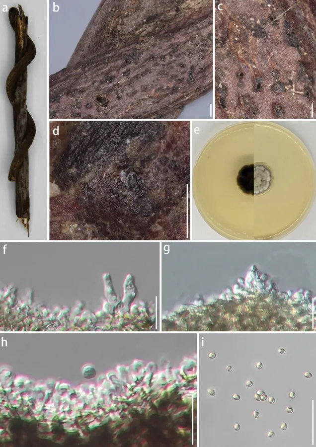
Xenoroussoella triseptata (GUCC 24-0202, new geographical record) a appearance on host surface e cultures on PDA from above and below b, c, d black ascostromata on host surface f–h conidiophores, conidiogenous cells, and conidia (i) conidia. Scale bars: 500 µm (b); 250 µm (c); 100 µm (d); 12.5 µm (f–h); 25 µm (i).

Jenis
Pelat ilmiah
Kredit
Shi-Qi Guo, Chada Norphanphoun, Kevin D. Hyde, Sha-Min Fu, Jing-E Sun, Xing-Chang Wang, Jiao-Jiao Wu, Fatimah Al-Otibi, Yong Wang
Lisensi
CC BY 4.0
Sumber media
Buka entri sumber

Gambar menunjukkan objek menurut identifikasi sumber, bukan verifikasi independen spesimen oleh Fungi. Tidak ada skala baru yang ditambahkan.

06 / Rujukan

Sumber profil

  1. [1] Three novel species and a new record of Pleosporales (Didymellaceae, Roussoellaceae) from China — Xenoroussoella triseptataBagian sumber: PMC11840433_section_33
    Tanggal penghimpunan sumber: 2026-10-12
    Teks sumber: CC BY 4.0 · Guo SQ, Norphanphoun C, Hyde KD, Fu SM, Sun JE, Wang XC, Wu JJ, Al-Otibi F, Wang Y.
  2. [2] Catalogue of Life Base2026-09-11 — Xenoroussoella triseptataBagian sumber: ColDP NameUsage.tsv ID 9ZHC9; accepted species; source 2073
    Tanggal penghimpunan sumber: 2026-10-12
  3. [3] Dokumentasi visual — Xenoroussoella triseptataBagian sumber: Individual media record; identification follows the source, not independent determination
    Tanggal penghimpunan sumber: 2026-10-12
    Media sumber: CC BY 4.0 · Shi-Qi Guo, Chada Norphanphoun, Kevin D. Hyde, Sha-Min Fu, Jing-E Sun, Xing-Chang Wang, Jiao-Jiao Wu, Fatimah Al-Otibi, Yong Wang

Adaptasi teks: CC BY 4.0. Guo SQ, Norphanphoun C, Hyde KD, Fu SM, Sun JE, Wang XC, Wu JJ, Al-Otibi F, Wang Y. — Three novel species and a new record of Pleosporales (Didymellaceae, Roussoellaceae) from China — Xenoroussoella triseptata

Uraian asli dan riwayat sumber

Riwayat atau halaman sumber

Ringkasan sumber

Saprobic on dead wood culms. Sexual morph: see Mapook et al. (2020). Asexual morph: Pycnidia on natural host, stromatic, developing above the epidermis and becoming raised when mature. Stromata 60–786 × 43–306 µm (x̄ = 320.8 × 172.0 µm, n = 10), with a color ranging from dark brown to black, rough surface with tiny protrusions, some exhibit a certain degree of gloss. and exhibit circular, oval, or irregular shapes, adhering tightly to the surface of decaying wood. On PDA medium, Conidiophores reduced to conidiogenous cells.

Revisi sumber

Morfologi sumber

Saprobic on dead wood culms. Sexual morph: see Mapook et al. (2020). Asexual morph: Pycnidia on natural host, stromatic, developing above the epidermis and becoming raised when mature. Stromata 60–786 × 43–306 µm (x̄ = 320.8 × 172.0 µm, n = 10), with a color ranging from dark brown to black, rough surface with tiny protrusions, some exhibit a certain degree of gloss. and exhibit circular, oval, or irregular shapes, adhering tightly to the surface of decaying wood. On PDA medium, Conidiophores reduced to conidiogenous cells. Conidiogenous cell 8–17 × 2–5 µm (x̄ = 11.5 × 3 µm, n = 10), enteroblastic, indeterminate, discrete, unbranched, clavate or ampulliform, aseptate, hyaline, smooth-walled. Conidia (3.3–) 3.7–3.8 (–4.7) × (2.8–) 3.1–3.2 (3.6) µm (x̄ = 3.8 × 3.2 µm, n = 50), oblong to ellipsoid or rounded to obtuse ends, aseptate, hyaline to pale yellow, smooth-walled. Under dark conditions at 25 °C, the colony reaches a diameter of 15 mm within seven days on PDA medium. The colony is dense, smooth, and irregularly shaped, with a lace-like margin and compact structure. The center is grayish-white, bordered by a white lace-like margin, while the outer edge is grayish-black. The reverse side of the colony is black, producing a dark pigment.

Revisi sumber

Ekologi sumber

Saprobic on dead wood culms. Sexual morph: see Mapook et al. (2020). Asexual morph: Pycnidia on natural host, stromatic, developing above the epidermis and becoming raised when mature. Stromata 60–786 × 43–306 µm (x̄ = 320.8 × 172.0 µm, n = 10), with a color ranging from dark brown to black, rough surface with tiny protrusions, some exhibit a certain degree of gloss. and exhibit circular, oval, or irregular shapes, adhering tightly to the surface of decaying wood. On PDA medium, Conidiophores reduced to conidiogenous cells. Conidiogenous cell 8–17 × 2–5 µm (x̄ = 11.5 × 3 µm, n = 10), enteroblastic, indeterminate, discrete, unbranched, clavate or ampulliform, aseptate, hyaline, smooth-walled. Conidia (3.3–) 3.7–3.8 (–4.7) × (2.8–) 3.1–3.2 (3.6) µm (x̄ = 3.8 × 3.2 µm, n = 50), oblong to ellipsoid or rounded to obtuse ends, aseptate, hyaline to pale yellow, smooth-walled. Under dark conditions at 25 °C, the colony reaches a diameter of 15 mm within seven days on PDA medium. The colony is dense, smooth, and irregularly shaped, with a lace-like margin and compact structure. The center is grayish-white, bordered by a white lace-like margin, while the outer edge is grayish-black. The reverse side of the colony is black, producing a dark pigment.

Revisi sumber

Struktur reproduksi sumber

Saprobic on dead wood culms. Sexual morph: see Mapook et al. (2020). Asexual morph: Pycnidia on natural host, stromatic, developing above the epidermis and becoming raised when mature. Stromata 60–786 × 43–306 µm (x̄ = 320.8 × 172.0 µm, n = 10), with a color ranging from dark brown to black, rough surface with tiny protrusions, some exhibit a certain degree of gloss. and exhibit circular, oval, or irregular shapes, adhering tightly to the surface of decaying wood. On PDA medium, Conidiophores reduced to conidiogenous cells. Conidiogenous cell 8–17 × 2–5 µm (x̄ = 11.5 × 3 µm, n = 10), enteroblastic, indeterminate, discrete, unbranched, clavate or ampulliform, aseptate, hyaline, smooth-walled.

Revisi sumber

Rujukan halaman ini

Fungi. Xenoroussoella triseptata. Rilis data fungi-2026-10-12-expansion-2. https://fungi-atlas.mgadib.chatgpt.site/jamur/xenoroussoella-triseptata/.

Status: impor sumber; pemeriksaan editorial per profil belum selesai. Untuk klaim biologis, gunakan rujukan pada bagian terkait dan periksa konteks sumber aslinya.