Ciri dan struktur
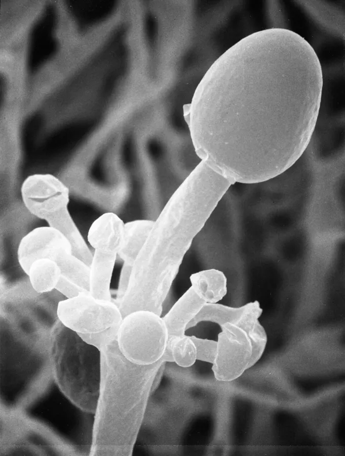
Mycelial growths of A. elegans have a high number of rhizoids branching out of each individual growth. On portions of growth that lack opposite rhizoids, aseptate hyphal growths with clear sporangiophores that are found with extreme variability in length and width. These hyphal structures grow out in whorled structures with growth terminating in the development of sporangiophores. The sporangia are oval to spherical in shape and 17–50 μm in diameter. The walls of the sporangia possess prominent spine-like projections, which is a major identifier of this specific fungus. The coloration of colonies of this fungi is white to cream-colored with an abundance of aerial mycelium. Cultures allowed to develop for a longer period of time (greater than 48 hours) change to become yellowish to buff color with increased aerial mycelium development and tight interweaving of these mycelia. When this fungus is found in a human host the structure is explained to be similar to the genus Mucor, but with unique ribbon-like hyphal structures and irregular branching and thickness.
Habitat dan peran ekologis
Actinomucor elegans was originally described by Schostakowitsch in Siberia in 1898 and reevaluated by Benjamin and Hesseltine in 1957. Commonly found in soil and used for the commercial production of fermented tofu and other products made by soy fermentation. Its major identifying features are its spine-like projections on the sporangiophore and its ribbon-like hyphal structure when found in the tissue of a host. Mycelial growths of A. elegans have a high number of rhizoids branching out of each individual growth. On portions of growth that lack opposite rhizoids, aseptate hyphal growths with clear sporangiophores that are found with extreme variability in length and width. These hyphal structures grow out in whorled structures with growth terminating in the development of sporangiophores. The sporangia are oval to spherical in shape and 17–50 μm in diameter. The walls of the sporangia possess prominent spine-like projections, which is a major identifier of this specific fungus. The coloration of colonies of this fungi is white to cream-colored with an abundance of aerial mycelium. Cultures allowed to develop for a longer period of time (greater than 48 hours) change to become yellowish to buff color with increased aerial mycelium development and tight interweaving of these mycelia.
- Habitat atau konteks temuan
- kultur yang didokumentasikan
- Substrat
- tanah
- Peran atau konteks
- Belum dikategorikan pada rilis ini.
Reproduksi
Mycelial growths of A. elegans have a high number of rhizoids branching out of each individual growth. On portions of growth that lack opposite rhizoids, aseptate hyphal growths with clear sporangiophores that are found with extreme variability in length and width. These hyphal structures grow out in whorled structures with growth terminating in the development of sporangiophores. The sporangia are oval to spherical in shape and 17–50 μm in diameter. The walls of the sporangia possess prominent spine-like projections, which is a major identifier of this specific fungus. The coloration of colonies of this fungi is white to cream-colored with an abundance of aerial mycelium. Cultures allowed to develop for a longer period of time (greater than 48 hours) change to become yellowish to buff color with increased aerial mycelium development and tight interweaving of these mycelia.
Catatan identitas dan cakupan
- Nama diterima mengikuti Catalogue of Life Base2026-09-11; uraian mempertahankan konsep dan nama sumber aslinya.
- Original geographic, specimen and life-cycle scope retained; source import awaits individual editorial review.
- Impor dan terjemahan otomatis belum diperiksa editorial per profil. Struktur reproduktif yang disebutkan tidak membuktikan seluruh siklus hidup diketahui.
- Pengukuran pada uraian mengikuti spesimen, kultur, dan metode sumber; angka tidak diperluas ke semua populasi.
Catatan distribusi dalam narasi mengikuti sumbernya. Dokumentasi spesimen atau foto mengikuti catatan sumber. Bukti wilayah Indonesia dan penilaian konservasi tidak disimpulkan dari gambar.
Melihat dengan konteks
Dokumentasi Actinomucor elegans dari Wikimedia Commons. Actinomucor elegans Identifikasi mengikuti record berkas sumber.
- Jenis
- Fotografi
- Kredit
- zygolife
- Lisensi
- CC BY-SA 2.0
- Sumber media
- Buka entri sumber
Sumber profil
- [1] Actinomucor elegans — Wikipedia (en)
- [2] Catalogue of Life Base2026-09-11 — Actinomucor elegans
- [3] Dokumentasi visual — Actinomucor elegans
Rujukan halaman ini
Fungi. Actinomucor elegans. Rilis data fungi-2026-10-12-expansion-2. https://fungi-atlas.mgadib.chatgpt.site/jamur/actinomucor-elegans/.
Status: impor sumber; pemeriksaan editorial per profil belum selesai. Untuk klaim biologis, gunakan rujukan pada bagian terkait dan periksa konteks sumber aslinya.